|
Habenula
The habenula (diminutive of Latin meaning rein) is a small bilateral neuronal structure in the brain of vertebrates, that has also been called a microstructure since it is no bigger than a pea. The naming as little rein describes its elongated shape in the epithalamus, where it borders the third ventricle, and lies in front of the pineal gland. Although it is a microstructure each habenular nucleus is divided into two distinct regions of nuclei, a medial habenula (MHb), and a lateral habenula (LHb) both having different neuronal populations, inputs, and outputs. The medial habenula can be subdivided into five subnuclei, the lateral habenula into four subnuclei. Research has shown morphological complexity in the MHb and LHb. Different inputs to the MHb are discriminated between the different subnuclei. In the two regions of nuclei there is a difference in gene expression giving different functions to each. The habenula is a conserved structure across vertebrates. In mammals it ... [...More Info...] [...Related Items...] OR: [Wikipedia] [Google] [Baidu] |
Habenular Commissure
The habenular commissure is a nerve tract of commissural fibers that connects the habenular nuclei on both sides of the habenular trigone in the epithalamus. The habenular commissure is part of the habenular trigone (a small depressed triangular area situated in front of the superior colliculus and on the lateral aspect of the posterior part of the taenia thalami). The habenulum trigone also contains the habenular nuclei. Fibers enter the habenular trigone from the stalk of the pineal gland, and the habenular commissure. Most of the habenular trigone's fibers are, however, directed downward and form a bundle, the fasciculus retroflexus, which passes medial to the red nucleus, and, after decussating with the corresponding fasciculus of the opposite side, ends in the interpeduncular nucleus. External links NIF Search - Habenular commissurevia the Neuroscience Information Framework The Neuroscience Information Framework is a repository of global neuroscience web resources, i ... [...More Info...] [...Related Items...] OR: [Wikipedia] [Google] [Baidu] |
Interpeduncular Nucleus
The interpeduncular nucleus (IPN) is an unpaired, ovoid group of neurons at the base of the midbrain tegmentum. In the midbrain it lies below the interpeduncular fossa. As the name suggests, the interpeduncular nucleus lies ''in between'' the cerebral peduncles. Composition The Interpeduncular nucleus is primarily GABAergic and contains at least two neuron clusters of different morphologies. The region is divided into 7 paired and unpaired subnuclei Subdivisions The presence of non-homologous subdivisions of the Interpeduncular nucleus was first noticed by Santiago Ramón y Cajal, Cajal over a hundred years ago. The currently recognized standard subdivision notation was mostly established by Hammill and Lenn in 1984 by combining the work and notations of four groups. Although most of their proposed convention stuck, at some point the proposed "rostral lateral" sub-nucleus was renamed "dorsomedial" and became immortalized in brain atlases. * Apical sub-nucleus (IPA) Unpaired sub-nu ... [...More Info...] [...Related Items...] OR: [Wikipedia] [Google] [Baidu] |
Dorsal Diencephalic Conduction System
The epithalamus (: epithalami) is a posterior (dorsal) segment of the diencephalon. The epithalamus includes the habenular nuclei, the stria medullaris, the anterior and posterior paraventricular nuclei, the posterior commissure, and the pineal gland. Functions The function of the epithalamus is to connect the limbic system to other parts of the brain. The epithalamus also serves as a connecting point for the dorsal diencephalic conduction system, which is responsible for carrying information from the limbic forebrain to limbic midbrain structures. Some functions of its components include the secretion of melatonin from the pineal gland (circadian rhythms), regulation of motor pathways and emotions, and how energy is conserved in the body. A study has shown that the lateral habenula, in the epithalamus, produces spontaneous theta oscillatory activity that was correlated with theta oscillation in the hippocampus. The same study also found that the increase in theta waves in ... [...More Info...] [...Related Items...] OR: [Wikipedia] [Google] [Baidu] |
Epithalamus
The epithalamus (: epithalami) is a posterior (dorsal) segment of the diencephalon. The epithalamus includes the habenular nuclei, the stria medullaris, the anterior and posterior paraventricular nuclei, the posterior commissure, and the pineal gland. Functions The function of the epithalamus is to connect the limbic system to other parts of the brain. The epithalamus also serves as a connecting point for the dorsal diencephalic conduction system, which is responsible for carrying information from the limbic forebrain to limbic midbrain structures. Some functions of its components include the secretion of melatonin from the pineal gland (circadian rhythms), regulation of motor pathways and emotions, and how energy is conserved in the body. A study has shown that the lateral habenula, in the epithalamus, produces spontaneous theta oscillatory activity that was correlated with theta oscillation in the hippocampus. The same study also found that the increase in theta waves in ... [...More Info...] [...Related Items...] OR: [Wikipedia] [Google] [Baidu] |
Fasciculus Retroflexus
The fasciculus retroflexus (FR) also known as the habenulointerpeduncular tract is a bundle of fibers located at the base of the midbrain in vertebrates. Connected to the habenula (Hbn) and the interpeduncular nucleus (IPN), the fasciculus retroflexus is involved in a variety of bodily phenomena, some being sleep retention. and drug addiction. It acts as a channel through which messages are sent between the stria medullaris and the mid- and hindbrain. The fasciculus retroflexus, along with the stria medullaris, the habenula, and the medial forebrain bundle forms a unit for the transfer of neurological impulses. In this unit, the fasciculus retroflexus mediates the transfer of information for processes such as pain, pleasure, and motor control Anatomy The fasciculus retroflexus is the main efferent track of the habenula. The FR is an extremely condensed bundle of fibers which consists of two concentric regions. The first of these is the inner fibers of the FR, beginning at the ... [...More Info...] [...Related Items...] OR: [Wikipedia] [Google] [Baidu] |
Internal Globus Pallidus
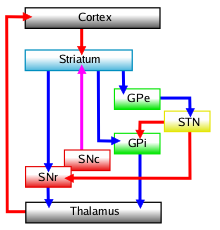
The internal globus pallidus (GPi or medial globus pallidus) is one of the two subcortical nuclei that provides inhibitory output in the basal ganglia, the other being the substantia nigra pars reticulata. Together with the external globus pallidus (GPe), it makes up one of the two segments of the globus pallidus, a structure that can decay with certain neurodegenerative disorders and is a target for medical and neurosurgical therapies. The GPi, along with the substantia nigra pars reticulata, comprise the primary output of the basal ganglia, with its outgoing GABAergic neurons having an inhibitory function in the thalamus, the centromedian complex and the pedunculopontine complex. Anatomy The efferent bundle is constituted first of the ansa and lenticular fasciculus, then crosses the internal capsule within and in parallel to the Edinger's comb system then arrives at the laterosuperior corner of the subthalamic nucleus and constitutes the field H2 of Forel, then H, and sudd ... [...More Info...] [...Related Items...] OR: [Wikipedia] [Google] [Baidu] |
Diagonal Band Of Broca
The diagonal band of Broca interconnects the amygdala and the septal area. It is one of the olfactory structures. It is situated upon the inferior aspect of the brain. It forms the medial margin of the anterior perforated substance. It was described by the French neuroanatomist Paul Broca. Structure It consists of fibers that are said to arise in the parolfactory area, the gyrus subcallosus and the anterior perforated substance, and course backward in the longitudinal striae to the dentate gyrus and the hippocampal region. This is a cholinergic bundle of nerve fibers posterior to the anterior perforated substance. It interconnects the subcallosal gyrus in the septal area with the hippocampus and lateral olfactory area. Nuclei Two structures are often described in this brain regions, namely the nuclei of the vertical and horizontal limbs of the diagonal band of Broca (nvlDBB and nhlDBB, respectively). nvlDBB projects to the hippocampal formation through the fornix and ... [...More Info...] [...Related Items...] OR: [Wikipedia] [Google] [Baidu] |
Hypothalamus
The hypothalamus (: hypothalami; ) is a small part of the vertebrate brain that contains a number of nucleus (neuroanatomy), nuclei with a variety of functions. One of the most important functions is to link the nervous system to the endocrine system via the pituitary gland. The hypothalamus is located below the thalamus and is part of the limbic system. It forms the Basal (anatomy), basal part of the diencephalon. All vertebrate brains contain a hypothalamus. In humans, it is about the size of an Almond#Nut, almond. The hypothalamus has the function of regulating certain metabolic biological process, processes and other activities of the autonomic nervous system. It biosynthesis, synthesizes and secretes certain neurohormones, called releasing hormones or hypothalamic hormones, and these in turn stimulate or inhibit the secretion of hormones from the pituitary gland. The hypothalamus controls thermoregulation, body temperature, hunger (physiology), hunger, important aspects o ... [...More Info...] [...Related Items...] OR: [Wikipedia] [Google] [Baidu] |
Nucleus Accumbens
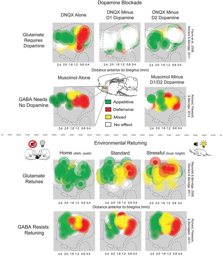
The nucleus accumbens (NAc or NAcc; also known as the accumbens nucleus, or formerly as the ''nucleus accumbens septi'', Latin for ' nucleus adjacent to the septum') is a region in the basal forebrain rostral to the preoptic area of the hypothalamus. The nucleus accumbens and the olfactory tubercle collectively form the ventral striatum. The ventral striatum and dorsal striatum collectively form the striatum, which is the main component of the basal ganglia. The dopaminergic neurons of the mesolimbic pathway project onto the GABAergic medium spiny neurons of the nucleus accumbens and olfactory tubercle. Each cerebral hemisphere has its own nucleus accumbens, which can be divided into two structures: the nucleus accumbens core and the nucleus accumbens shell. These substructures have different morphology and functions. Different NAcc subregions (core vs shell) and neuron subpopulations within each region ( D1-type vs D2-type medium spiny neurons) are responsible fo ... [...More Info...] [...Related Items...] OR: [Wikipedia] [Google] [Baidu] |
Human Brain
The human brain is the central organ (anatomy), organ of the nervous system, and with the spinal cord, comprises the central nervous system. It consists of the cerebrum, the brainstem and the cerebellum. The brain controls most of the activities of the human body, body, processing, integrating, and coordinating the information it receives from the sensory nervous system. The brain integrates sensory information and coordinates instructions sent to the rest of the body. The cerebrum, the largest part of the human brain, consists of two cerebral hemispheres. Each hemisphere has an inner core composed of white matter, and an outer surface – the cerebral cortex – composed of grey matter. The cortex has an outer layer, the neocortex, and an inner allocortex. The neocortex is made up of six Cerebral cortex#Layers of neocortex, neuronal layers, while the allocortex has three or four. Each hemisphere is divided into four lobes of the brain, lobes – the frontal lobe, frontal, pa ... [...More Info...] [...Related Items...] OR: [Wikipedia] [Google] [Baidu] |
Ventral Pallidum
The ventral pallidum (VP) is a structure within the basal ganglia of the brain. It is an output nucleus whose fibres project to thalamic nuclei, such as the ventral anterior nucleus, the ventral lateral nucleus, and the medial dorsal nucleus. The VP is a core component of the reward system which forms part of the limbic loop of the basal ganglia, a pathway involved in the regulation of motivational salience, behavior, and emotions. It is involved in addiction. The VP contains one of the brain's pleasure centers, which mediates the subjective perception of pleasure that results from "consuming" certain rewarding stimuli (e.g., palatable food). Anatomy The ventral pallidum lies within the basal ganglia, a group of subcortical nuclei. Along with the external globus pallidus, it is separated from other basal ganglia nuclei by the anterior commissure. The ventral pallidum contains GABAergic neurons, glutamatergic neurons, and cholinergic neurons that are well conserved ac ... [...More Info...] [...Related Items...] OR: [Wikipedia] [Google] [Baidu] |
Addiction
Addiction is a neuropsychological disorder characterized by a persistent and intense urge to use a drug or engage in a behavior that produces natural reward, despite substantial harm and other negative consequences. Repetitive drug use can alter brain function in synapses similar to natural rewards like food or falling in love in ways that perpetuate craving and weakens self-control for people with pre-existing vulnerabilities. This phenomenon – drugs reshaping brain function – has led to an understanding of addiction as a brain disorder with a complex variety of psychosocial as well as neurobiological factors that are implicated in the development of addiction. While mice given cocaine showed the compulsive and involuntary nature of addiction, for humans this is more complex, related to behavior or personality traits. Classic signs of addiction include compulsive engagement in rewarding stimuli, ''preoccupation'' with substances or behavior, and continued use des ... [...More Info...] [...Related Items...] OR: [Wikipedia] [Google] [Baidu] |


