|
HRTEM
High-resolution transmission electron microscopy is an imaging mode of specialized transmission electron microscopes that allows for direct imaging of the atomic structure of samples. It is a powerful tool to study properties of materials on the atomic scale, such as semiconductors, metals, nanoparticles and sp2-bonded carbon (e.g., graphene, C nanotubes). While this term is often also used to refer to high resolution scanning transmission electron microscopy, mostly in high angle annular dark field mode, this article describes mainly the imaging of an object by recording the two-dimensional spatial wave amplitude distribution in the image plane, in analogy to a "classic" light microscope. For disambiguation, the technique is also often referred to as phase contrast transmission electron microscopy. At present, the highest point resolution realised in phase contrast transmission electron microscopy is around . At these small scales, individual atoms of a crystal and its defects can ... [...More Info...] [...Related Items...] OR: [Wikipedia] [Google] [Baidu] |
Contrast Transfer Function
The contrast transfer function (CTF) mathematically describes how aberrations in a transmission electron microscope (TEM) modify the image of a sample.Spence, John C. H. (1988 2nd ed) ''Experimental high-resolution electron microscopy'' (Oxford U. Press, NY) .Ludwig Reimer (1997 4th ed) ''Transmission electron microscopy: Physics of image formation and microanalysis'' (Springer, BerlinpreviewEarl J. Kirkland (1998) ''Advanced computing in electron microscopy'' (Plenum Press, NY). This contrast transfer function (CTF) sets the resolution of high-resolution transmission electron microscopy (HRTEM), also known as phase contrast TEM. By considering the recorded image as a CTF-degraded true object, describing the CTF allows the true object to be reverse-engineered. This is typically denoted CTF-correction, and is vital to obtain high resolution structures in three-dimensional electron microscopy, especially electron cryo-microscopy. Its equivalent in light-based optics is the optical t ... [...More Info...] [...Related Items...] OR: [Wikipedia] [Google] [Baidu] |
Transmission Electron Microscopy
Transmission electron microscopy (TEM) is a microscopy technique in which a beam of electrons is transmitted through a specimen to form an image. The specimen is most often an ultrathin section less than 100 nm thick or a suspension on a grid. An image is formed from the interaction of the electrons with the sample as the beam is transmitted through the specimen. The image is then magnified and focused onto an imaging device, such as a fluorescent screen, a layer of photographic film, or a sensor such as a scintillator attached to a charge-coupled device. Transmission electron microscopes are capable of imaging at a significantly higher resolution than light microscopes, owing to the smaller de Broglie wavelength of electrons. This enables the instrument to capture fine detail—even as small as a single column of atoms, which is thousands of times smaller than a resolvable object seen in a light microscope. Transmission electron microscopy is a major analytical method ... [...More Info...] [...Related Items...] OR: [Wikipedia] [Google] [Baidu] |
Phase-contrast Imaging
Phase-contrast imaging is a method of imaging that has a range of different applications. It exploits differences in the refractive index of different materials to differentiate between structures under analysis. In conventional light microscopy, phase contrast can be employed to distinguish between structures of similar transparency, and to examine crystals on the basis of their double refraction. This has uses in biological, medical and geological science. In X-ray tomography, the same physical principles can be used to increase image contrast by highlighting small details of differing refractive index within structures that are otherwise uniform. In transmission electron microscopy (TEM), phase contrast enables very high resolution (HR) imaging, making it possible to distinguish features a few Angstrom apart (at this point highest resolution is 40 pm). Light microscopy Phase contrast takes advantage of the fact that different structures have different refractive indices, and eit ... [...More Info...] [...Related Items...] OR: [Wikipedia] [Google] [Baidu] |
Transmission Electron Microscope
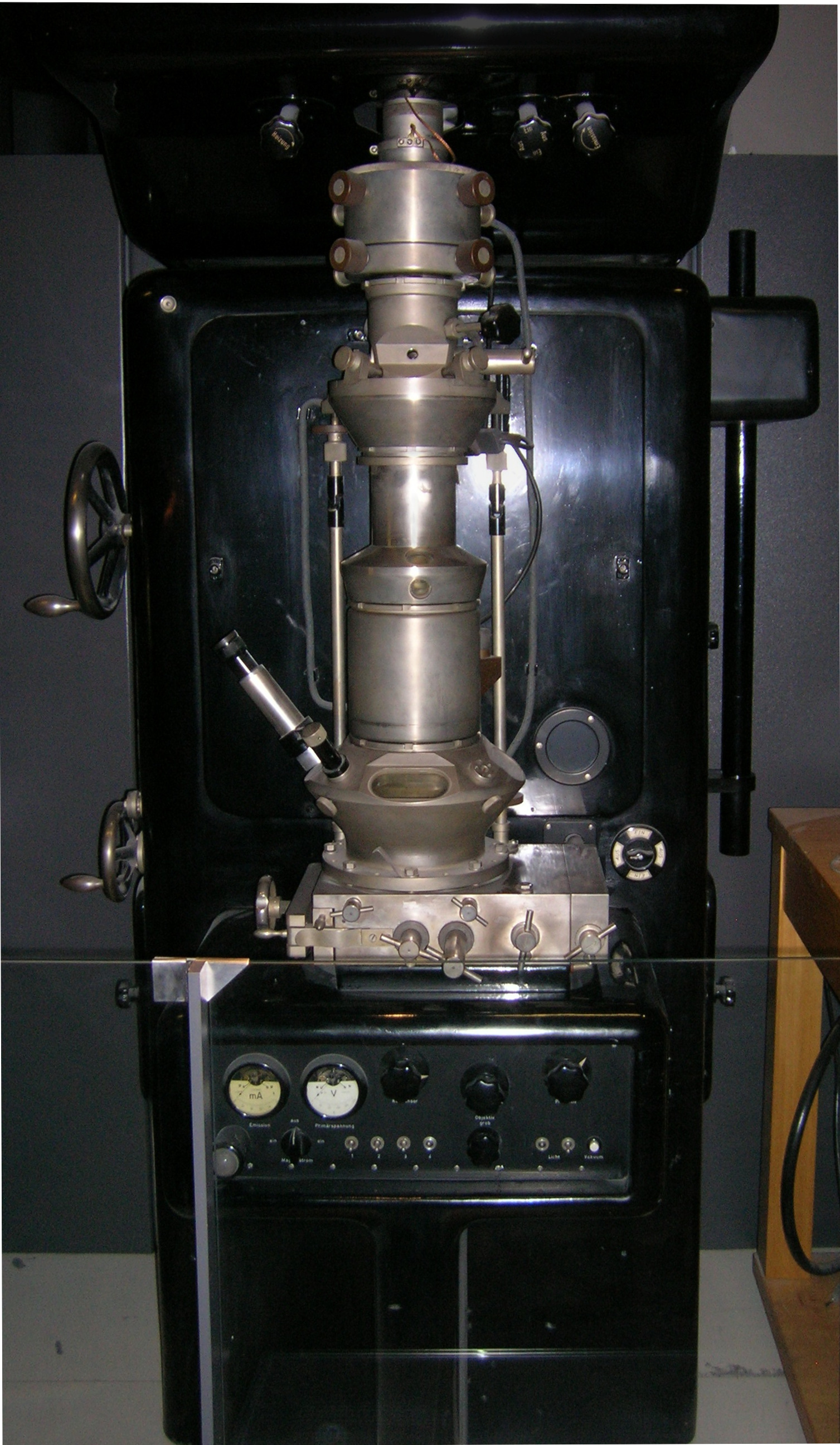
Transmission electron microscopy (TEM) is a microscopy technique in which a beam of electrons is transmitted through a specimen to form an image. The specimen is most often an ultrathin section less than 100 nm thick or a suspension on a grid. An image is formed from the interaction of the electrons with the sample as the beam is transmitted through the specimen. The image is then magnified and focused onto an imaging device, such as a fluorescent screen, a layer of photographic film, or a sensor such as a scintillator attached to a charge-coupled device. Transmission electron microscopes are capable of imaging at a significantly higher resolution than light microscopes, owing to the smaller de Broglie wavelength of electrons. This enables the instrument to capture fine detail—even as small as a single column of atoms, which is thousands of times smaller than a resolvable object seen in a light microscope. Transmission electron microscopy is a major analytical method i ... [...More Info...] [...Related Items...] OR: [Wikipedia] [Google] [Baidu] |
Electron Crystallography
Electron crystallography is a method to determine the arrangement of atoms in solids using a transmission electron microscope (TEM). Comparison with X-ray crystallography It can complement X-ray crystallography for studies of very small crystals ( 1 micrometer) crystals impervious to electrons, which only penetrate short distances. One of the main difficulties in X-ray crystallography is determining phases in the diffraction pattern. Because of the complexity of X-ray lenses, it is difficult to form an image of the crystal being diffracted, and hence phase information is lost. Fortunately, electron microscopes can resolve atomic structure in real space and the crystallographic structure factor phase information can be experimentally determined from an image's Fourier transform. The Fourier transform of an atomic resolution image is similar, but different, to a diffraction pattern—with reciprocal lattice spots reflecting the symmetry and spacing of a crystal. Aaron Klug wa ... [...More Info...] [...Related Items...] OR: [Wikipedia] [Google] [Baidu] |
Electron Microscope
An electron microscope is a microscope that uses a beam of accelerated electrons as a source of illumination. As the wavelength of an electron can be up to 100,000 times shorter than that of visible light photons, electron microscopes have a higher resolving power than light microscopes and can reveal the structure of smaller objects. A scanning transmission electron microscope has achieved better than 50 pm resolution in annular dark-field imaging mode and magnifications of up to about 10,000,000× whereas most light microscopes are limited by diffraction to about 200 nm resolution and useful magnifications below 2000×. Electron microscopes use shaped magnetic fields to form electron optical lens systems that are analogous to the glass lenses of an optical light microscope. Electron microscopes are used to investigate the ultrastructure of a wide range of biological and inorganic specimens including microorganisms, cells, large molecules, biopsy samples, ... [...More Info...] [...Related Items...] OR: [Wikipedia] [Google] [Baidu] |
Scanning Transmission Electron Microscope
A scanning transmission electron microscope (STEM) is a type of transmission electron microscope (TEM). Pronunciation is tɛmor �sti:i:ɛm As with a conventional transmission electron microscope (CTEM), images are formed by electrons passing through a sufficiently thin specimen. However, unlike CTEM, in STEM the electron beam is focused to a fine spot (with the typical spot size 0.05 – 0.2 nm) which is then scanned over the sample in a raster illumination system constructed so that the sample is illuminated at each point with the beam parallel to the optical axis. The rastering of the beam across the sample makes STEM suitable for analytical techniques such as Z-contrast annular dark-field imaging, and spectroscopic mapping by energy dispersive X-ray (EDX) spectroscopy, or electron energy loss spectroscopy (EELS). These signals can be obtained simultaneously, allowing direct correlation of images and spectroscopic data. A typical STEM is a conventional transmission el ... [...More Info...] [...Related Items...] OR: [Wikipedia] [Google] [Baidu] |
Scanning Confocal Electron Microscopy
Scanning confocal electron microscopy (SCEM) is an electron microscopy technique analogous to scanning confocal optical microscopy (SCOM). In this technique, the studied sample is illuminated by a focussed electron beam, as in other scanning microscopy techniques, such as scanning transmission electron microscopy or scanning electron microscopy. However, in SCEM, the collection optics is arranged symmetrically to the illumination optics to gather only the electrons that pass the beam focus. This results in superior depth resolution of the imaging. The technique is relatively new and is being actively developed. History The idea of SCEM logically follows from SCOM and thus is rather old. However, practical design and construction of scanning confocal electron microscope is a complex problem first solved by Nestor J. Zaluzec. His first scanning confocal electron microscope demonstrated the 3D properties of the SCEM, but have not realized the sub-nanometer lateral spatial resoluti ... [...More Info...] [...Related Items...] OR: [Wikipedia] [Google] [Baidu] |
Focal Series Reconstruction
*
{{disambiguation ...
Focal or FOCAL may refer to: *Focal (lexicographical website), an Irish lexicographical website *FOCAL (programming language), a programming language for the PDP-8 and similar machines *Focal (HP-41), for programming HP calculators *FOCAL (spacecraft), a proposed space telescope *FOCAL International, a trade body representing the film archive industry *Focal-JMLab, a French manufacturer of audio equipment *Focal Radio, a radio station based in Stoke-on-Trent, England *Focal neurologic signs See also *Focal point (other) *Focus (other) Focus, or its plural form foci may refer to: Arts * Focus or Focus Festival, former name of the Adelaide Fringe arts festival in South Australia Film *''Focus'', a 1962 TV film starring James Whitmore * ''Focus'' (2001 film), a 2001 film based ... [...More Info...] [...Related Items...] OR: [Wikipedia] [Google] [Baidu] |
Electron Energy Loss Spectroscopy
In electron energy loss spectroscopy (EELS) a material is exposed to a beam of electrons with a known, narrow range of kinetic energies. Some of the electrons will undergo inelastic scattering, which means that they lose energy and have their paths slightly and randomly deflected. The amount of energy loss can be measured via an electron spectrometer and interpreted in terms of what caused the energy loss. Inelastic interactions include phonon excitations, inter- and intra-band transitions, plasmon excitations, inner shell ionizations, and Cherenkov radiation. The inner-shell ionizations are particularly useful for detecting the elemental components of a material. For example, one might find that a larger-than-expected number of electrons comes through the material with 285 eV less energy than they had when they entered the material. This is approximately the amount of energy needed to remove an inner-shell electron from a carbon atom, which can be taken as evidence t ... [...More Info...] [...Related Items...] OR: [Wikipedia] [Google] [Baidu] |
Talbot Effect
The Talbot effect is a diffraction effect first observed in 1836 by Henry Fox Talbot. When a plane wave is incident upon a periodic diffraction grating, the image of the grating is repeated at regular distances away from the grating plane. The regular distance is called the Talbot length, and the repeated images are called self images or Talbot images. Furthermore, at half the Talbot length, a self-image also occurs, but phase-shifted by half a period (the physical meaning of this is that it is laterally shifted by half the width of the grating period). At smaller regular fractions of the Talbot length, sub-images can also be observed. At one quarter of the Talbot length, the self-image is halved in size, and appears with half the period of the grating (thus twice as many images are seen). At one eighth of the Talbot length, the period and size of the images is halved again, and so forth creating a fractal pattern of sub images with ever-decreasing size, often referred to as a Talb ... [...More Info...] [...Related Items...] OR: [Wikipedia] [Google] [Baidu] |


