|
H5N1 Genetic Structure
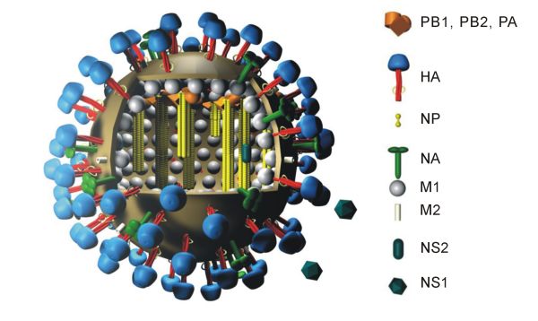
The genetic structure of H5N1, a highly pathogenic avian influenza virus ( influenza A virus subtype H5N1), is characterized by a segmented RNA genome consisting of eight gene segments that encode for various viral proteins essential for replication, host adaptation, and immune evasion. Virus Influenza A virus subtype H5N1 (A/H5N1) is a subtype of the influenza A virus, which causes influenza (flu), predominantly in birds. It is enzootic (maintained in the population) in many bird populations, and also panzootic (affecting animals of many species over a wide area). A/H5N1 virus can also infect mammals (including humans) that have been exposed to infected birds; in these cases, symptoms are frequently severe or fatal. All subtypes of the influenza A virus share the same genetic structure and are potentially able to exchange genetic material by means of reassortment A/H5N1 virus is shed in the saliva, mucus, and feces of infected birds; other infected animals may shed bird ... [...More Info...] [...Related Items...] OR: [Wikipedia] [Google] [Baidu] |
Influenza A Virus Subtype H5N1
Influenza A virus subtype H5N1 (A/H5N1) is a subtype of the influenza A virus, which causes the disease avian influenza (often referred to as "bird flu"). It is enzootic (maintained in the population) in many bird populations, and also panzootic (affecting animals of many species over a wide area). A/H5N1 virus can also infect mammals (including humans) that have been exposed to infected birds; in these cases, symptoms are frequently severe or fatal. A/H5N1 virus is shed in the saliva, mucus, and feces of infected birds; other infected animals may shed bird flu viruses in respiratory secretions and other body fluids (such as milk). The virus can spread rapidly through poultry flocks and among wild birds. An estimated half billion farmed birds have been slaughtered in efforts to contain the virus. Symptoms of A/H5N1 influenza vary according to both the strain of virus underlying the infection and on the species of bird or mammal affected. Classification as either Low Pathogeni ... [...More Info...] [...Related Items...] OR: [Wikipedia] [Google] [Baidu] |
Capsid
A capsid is the protein shell of a virus, enclosing its genetic material. It consists of several oligomeric (repeating) structural subunits made of protein called protomers. The observable 3-dimensional morphological subunits, which may or may not correspond to individual proteins, are called capsomeres. The proteins making up the capsid are called capsid proteins or viral coat proteins (VCP). The virus genomic component inside the capsid, along with occasionally present virus core protein, is called the virus core. The capsid and core together are referred to as a nucleocapsid (cf. also virion). Capsids are broadly classified according to their structure. The majority of the viruses have capsids with either helical or icosahedral structure. Some viruses, such as bacteriophages, have developed more complicated structures due to constraints of elasticity and electrostatics. The icosahedral shape, which has 20 equilateral triangular faces, approximates a sphere, while th ... [...More Info...] [...Related Items...] OR: [Wikipedia] [Google] [Baidu] |
Viral Matrix Protein
Viral matrix proteins are structural proteins linking the viral envelope with the virus core. They play a crucial role in virus assembly, and interact with the RNP complex as well as with the viral membrane. They are found in many enveloped viruses including paramyxoviruses, orthomyxoviruses, herpesviruses, retroviruses, filoviruses and other groups. An example is the M1 protein of the influenza virus, showing affinity to the glycoproteins inserted in the host cell membrane on one side and affinity for the RNP complex molecules on the other side, which allows formation at the membrane of a complex made of the viral ribonucleoprotein at the inner side indirectly connected to the viral glycoproteins protruding from the membrane. This assembly complex will now bud In botany, a bud is an undeveloped or Plant embryogenesis, embryonic Shoot (botany), shoot and normally occurs in the axil of a leaf or at the tip of a Plant stem, stem. Once formed, a bud may remain for some ti ... [...More Info...] [...Related Items...] OR: [Wikipedia] [Google] [Baidu] |
Neuraminidase
Exo-α-sialidase (, sialidase, neuraminidase; systematic name acetylneuraminyl hydrolase) is a glycoside hydrolase that cleaves the glycosidic linkages of neuraminic acids: : Hydrolysis of α-(2→3)-, α-(2→6)-, α-(2→8)- glycosidic linkages of terminal sialic acid residues in oligosaccharides, glycoproteins, glycolipids, colominic acid and synthetic substrates Neuraminidase Enzyme, enzymes are a large family, found in a range of organisms. The best-known neuraminidase is the viral neuraminidase, a drug target for the prevention of the spread of influenza infection. Viral neuraminidase was the first neuraminidase to be identified. It was discovered in 1957 by Alfred Gottschalk (biochemist), Alfred Gottschalk at the WEHI, Walter and Eliza Hall Institute in Melbourne. The viral neuraminidases are frequently used as antigenic determinants found on the surface of the influenza virus. Some variants of the influenza neuraminidase confer more virulence to the virus than others. Ot ... [...More Info...] [...Related Items...] OR: [Wikipedia] [Google] [Baidu] |
Importin α
Importin alpha, or karyopherin alpha refers to a class of adaptor proteins that are involved in the import of proteins into the cell nucleus. They are a sub-family of karyopherin proteins. Importin α is known to bind to the nuclear localization signal A nuclear localization signal ''or'' sequence (NLS) is an amino acid sequence that 'tags' a protein for import into the cell nucleus by nuclear transport. Typically, this signal consists of one or more short sequences of positively charged lysin ... (NLS) sequence of nucleus targeted proteins. After this recognition, importin α links the protein to importin β, which transports the NLS-containing protein across the nuclear envelope to its destination. Because of their complementary functional relationship, importin α and importin β are often referred to as the importin α/β heterodimer, but they are functionally separate, and do not normally exist in conjugation with each other, but only associate for cellular import proces ... [...More Info...] [...Related Items...] OR: [Wikipedia] [Google] [Baidu] |
Nucleoprotein
Nucleoproteins are proteins conjugated with nucleic acids (either DNA or RNA). Typical nucleoproteins include ribosomes, nucleosomes and viral nucleocapsid proteins. Structures Nucleoproteins tend to be positively charged, facilitating interaction with the negatively charged nucleic acid chains. The tertiary structures and biological functions of many nucleoproteins are understood.Graeme K. Hunter G. K. (2000): Vital Forces. The discovery of the molecular basis of life. Academic Press, London 2000, . Important techniques for determining the structures of nucleoproteins include X-ray diffraction, nuclear magnetic resonance and cryo-electron microscopy. Viruses Virus genomes (either DNA or RNA) are extremely tightly packed into the viral capsid. Many viruses are therefore little more than an organised collection of nucleoproteins with their binding sites pointing inwards. Structurally characterised viral nucleoproteins include influenza, rabies, Ebola, Bunyamwera, Schm ... [...More Info...] [...Related Items...] OR: [Wikipedia] [Google] [Baidu] |
Hemagglutinin (influenza)
Influenza hemagglutinin (HA) or haemagglutinin#Notes, [p] (British English) is a homotrimeric glycoprotein found on the surface of influenza viruses and is integral to its infectivity. Hemagglutinin is a class I fusion protein, having multifunctional activity as both an attachment factor and membrane fusion protein. Therefore, HA is responsible for binding influenza viruses to sialic acid on the Cell membrane, surface of target cells, such as cells in the Respiratory tract, upper respiratory tract or erythrocytes, resulting in the Receptor-mediated endocytosis, internalization of the virus. Additionally, HA is responsible for the fusion of the viral envelope with the late endosome, endosomal membrane once exposed to low pH (5.0–5.5). The name "hemagglutinin" comes from the protein's ability to cause red blood cells (i.e., erythrocytes) to clump together (i.e., Agglutination (biology), agglutinate) ''in vitro''. Subtypes Hemagglutinin (HA) in influenza A virus (IAV) has at lea ... [...More Info...] [...Related Items...] OR: [Wikipedia] [Google] [Baidu] |
Interferon
Interferons (IFNs, ) are a group of signaling proteins made and released by host cells in response to the presence of several viruses. In a typical scenario, a virus-infected cell will release interferons causing nearby cells to heighten their anti-viral defenses. IFNs belong to the large class of proteins known as cytokines, molecules used for communication between cells to trigger the protective defenses of the immune system that help eradicate pathogens. Interferons are named for their ability to "interfere" with viral replication by protecting cells from virus infections. However, virus-encoded genetic elements have the ability to antagonize the IFN response, contributing to viral pathogenesis and viral diseases. IFNs also have various other functions: they activate immune cells, such as natural killer cells and macrophages, and they increase host defenses by up-regulating antigen presentation by virtue of increasing the expression of major histocompatibility compl ... [...More Info...] [...Related Items...] OR: [Wikipedia] [Google] [Baidu] |
Cytokine
Cytokines () are a broad and loose category of small proteins (~5–25 kDa) important in cell signaling. Cytokines are produced by a broad range of cells, including immune cells like macrophages, B cell, B lymphocytes, T cell, T lymphocytes and mast cells, as well as Endothelium, endothelial cells, fibroblasts, and various stromal cells; a given cytokine may be produced by more than one type of cell. Due to their size, cytokines cannot cross the lipid bilayer of cells to enter the cytoplasm and therefore typically exert their functions by interacting with specific cytokine receptor, cytokine receptors on the target cell surface. Cytokines are especially important in the immune system; cytokines modulate the balance between humoral immunity, humoral and cell-mediated immunity, cell-based immune responses, and they regulate the maturation, growth, and responsiveness of particular cell populations. Some cytokines enhance or inhibit the action of other cytokines in complex way ... [...More Info...] [...Related Items...] OR: [Wikipedia] [Google] [Baidu] |
Ribosomal Frameshift
Ribosomal frameshifting, also known as translational frameshifting or translational recoding, is a biological phenomenon that occurs during translation that results in the production of multiple, unique proteins from a single mRNA. The process can be programmed by the nucleotide sequence of the mRNA and is sometimes affected by the secondary, 3-dimensional mRNA structure. It has been described mainly in viruses (especially retroviruses), retrotransposons and bacterial insertion elements, and also in some cellular genes. Small molecules, proteins, and nucleic acids have also been found to stimulate levels of frameshifting. In December 2023, it was reported that ''in vitro''-transcribed (IVT) mRNAs in response to BNT162b2 (Pfizer–BioNTech) anti-COVID-19 vaccine caused ribosomal frameshifting. Process overview Proteins are translated by reading tri-nucleotides on the mRNA strand, also known as codons, from one end of the mRNA to the other (from the 5' to the 3' end) starting wi ... [...More Info...] [...Related Items...] OR: [Wikipedia] [Google] [Baidu] |
Mitochondrial Antiviral-signaling Protein
Mitochondrial antiviral-signaling protein (MAVS) is a protein that is essential for antiviral innate immunity. MAVS is located in the outer membrane of the mitochondria, peroxisomes, and mitochondrial-associated endoplasmic reticulum membrane (MAM). Upon viral infection, a group of cytosolic proteins will detect the presence of the virus and bind to MAVS, thereby activating MAVS. The activation of MAVS leads the virally infected cell to secrete cytokines. This induces an immune response which kills the host's virally infected cells, resulting in clearance of the virus. Structure MAVS is also known as IFN-β promoter stimulator I (IPS-1), caspase activation recruitment domain adaptor inducing IFN-β (CARDIF), or virus induced signaling adaptor (VISA). MAVS is encoded by a ''MAVS'' gene. MAVS is a 540 amino acid protein that consists of three components, a N terminal caspase activation recruitment domain (CARD), a proline rich domain, and a transmembrane C terminal domain (TM). ... [...More Info...] [...Related Items...] OR: [Wikipedia] [Google] [Baidu] |
JAK-STAT Signaling Pathway
The JAK-STAT signaling pathway is a chain of interactions between proteins in a cell, and is involved in processes such as immunity, cell division, cell death, and tumor formation. The pathway communicates information from chemical signals outside of a cell to the cell nucleus, resulting in the activation of genes through the process of transcription. There are three key parts of JAK-STAT signalling: Janus kinases (JAKs), signal transducer and activator of transcription proteins (STATs), and receptors (which bind the chemical signals). Disrupted JAK-STAT signalling may lead to a variety of diseases, such as skin conditions, cancers, and disorders affecting the immune system. Structure of JAKs and STATs ''Main articles: JAKs and STATs'' There are four JAK proteins: JAK1, JAK2, JAK3 and TYK2. JAKs contains a FERM domain (approximately 400 residues), an SH2-related domain (approximately 100 residues), a kinase domain (approximately 250 residues) and a pseudokinase domai ... [...More Info...] [...Related Items...] OR: [Wikipedia] [Google] [Baidu] |


