|
Gonadotropin-releasing Hormone Family
The gonadotropin-releasing hormones (GnRH) (gonadoliberin) are a family of peptides that play a pivotal role in reproduction. The main function of GnRH is to act on the pituitary to stimulate the synthesis and secretion of luteinizing and follicle-stimulating hormones, but GnRH also acts on the brain, retina, sympathetic nervous system, gonads, and placenta in certain species. There seems to be at least three forms of GnRH. The second form is expressed in midbrain and seems to be widespread. The third form has been found so far only in fish. GnRH is a C-terminal amidated decapeptide processed from a larger precursor protein. Four of the ten residues are perfectly conserved in all species where GnRH has been sequenced. Subfamilies * Gonadoliberin I Human proteins containing this domain GNRH1 Gonadotropin-releasing hormone (GnRH) is a releasing hormone responsible for the release of follicle-stimulating hormone (FSH) and luteinizing hormone (LH) from the anterior pituitary ... [...More Info...] [...Related Items...] OR: [Wikipedia] [Google] [Baidu] |
Luteinizing
Luteinizing hormone (LH, also known as luteinising hormone, lutropin and sometimes lutrophin) is a hormone produced by gonadotropic cells in the anterior pituitary gland. The production of LH is regulated by gonadotropin-releasing hormone (GnRH) from the hypothalamus. In females, an acute rise of LH known as an LH surge, triggers ovulation and development of the corpus luteum. In males, where LH had also been called interstitial cell–stimulating hormone (ICSH), it stimulates Leydig cell production of testosterone. It acts synergistically with follicle-stimulating hormone ( FSH). Etymology The term luteinizing comes from the Latin "luteus", meaning "yellow". This is in reference to the corpus luteum, which is a mass of cells that forms in an ovary after an ovum (egg) has been discharged. The corpus luteum is so named because it often has a distinctive yellow color. The process of forming the corpus luteum is known as " luteinization", and thus the hormone that triggers this ... [...More Info...] [...Related Items...] OR: [Wikipedia] [Google] [Baidu] |
Follicle-stimulating Hormone
Follicle-stimulating hormone (FSH) is a gonadotropin, a glycoprotein polypeptide hormone. FSH is synthesized and secreted by the gonadotropic cells of the anterior pituitary gland and regulates the development, growth, puberty, pubertal maturation, and reproductive processes of the body. FSH and luteinizing hormone (LH) work together in the reproductive system. Structure FSH is a 35.5 kDa glycoprotein protein dimer, heterodimer, consisting of two polypeptide units, alpha and beta. Its structure is similar to those of luteinizing hormone (LH), thyroid-stimulating hormone (TSH), and human chorionic gonadotropin (hCG). The chorionic gonadotropin alpha, alpha subunits of the glycoproteins LH, FSH, TSH, and hCG are identical and consist of 96 amino acids, while the beta subunits vary. Both subunits are required for biological activity. FSH has a beta subunit of 111 amino acids (FSH β), which confers its specific biologic action, and is responsible for interaction with the follicl ... [...More Info...] [...Related Items...] OR: [Wikipedia] [Google] [Baidu] |
Brain
The brain is an organ (biology), organ that serves as the center of the nervous system in all vertebrate and most invertebrate animals. It consists of nervous tissue and is typically located in the head (cephalization), usually near organs for special senses such as visual perception, vision, hearing, and olfaction. Being the most specialized organ, it is responsible for receiving information from the sensory nervous system, processing that information (thought, cognition, and intelligence) and the coordination of motor control (muscle activity and endocrine system). While invertebrate brains arise from paired segmental ganglia (each of which is only responsible for the respective segmentation (biology), body segment) of the ventral nerve cord, vertebrate brains develop axially from the midline dorsal nerve cord as a brain vesicle, vesicular enlargement at the rostral (anatomical term), rostral end of the neural tube, with centralized control over all body segments. All vertebr ... [...More Info...] [...Related Items...] OR: [Wikipedia] [Google] [Baidu] |
Retina
The retina (; or retinas) is the innermost, photosensitivity, light-sensitive layer of tissue (biology), tissue of the eye of most vertebrates and some Mollusca, molluscs. The optics of the eye create a focus (optics), focused two-dimensional image of the visual world on the retina, which then processes that image within the retina and sends nerve impulses along the optic nerve to the visual cortex to create visual perception. The retina serves a function which is in many ways analogous to that of the photographic film, film or image sensor in a camera. The neural retina consists of several layers of neurons interconnected by Chemical synapse, synapses and is supported by an outer layer of pigmented epithelial cells. The primary light-sensing cells in the retina are the photoreceptor cells, which are of two types: rod cell, rods and cone cell, cones. Rods function mainly in dim light and provide monochromatic vision. Cones function in well-lit conditions and are responsible fo ... [...More Info...] [...Related Items...] OR: [Wikipedia] [Google] [Baidu] |
Sympathetic Nervous System
The sympathetic nervous system (SNS or SANS, sympathetic autonomic nervous system, to differentiate it from the somatic nervous system) is one of the three divisions of the autonomic nervous system, the others being the parasympathetic nervous system and the enteric nervous system. The enteric nervous system is sometimes considered part of the autonomic nervous system, and sometimes considered an independent system. The autonomic nervous system functions to regulate the body's unconscious actions. The sympathetic nervous system's primary process is to stimulate the body's fight or flight response. It is, however, constantly active at a basic level to maintain homeostasis. The sympathetic nervous system is described as being antagonistic to the parasympathetic nervous system. The latter stimulates the body to "feed and breed" and to (then) "rest-and-digest". The SNS has a major role in various physiological processes such as blood glucose levels, body temperature, cardiac output, ... [...More Info...] [...Related Items...] OR: [Wikipedia] [Google] [Baidu] |
Gonads
A gonad, sex gland, or reproductive gland is a mixed gland and sex organ that produces the gametes and sex hormones of an organism. Female reproductive cells are egg cells, and male reproductive cells are sperm. The male gonad, the testicle, produces sperm in the form of Spermatozoon, spermatozoa. The female gonad, the ovary, produces egg cells. Both of these gametes are haploid cells. Some hermaphroditic animals (and some humanssee Ovotesticular syndrome) have a type of gonad called an ovotestis. Evolution It is hard to find a common origin for gonads, but gonads most likely evolved independently several times. Regulation The gonads are controlled by luteinizing hormone (LH) and follicle-stimulating hormone (FSH), produced and secreted by gonadotropic cell, gonadotropes or gonadotrophins in the anterior pituitary gland. This secretion is regulated by gonadotropin-releasing hormone (GnRH) produced in the hypothalamus. Development The gonads develop from three sources ... [...More Info...] [...Related Items...] OR: [Wikipedia] [Google] [Baidu] |
Placenta
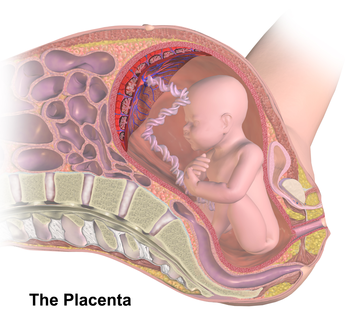
The placenta (: placentas or placentae) is a temporary embryonic and later fetal organ that begins developing from the blastocyst shortly after implantation. It plays critical roles in facilitating nutrient, gas, and waste exchange between the physically separate maternal and fetal circulations, and is an important endocrine organ, producing hormones that regulate both maternal and fetal physiology during pregnancy. The placenta connects to the fetus via the umbilical cord, and on the opposite aspect to the maternal uterus in a species-dependent manner. In humans, a thin layer of maternal decidual ( endometrial) tissue comes away with the placenta when it is expelled from the uterus following birth (sometimes incorrectly referred to as the 'maternal part' of the placenta). Placentas are a defining characteristic of placental mammals, but are also found in marsupials and some non-mammals with varying levels of development. Mammalian placentas probably first evolved abou ... [...More Info...] [...Related Items...] OR: [Wikipedia] [Google] [Baidu] |
Midbrain
The midbrain or mesencephalon is the uppermost portion of the brainstem connecting the diencephalon and cerebrum with the pons. It consists of the cerebral peduncles, tegmentum, and tectum. It is functionally associated with vision, hearing, motor control, sleep and wakefulness, arousal (alertness), and temperature regulation.Breedlove, Watson, & Rosenzweig. Biological Psychology, 6th Edition, 2010, pp. 45-46 The name ''mesencephalon'' comes from the Greek ''mesos'', "middle", and ''enkephalos'', "brain". Structure The midbrain is the shortest segment of the brainstem, measuring less than 2cm in length. It is situated mostly in the posterior cranial fossa, with its superior part extending above the tentorial notch. The principal regions of the midbrain are the tectum, the cerebral aqueduct, tegmentum, and the cerebral peduncles. Rostral and caudal, Rostrally the midbrain adjoins the diencephalon (thalamus, hypothalamus, etc.), while Rostral and caudal, cau ... [...More Info...] [...Related Items...] OR: [Wikipedia] [Google] [Baidu] |
Gonadoliberin I
Gonadotropin-releasing hormone (GnRH) is a releasing hormone responsible for the release of follicle-stimulating hormone (FSH) and luteinizing hormone (LH) from the anterior pituitary. GnRH is a tropic peptide hormone synthesized and released from GnRH neurons within the hypothalamus. GnRH is inhibited by testosterone. The peptide belongs to gonadotropin-releasing hormone family. It constitutes the initial step in the hypothalamic–pituitary–gonadal axis. Structure The identity of GnRH was clarified by the 1977 Nobel Laureates Roger Guillemin and Andrew V. Schally: pyroGlu-His-Trp-Ser-Tyr-Gly-Leu-Arg-Pro-Gly-NH2 As is standard for peptide representation, the sequence is given from amino terminus to carboxyl terminus; also standard is omission of the designation of chirality, with assumption that all amino acids are in their L- form. The abbreviations are the standard abbreviations for the corresponding proteinogenic amino acids, except for ''pyroGlu'', which refers to p ... [...More Info...] [...Related Items...] OR: [Wikipedia] [Google] [Baidu] |
Gonadotropin-releasing Hormone
Gonadotropin-releasing hormone (GnRH) is a releasing hormone responsible for the release of follicle-stimulating hormone (FSH) and luteinizing hormone (LH) from the anterior pituitary. GnRH is a tropic peptide hormone synthesized and released from GnRH neurons within the hypothalamus. GnRH is inhibited by testosterone. The peptide belongs to gonadotropin-releasing hormone family. It constitutes the initial step in the hypothalamic–pituitary–gonadal axis. Structure The identity of GnRH was clarified by the 1977 Nobel Laureates Roger Guillemin and Andrew V. Schally: pyroGlu-His-Trp-Ser-Tyr-Gly-Leu-Arg-Pro-Gly-NH2 As is standard for peptide representation, the sequence is given from amino terminus to carboxyl terminus; also standard is omission of the designation of chirality, with assumption that all amino acids are in their L- form. The abbreviations are the standard abbreviations for the corresponding proteinogenic amino acids, except for ''pyroGlu'', whic ... [...More Info...] [...Related Items...] OR: [Wikipedia] [Google] [Baidu] |
GNRH2
Progonadoliberin-2 is a protein that in humans is encoded by the ''GNRH2'' gene. The protein encoded by this gene is a preproprotein that is cleaved to form a secreted 10 aa peptide hormone, QHWSHGWYPG. The secreted decapeptide regulates reproduction in females by stimulating the secretion of both luteinizing- and follicle-stimulating hormones. Three transcript variants that encode unique proproteins but the same peptide hormone have been found for this gene. The peptide belongs to gonadotropin-releasing hormone family The gonadotropin-releasing hormones (GnRH) (gonadoliberin) are a family of peptides that play a pivotal role in reproduction. The main function of GnRH is to act on the pituitary to stimulate the synthesis and secretion of luteinizing and folli .... References Further reading * * * * * * * * * * * * * * * Precursor proteins Decapeptides {{gene-20-stub ... [...More Info...] [...Related Items...] OR: [Wikipedia] [Google] [Baidu] |
Protein Domains
In molecular biology, a protein domain is a region of a protein's polypeptide chain that is self-stabilizing and that folds independently from the rest. Each domain forms a compact folded three-dimensional structure. Many proteins consist of several domains, and a domain may appear in a variety of different proteins. Molecular evolution uses domains as building blocks and these may be recombined in different arrangements to create proteins with different functions. In general, domains vary in length from between about 50 amino acids up to 250 amino acids in length. The shortest domains, such as zinc fingers, are stabilized by metal ions or disulfide bridges. Domains often form functional units, such as the calcium-binding EF hand domain of calmodulin. Because they are independently stable, domains can be "swapped" by genetic engineering between one protein and another to make chimeric proteins. Background The concept of the domain was first proposed in 1973 by Wetlaufer after ... [...More Info...] [...Related Items...] OR: [Wikipedia] [Google] [Baidu] |


