|
Gnarled Enamel
Gnarled enamel is a description of enamel seen in histologic sections of a tooth underneath a cusp. This is optical appearance of enamel. The enamel appears different and very complex under the cusp, but this is not due to a different arrangement of dental tissues. Instead, the enamel still has the same arrangement of enamel rod An enamel prism, or enamel rod, is the basic unit of tooth enamel. Measuring 3-6 μm in diameter, enamel prism are tightly packed hydroxyapatite crystals structures. The hydroxyapatite crystals are hexagonal in shape, providing rigidity to the pri ...s. The strange appearance results from the lines of enamel rods directed vertically under a cusp and from their orientation in a small circumference. Gnarled enamel can sometimes be a problem for dentists; although experienced dentists have no problem identifying gnarled enamel and making the correct preparations, some inexperienced dentists face problems when performing a standard filling or root canal. The ... [...More Info...] [...Related Items...] OR: [Wikipedia] [Google] [Baidu] |
Tooth Enamel
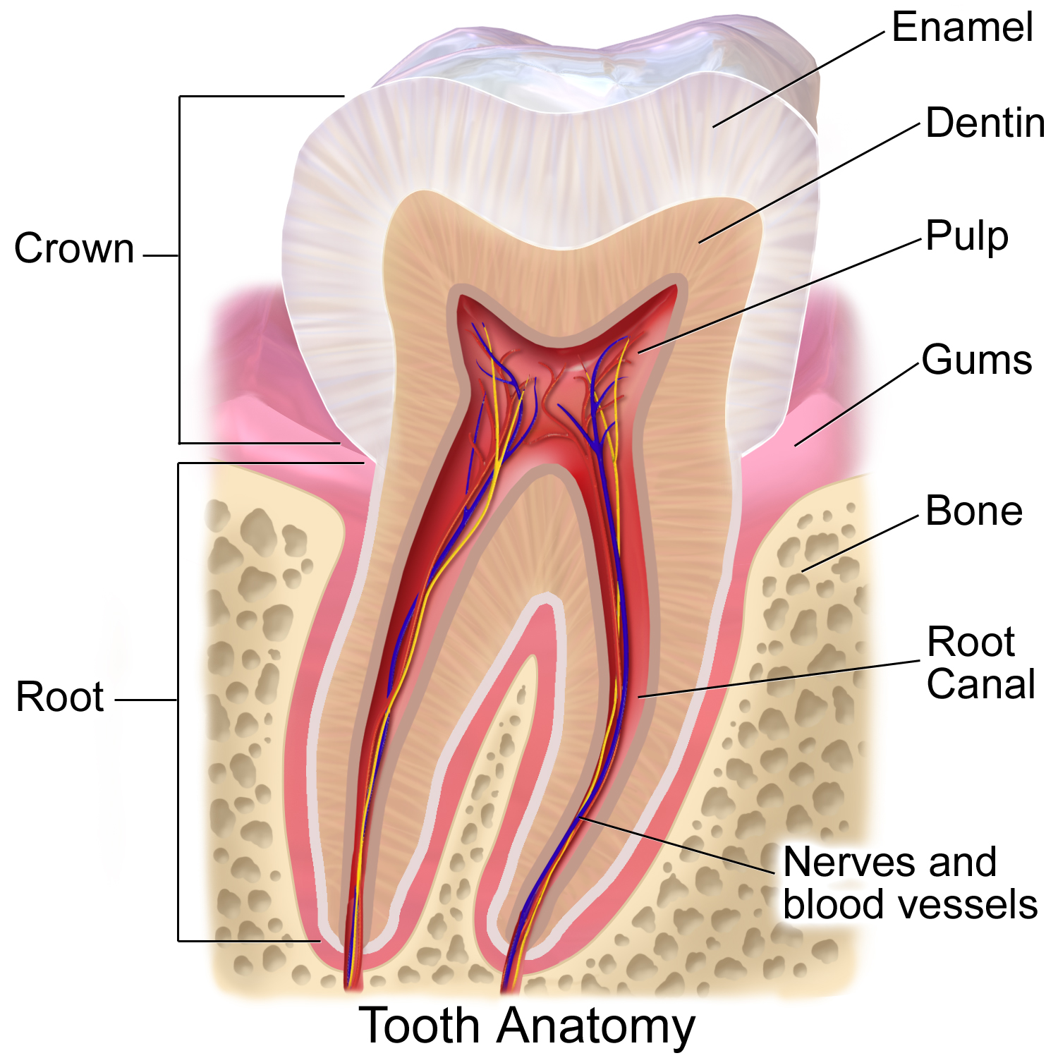
Tooth enamel is one of the four major tissues that make up the tooth in humans and many other animals, including some species of fish. It makes up the normally visible part of the tooth, covering the crown. The other major tissues are dentin, cementum, and dental pulp. It is a very hard, white to off-white, highly mineralised substance that acts as a barrier to protect the tooth but can become susceptible to degradation, especially by acids from food and drink. Calcium hardens the tooth enamel. In rare circumstances enamel fails to form, leaving the underlying dentin exposed on the surface. Features Enamel is the hardest substance in the human body and contains the highest percentage of minerals (at 96%),Ross ''et al.'', p. 485 with water and organic material composing the rest.Ten Cate's Oral Histology, Nancy, Elsevier, pp. 70–94 The primary mineral is hydroxyapatite, which is a crystalline calcium phosphate. Enamel is formed on the tooth while the tooth develops wit ... [...More Info...] [...Related Items...] OR: [Wikipedia] [Google] [Baidu] |
Histology
Histology, also known as microscopic anatomy or microanatomy, is the branch of biology which studies the microscopic anatomy of biological tissues. Histology is the microscopic counterpart to gross anatomy, which looks at larger structures visible without a microscope. Although one may divide microscopic anatomy into ''organology'', the study of organs, ''histology'', the study of tissues, and '' cytology'', the study of cells, modern usage places all of these topics under the field of histology. In medicine, histopathology is the branch of histology that includes the microscopic identification and study of diseased tissue. In the field of paleontology, the term paleohistology refers to the histology of fossil organisms. Biological tissues Animal tissue classification There are four basic types of animal tissues: muscle tissue, nervous tissue, connective tissue, and epithelial tissue. All animal tissues are considered to be subtypes of these four principal tissue type ... [...More Info...] [...Related Items...] OR: [Wikipedia] [Google] [Baidu] |
Tooth
A tooth ( : teeth) is a hard, calcified structure found in the jaws (or mouths) of many vertebrates and used to break down food. Some animals, particularly carnivores and omnivores, also use teeth to help with capturing or wounding prey, tearing food, for defensive purposes, to intimidate other animals often including their own, or to carry prey or their young. The roots of teeth are covered by gums. Teeth are not made of bone, but rather of multiple tissues of varying density and hardness that originate from the embryonic germ layer, the ectoderm. The general structure of teeth is similar across the vertebrates, although there is considerable variation in their form and position. The teeth of mammals have deep roots, and this pattern is also found in some fish, and in crocodilians. In most teleost fish, however, the teeth are attached to the outer surface of the bone, while in lizards they are attached to the inner surface of the jaw by one side. In cartilaginous fi ... [...More Info...] [...Related Items...] OR: [Wikipedia] [Google] [Baidu] |
Cusp (dentistry)
A cusp is a pointed, projecting, or elevated feature. In animals, it is usually used to refer to raised points on the crowns of teeth. The concept is also used with regard to the leaflets of the four heart valves. The mitral valve, which has two cusps, is also known as the bicuspid valve, and the tricuspid valve has three cusps. In humans A cusp is an occlusal or incisal eminence on a tooth. Canine teeth, otherwise known as cuspids, each possess a single cusp, while premolars, otherwise known as bicuspids, possess two each. Molars normally possess either four or five cusps. In certain populations the maxillary molars, especially first molars, will possess a fifth cusp situated on the mesiolingual cusp known as the Cusp of Carabelli. Buccal Cusp- One other variation of the upper first premolar is the 'Uto-Aztecan' upper premolar. It is a bulge on the buccal cusp that is only found in Native American Indians, with highest frequencies of occurrence in Arizona. The name is n ... [...More Info...] [...Related Items...] OR: [Wikipedia] [Google] [Baidu] |
Enamel Rod
An enamel prism, or enamel rod, is the basic unit of tooth enamel. Measuring 3-6 μm in diameter, enamel prism are tightly packed hydroxyapatite crystals structures. The hydroxyapatite crystals are hexagonal in shape, providing rigidity to the prism and strengthening the enamel. In cross-section, it is best compared to a complex “keyhole” or a “fish-like” shape. The head, which is called the prism core, is oriented toward the tooth’s crown; The tail, which is called the prism sheath, is oriented toward the tooth cervical margin /sup>. The prism core has tightly packed hydroxyapatite crystals. On the other hand, the prism sheath has its crystals less tightly packed and has more space for organic components. These prism structures can usually be visualised within ground sections and/or with the use of a scanning electron microscope on enamel that has been acid etched ">/sup>. The number of enamel prisms range approximately from 5 million to 12 million in the number b ... [...More Info...] [...Related Items...] OR: [Wikipedia] [Google] [Baidu] |


