|
Gingivoplasty
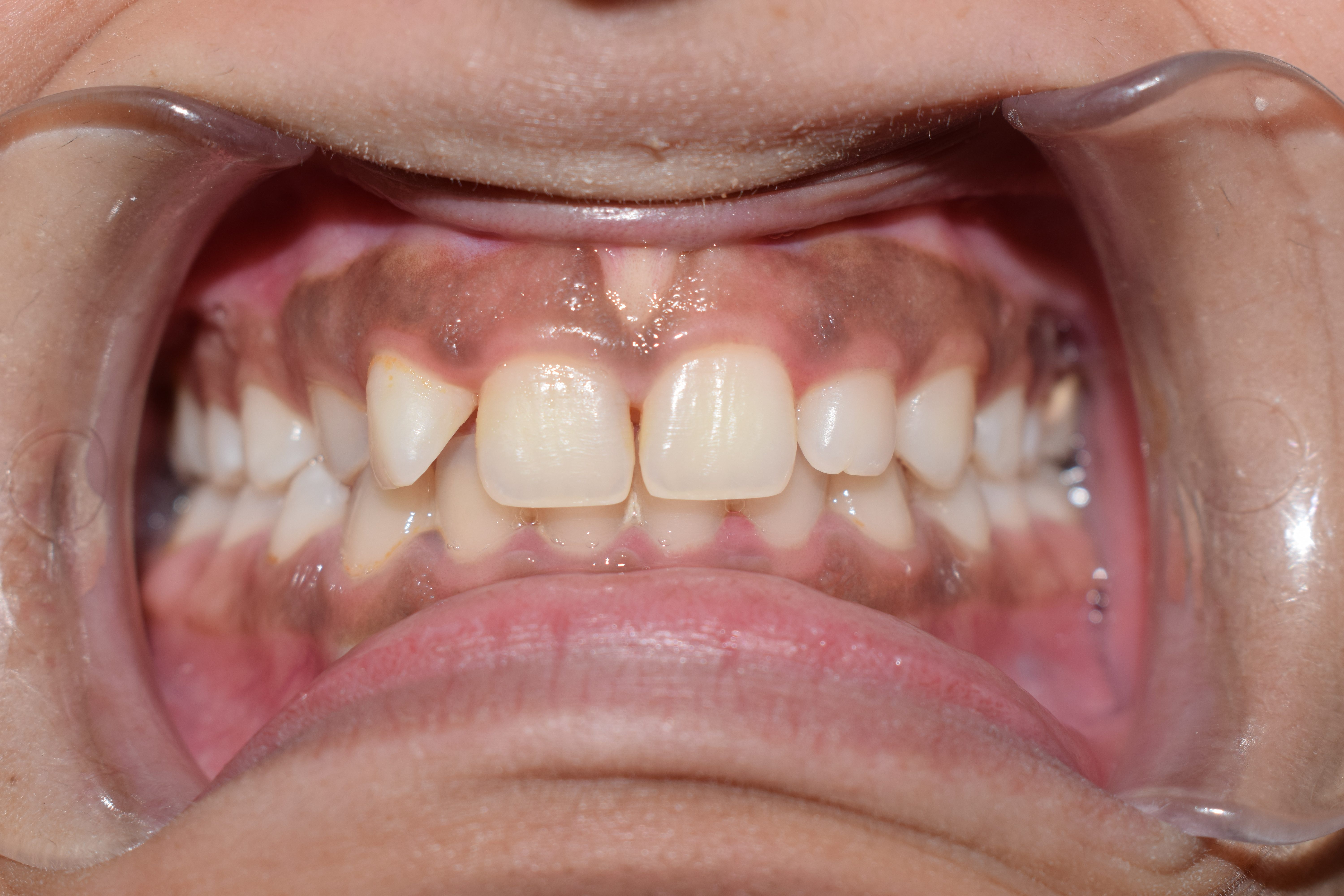
Gingivoplasty is the process by which the gingiva are reshaped to correct deformities. Gingivoplasty is similar to gingivectomy but with a different objective. This is a procedure performed to eliminate periodontal pockets along with the reshaping as part of the technique. This procedure is followed to create physiological gingival contours with the sole purpose of recontouring the gingiva in the absence of the pockets. Gingival and periodontal disease often produces deformities in the gingiva that are conducive to the accumulation of plaque and food debris, which prolong and aggregate the disease process. Such deformities include the following:- # Gingival clefts and craters # Crater- like interdental papilla caused by acute necrotizing ulcerative gingivitis # Gingival enlargements Gingivoplasty is accomplished with a periodontal knife, a scalpel, rotary coarse diamond stones, electrodes or laser A laser is a device that emits light through a process of optical amp ... [...More Info...] [...Related Items...] OR: [Wikipedia] [Google] [Baidu] |
Gingiva
The gums or gingiva (: gingivae) consist of the mucosal tissue that lies over the mandible and maxilla inside the mouth. Gum health and disease can have an effect on general health. Structure The gums are part of the soft tissue lining of the mouth. They surround the teeth and provide a seal around them. Unlike the soft tissue linings of the lips and cheeks, most of the gums are tightly bound to the underlying bone which helps resist the friction of food passing over them. Thus when healthy, it presents an effective barrier to the barrage of periodontal insults to deeper tissue. Healthy gums are usually coral pink in light skinned people, and may be naturally darker with melanin pigmentation. Changes in color, particularly increased redness, together with swelling and an increased tendency to bleed, suggest an inflammation that is possibly due to the accumulation of bacterial plaque. Overall, the clinical appearance of the tissue reflects the underlying histology, both in hea ... [...More Info...] [...Related Items...] OR: [Wikipedia] [Google] [Baidu] |
Gingivectomy
Gingivectomy is a dental procedure in which a dentist or oral surgeon cuts away part of the gums in the mouth (the ''gingiva''). It is the oldest surgical approach in periodontal therapy and is usually done for improvement of aesthetics or prognosis of teeth. By removing the pocket wall, gingivectomy provides visibility and accessibility for complete calculus removal and thorough smoothing of the roots, creating a favourable environment for gingival healing and restoration of a physiologic gingival contour. The procedure may also be carried out so that access to sub-gingival caries or crown margins is allowed. A common aesthetic reason for gingivectomy is a gummy smile due to gingival overgrowth. Indications Elimination of suprabony fibrous and firm pockets Gingivectomy is the primary treatment method available in reducing the pocket depths of patients with periodontitis and suprabony pockets. In a retrospective comparison between different treatment approach to periodont ... [...More Info...] [...Related Items...] OR: [Wikipedia] [Google] [Baidu] |
Periodontal Pocket
In dental anatomy, the gingival and periodontal pockets (also informally referred to as gum pockets) are dental terms indicating the presence of an abnormal depth of the gingival sulcus near the point at which the gingival (gum) tissue contacts the tooth. Tooth gingival interface The interface between a tooth and the surrounding gingival tissue is a dynamic structure. The gingival tissue forms a crevice surrounding the tooth, similar to a miniature, fluid-filled moat, wherein food debris, endogenous and exogenous cells, and chemicals float. The depth of this crevice, known as a sulcus, is in a constant state of flux due to microbial invasion and subsequent immune response. Located at the depth of the sulcus is the epithelial attachment, consisting of approximately 1 mm of junctional epithelium and another 1 mm of gingival fiber attachment, comprising the 2 mm of biologic width naturally found in the oral cavity. The sulcus is literally the area of separation ... [...More Info...] [...Related Items...] OR: [Wikipedia] [Google] [Baidu] |
Gingival Disease
Gingival disease is a term used to group the diseases that affect the gingiva(gums). The most common gingival disease is gingivitis, the earliest stage of gingival-related diseases. Gingival disease encompasses all the conditions surrounding the gums; this includes plaque-induced gingivitis, non-dental biofilm plaque-induced gingivitis, and periodontal diseases. Types Gingival health that is not well cared for is usually connected with inflammation of the gums. This leads to gingivitis, which is linked to two categories: * Dental plaque biofilm-induced gingivitis * Non-dental-plaque-induced gingival disease Dental plaque biofilm-induced gingivitis is often called "localized inflammation initiated by microbial biofilm accumulation on teeth,". /sup>Non-dental-plaque-induced gingival diseases are the most uncommon bacterial infections of the gingiva. Here is each category classification based on the Classification of Periodontal Diseases and Conditions in 2017: Gingival Classifica ... [...More Info...] [...Related Items...] OR: [Wikipedia] [Google] [Baidu] |
Periodontal Disease
Periodontal disease, also known as gum disease, is a set of inflammatory conditions affecting the tissues surrounding the teeth. In its early stage, called gingivitis, the gums become swollen and red and may bleed. It is considered the main cause of tooth loss for adults worldwide. In its more serious form, called periodontitis, the gums can pull away from the tooth, bone can be lost, and the teeth may loosen or fall out. Halitosis (bad breath) may also occur. Periodontal disease typically arises from the development of plaque biofilm, which harbors harmful bacteria such as ''Porphyromonas gingivalis'' and ''Treponema denticola''. These bacteria infect the gum tissue surrounding the teeth, leading to inflammation and, if left untreated, progressive damage to the teeth and gum tissue. Recent meta-analysis have shown that the composition of the oral microbiota and its response to periodontal disease differ between men and women. These differences are particularly notable in t ... [...More Info...] [...Related Items...] OR: [Wikipedia] [Google] [Baidu] |
Dental Plaque
Dental plaque is a biofilm of microorganisms (mostly bacteria, but also fungi) that grows on surfaces within the mouth. It is a sticky colorless deposit at first, but when it forms Calculus (dental), tartar, it is often brown or pale yellow. It is commonly found between the teeth, on the front of teeth, behind teeth, on chewing surfaces, along the gums, gumline (supragingival), or below the gumline cervical margins (subgingival). Dental plaque is also known as microbial plaque, oral biofilm, dental biofilm, dental plaque biofilm or bacterial plaque biofilm. Bacterial plaque is one of the major causes for dental decay and gum disease. It has been observed that differences in the composition of dental plaque microbiota exist between men and women, particularly in the presence of periodontal disease, periodontitis. Progression and build-up of dental plaque can give rise to tooth decay – the localised destruction of the tissues of the tooth by acid produced from the bacterial degrad ... [...More Info...] [...Related Items...] OR: [Wikipedia] [Google] [Baidu] |
Interdental Papilla
The interdental papilla, also known as the interdental gingiva, is the part of the gums (gingiva) that exists coronal to the free gingival margin on the mesial and distal surfaces of the teeth. The interdental papillae fill in the area between the teeth apical to their contact areas to prevent food impaction; they assume a conical shape for the anterior teeth and a blunted shape buccolingually for the posterior teeth. A missing papilla is often visible as a small triangular gap between adjacent teeth. Dentists will sometimes refer to this gap as a ' black triangle'. It can sometimes be corrected with orthodontic treatment. The relationship of interdental bone to the interproximal contact point between adjacent teeth is a determining factor in whether the interdental papilla will be present. If a distance greater than 8 mm exists between the interdental bone and the interproximal contact, usually no papilla will be present. If the distance is 5 mm or less, then a papil ... [...More Info...] [...Related Items...] OR: [Wikipedia] [Google] [Baidu] |
Acute Necrotizing Ulcerative Gingivitis
Necrotizing gingivitis (NG) is a common, non-contagious infection of the gums with sudden onset. The main features are painful, bleeding gums, and ulceration of interdental papillae (the sections of gum between adjacent teeth). This disease, along with necrotizing periodontitis (NP) and necrotizing stomatitis, is classified as a necrotizing periodontal disease, one of the three general types of gum disease caused by inflammation of the gums (periodontitis). The often severe gum pain that characterizes NG distinguishes it from the more common gingivitis or chronic periodontitis which is rarely painful. If NG is improperly treated or neglected, it may become chronic and/or recurrent. The causative organisms are mostly anaerobic bacteria, particularly Fusobacteriota and spirochete species. Predisposing factors include poor oral hygiene, smoking, poor nutrition, psychological stress, and a weakened immune system. When the attachments of the teeth to the bone are involved, the ... [...More Info...] [...Related Items...] OR: [Wikipedia] [Google] [Baidu] |
Gingival Enlargement
Gingival enlargement is an increase in the size of the gingiva (gums). It is a common feature of gingival disease. Gingival enlargement can be caused by a number of factors, including inflammatory conditions and the side effects of certain medications. The treatment is based on the cause. A closely related term is epulis, denoting a localized tumor (i.e. lump) on the gingiva. Classification The terms gingival hyperplasia and gingival hypertrophy have been used to describe this topic in the past. These are not precise descriptions of gingival enlargement because these terms are strictly histologic diagnoses, and such diagnoses require microscopic analysis of a tissue sample. Hyperplasia refers to an increased number of cells, and hypertrophy refers to an increase in the size of individual cells. As these identifications cannot be performed with a clinical examination and evaluation of the tissue, the term ''gingival enlargement'' is more properly applied. Gingival enlargement ha ... [...More Info...] [...Related Items...] OR: [Wikipedia] [Google] [Baidu] |
Dental Laser
A dental laser is a type of laser designed specifically for use in oral surgery or dentistry. In the United States, the use of lasers on the gums was first approved by the Food and Drug Administration in the early 1990s, and use on hard tissue like teeth or the bone of the mandible gained approval in 1996. Several variants of dental lasers are in use with different wavelengths and these mean they are better suited for different applications. Soft tissue lasers * Diode lasers *Carbon dioxide lasers * Nd:YAG laser Diode lasers wavelengths in the 810–1,100 nm range are poorly absorbed by the soft tissues such as the gingivae, and cannot be used for soft tissue cutting or ablation. Instead, the distal end of diode's glass fiber is charred (by burned ink or by burned corkwood, etc.) and the char is heated by the 810-1,100 nm laser beam, which in turn heats up the glass fiber's tip. The soft tissue is cut, on contact, by the hot charred glass tip and not by the laser beam itself ... [...More Info...] [...Related Items...] OR: [Wikipedia] [Google] [Baidu] |
Gingival Margin
In dental anatomy, the free gingival margin is the interface between the sulcular epithelium and the epithelium of the oral cavity. This interface exists at the most coronal point of the gingiva, otherwise known as the crest of the marginal gingiva. Because the short part of gingiva existing above the height of the underlying alveolar process of maxilla, known as the free gingiva, is not bound down to the periosteum that envelops the bone, it is moveable. However, due to the presence of gingival fibers such as the dentogingival and circular fibers, the free gingiva remains pulled up against the surface of the tooth unless being pushed away by, for example, a periodontal probe or the bristles of a toothbrush. Gingival retraction or recession ''Gingival retraction'' or ''gingival recession'' is when there is lateral movement of the gingival margin away from the tooth surface. It is usually termed ''gingival retraction'' as an intentional procedure, and in such cases it is perfor ... [...More Info...] [...Related Items...] OR: [Wikipedia] [Google] [Baidu] |


