|
Generalized Arterial Calcification Of Infancy
Generalized arterial calcification of infancy (GACI) is an extremely rare genetic disorder. It is caused by mutations in the ENPP1 gene in 75% of the subjects or in mutations in the ABCC6 genes in 10% of patients. However, sometimes individuals affected with GACI do not have mutations in the ''ENPP1'' or ''ABCC6'' gene and in those cases the cause of the disorder is unknown. The condition usually affects infants during the first 6 months of life. This condition is inherited as an autosomal recessive pattern. It is characterized by generalized calcification of the arterial internal elastic lamina, leading to rupture of the lamina and occlusive changes in the tunica intima with stenosis and decreased elasticity of the vessel wall. Unfortunately, many infants die of vaso-occlusive disease, especially of the coronary arteries. There are 2 forms of GACI that can be indicated on a genetic test: GACI Type 1 is caused by mutations in the ''ENPP1'' gene. It is called ''ENPP1'' Deficienc ... [...More Info...] [...Related Items...] OR: [Wikipedia] [Google] [Baidu] |
Medical Genetics
Medical genetics is the branch tics in that human genetics is a field of scientific research that may or may not apply to medicine, while medical genetics refers to the application of genetics to medical care. For example, research on the causes and inheritance of genetic disorders would be considered within both human genetics and medical genetics, while the diagnosis, management, and counselling people with genetic disorders would be considered part of medical genetics. In contrast, the study of typically non-medical phenotypes such as the genetics of eye color would be considered part of human genetics, but not necessarily relevant to medical genetics (except in situations such as albinism). ''Genetic medicine'' is a newer term for medical genetics and incorporates areas such as gene therapy, personalized medicine, and the rapidly emerging new medical specialty, predictive medicine. Scope Medical genetics encompasses many different areas, including clinical practice o ... [...More Info...] [...Related Items...] OR: [Wikipedia] [Google] [Baidu] |
Autorecessive En 01
In genetics, dominance is the phenomenon of one variant (allele) of a gene on a chromosome masking or overriding the effect of a different variant of the same gene on the other copy of the chromosome. The first variant is termed dominant and the second recessive. This state of having two different variants of the same gene on each chromosome is originally caused by a mutation in one of the genes, either new (''de novo'') or inherited. The terms autosomal dominant or autosomal recessive are used to describe gene variants on non-sex chromosomes (autosomes) and their associated traits, while those on sex chromosomes (allosomes) are termed X-linked dominant, X-linked recessive or Y-linked; these have an inheritance and presentation pattern that depends on the sex of both the parent and the child (see Sex linkage). Since there is only one copy of the Y chromosome, Y-linked traits cannot be dominant or recessive. Additionally, there are other forms of dominance such as incomplete do ... [...More Info...] [...Related Items...] OR: [Wikipedia] [Google] [Baidu] |
Ascites
Ascites is the abnormal build-up of fluid in the abdomen. Technically, it is more than 25 ml of fluid in the peritoneal cavity, although volumes greater than one liter may occur. Symptoms may include increased abdominal size, increased weight, abdominal discomfort, and shortness of breath. Complications can include spontaneous bacterial peritonitis. In the developed world, the most common cause is liver cirrhosis. Other causes include cancer, heart failure, tuberculosis, pancreatitis, and blockage of the hepatic vein. In cirrhosis, the underlying mechanism involves high blood pressure in the portal system and dysfunction of blood vessels. Diagnosis is typically based on an examination together with ultrasound or a CT scan. Testing the fluid can help in determining the underlying cause. Treatment often involves a low-salt diet, medication such as diuretics, and draining the fluid. A transjugular intrahepatic portosystemic shunt (TIPS) may be placed but is associated with ... [...More Info...] [...Related Items...] OR: [Wikipedia] [Google] [Baidu] |
Hepatosplenomegaly
Hepatosplenomegaly (commonly abbreviated HSM) is the simultaneous enlargement of both the liver (hepatomegaly) and the spleen (splenomegaly). Hepatosplenomegaly can occur as the result of acute viral hepatitis, infectious mononucleosis, and histoplasmosis or it can be the sign of a serious and life-threatening lysosomal storage disease. Systemic venous hypertension can also increase the risk for developing hepatosplenomegaly, which may be seen in those patients with right-sided heart failure. Common causes Rare disorders * Lipoproteinlipase deficiency * Multiple sulfatase deficiency Multiple sulfatase deficiency (MSD), also known as Austin disease, or mucosulfatidosis, is a very rare autosomal recessiveJames, William; Berger, Timothy; Elston, Dirk (2005). ''Andrews' Diseases of the Skin: Clinical Dermatology''. (10th ed.). Sau ... * Osteopetrosis * Adult-onset Still's disease (AOSD) References External links Symptoms and signs: Digestive system and abdomen Med ... [...More Info...] [...Related Items...] OR: [Wikipedia] [Google] [Baidu] |
Ascending Aorta
The ascending aorta (AAo) is a portion of the aorta commencing at the upper part of the base of the left ventricle, on a level with the lower border of the third costal cartilage behind the left half of the sternum. Structure It passes obliquely upward, forward, and to the right, in the direction of the heart's axis, as high as the upper border of the second right costal cartilage, describing a slight curve in its course, and being situated, about behind the posterior surface of the sternum. The total length is about . Components The aortic root is the portion of the aorta beginning at the aortic annulus and extending to the sinotubular junction. It is sometimes regarded as a part of the ascending aorta, and sometimes regarded as a separate entity from the rest of the ascending aorta. Between each commissure of the aortic valve and opposite the cusps of the aortic valve, three small dilatations called the aortic sinuses. The sinotubular junction is the point in the ascend ... [...More Info...] [...Related Items...] OR: [Wikipedia] [Google] [Baidu] |
Aortic Annulus
In cardiology, the cardiac skeleton, also known as the fibrous skeleton of the heart, is a high-density homogeneous structure of connective tissue that forms and anchors the valves of the heart, and influences the forces exerted by and through them. The cardiac skeleton separates and partitions the atria (the smaller, upper two chambers) from the ventricles (the larger, lower two chambers). The unique matrix of connective tissue within the cardiac skeleton isolates electrical influence within these defined chambers. In normal anatomy, there is only one conduit for electrical conduction from the upper chambers to the lower chambers, known as the atrioventricular node. The physiologic cardiac skeleton forms a firewall governing autonomic/electrical influence until bordering the bundle of His which further governs autonomic flow to the bundle branches of the ventricles. Understood as such, the cardiac skeleton efficiently centers and robustly funnels electrical energy from th ... [...More Info...] [...Related Items...] OR: [Wikipedia] [Google] [Baidu] |
Pulmonary Plethora
The lungs are the primary organs of the respiratory system in humans and most other animals, including some snails and a small number of fish. In mammals and most other vertebrates, two lungs are located near the backbone on either side of the heart. Their function in the respiratory system is to extract oxygen from the air and transfer it into the bloodstream, and to release carbon dioxide from the bloodstream into the Atmosphere of Earth, atmosphere, in a process of gas exchange. Respiration (physiology), Respiration is driven by different muscle, muscular systems in different species. Mammals, reptiles and birds use their different muscles to support and foster breathing. In earlier tetrapods, air was driven into the lungs by the pharyngeal muscles via buccal pumping, a mechanism still seen in amphibians. In humans, the main muscles of respiration, muscle of respiration that drives breathing is the thoracic diaphragm, diaphragm. The lungs also provide airflow that makes voc ... [...More Info...] [...Related Items...] OR: [Wikipedia] [Google] [Baidu] |
Cardiomegaly
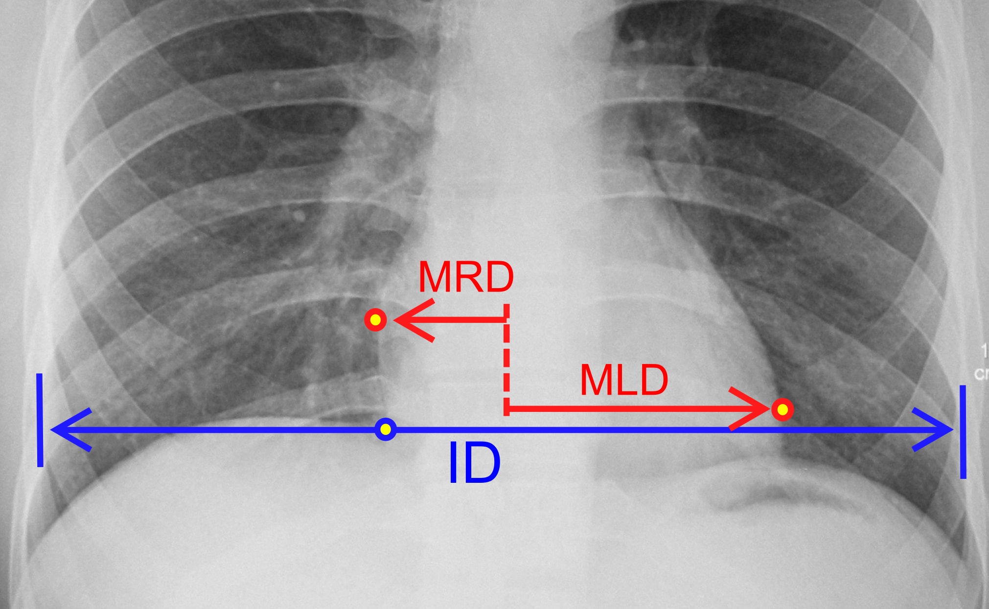
Cardiomegaly (sometimes megacardia or megalocardia) is a medical condition in which the heart is enlarged. As such, it is more commonly referred to simply as "having an enlarged heart". It is usually the result of underlying conditions that make the heart work harder, such as obesity, heart valve disease, high blood pressure ( hypertension), and coronary artery disease. Cardiomyopathy is also associated with cardiomegaly. Cardiomegaly can be serious depending on what part of the heart is enlarged, and can result in congestive heart failure. Recent studies suggest that cardiomegaly is associated with a higher risk of sudden cardiac death. Cardiomegaly may improve over time, but many people with an enlarged heart (dilated cardiomyopathy) need lifelong treatment with medication. Having an immediate family member who has or had cardiomegaly may indicate that a person is more susceptible to getting this condition. Signs and symptoms For many people, cardiomegaly is asymptomatic. For ... [...More Info...] [...Related Items...] OR: [Wikipedia] [Google] [Baidu] |
Myocardium
Cardiac muscle (also called heart muscle, myocardium, cardiomyocytes and cardiac myocytes) is one of three types of vertebrate muscle tissues, with the other two being skeletal muscle and smooth muscle. It is an involuntary, striated muscle that constitutes the main tissue of the wall of the heart. The cardiac muscle (myocardium) forms a thick middle layer between the outer layer of the heart wall (the pericardium) and the inner layer (the endocardium), with blood supplied via the coronary circulation. It is composed of individual cardiac muscle cells joined by intercalated discs, and encased by collagen fibers and other substances that form the extracellular matrix. Cardiac muscle contracts in a similar manner to skeletal muscle, although with some important differences. Electrical stimulation in the form of a cardiac action potential triggers the release of calcium from the cell's internal calcium store, the sarcoplasmic reticulum. The rise in calcium causes the c ... [...More Info...] [...Related Items...] OR: [Wikipedia] [Google] [Baidu] |
Sonography
Medical ultrasound includes diagnostic techniques (mainly imaging techniques) using ultrasound, as well as therapeutic applications of ultrasound. In diagnosis, it is used to create an image of internal body structures such as tendons, muscles, joints, blood vessels, and internal organs, to measure some characteristics (e.g. distances and velocities) or to generate an informative audible sound. Its aim is usually to find a source of disease or to exclude pathology. The usage of ultrasound to produce visual images for medicine is called medical ultrasonography or simply sonography. The practice of examining pregnant women using ultrasound is called obstetric ultrasonography, and was an early development of clinical ultrasonography. Ultrasound is composed of sound waves with frequencies which are significantly higher than the range of human hearing (>20,000 Hz). Ultrasonic images, also known as sonograms, are created by sending pulses of ultrasound into tissue using a pro ... [...More Info...] [...Related Items...] OR: [Wikipedia] [Google] [Baidu] |
Radiography
Radiography is an imaging technique using X-rays, gamma rays, or similar ionizing radiation and non-ionizing radiation to view the internal form of an object. Applications of radiography include medical radiography ("diagnostic" and "therapeutic") and industrial radiography. Similar techniques are used in airport security (where "body scanners" generally use backscatter X-ray). To create an image in conventional radiography, a beam of X-rays is produced by an X-ray generator and is projected toward the object. A certain amount of the X-rays or other radiation is absorbed by the object, dependent on the object's density and structural composition. The X-rays that pass through the object are captured behind the object by a detector (either photographic film or a digital detector). The generation of flat two dimensional images by this technique is called projectional radiography. In computed tomography (CT scanning) an X-ray source and its associated detectors rotate around ... [...More Info...] [...Related Items...] OR: [Wikipedia] [Google] [Baidu] |

_(14582377398).jpg)


