|
Gastrocnemius Muscle
The gastrocnemius muscle (plural ''gastrocnemii'') is a superficial two-headed muscle that is in the back part of the lower leg of humans. It is located superficial to the soleus in the posterior (back) compartment of the leg. It runs from its two heads just above the knee to the heel, extending across a total of three joints (knee, ankle and subtalar joints). The muscle is named via Latin, from Greek γαστήρ (''gaster'') 'belly' or 'stomach' and κνήμη (''knḗmē'') 'leg', meaning 'stomach of the leg' (referring to the bulging shape of the calf). Structure Origin/proximal attachment The lateral head originates from the lateral condyle of the femur, while the medial head originates from the medial condyle of the femur. Insertion/distal attachment Its other end forms a common tendon with the soleus muscle; this tendon is known as the calcaneal tendon or Achilles tendon and inserts onto the posterior surface of the calcaneus, or heel bone. Relations The ga ... [...More Info...] [...Related Items...] OR: [Wikipedia] [Google] [Baidu] |
Lateral Condyle Of Femur
The lateral condyle is one of the two projections on the lower extremity of the femur. The other one is the medial condyle. The lateral condyle is the more prominent and is broader both in its front-to-back and transverse diameters. Clinical significance The most common injury to the lateral femoral condyle is an osteochondral fracture combined with a patellar dislocation. The osteochondral fracture occurs on the weight-bearing portion of the lateral condyle. Typically, the condyle will fracture (and the patella may dislocate) as a result of severe impaction from activities such as downhill skiing and parachuting. Open reduction and internal fixation surgery is typically used to repair an osteochondral fracture. For a AO Type B1 partial articular fracture of the lateral condyle, interfragmentary lag screws are used to secure the bone back together. Supplementation of buttress screws or a buttress plate is used if the fracture extends to the proximal metaphysis or distal diaphysi ... [...More Info...] [...Related Items...] OR: [Wikipedia] [Google] [Baidu] |
Calf (leg)
The calf (: calves; Latin: ''sura'') is the back portion of the lower leg in human anatomy. The muscles within the calf correspond to the posterior compartment of the leg. The two largest muscles within this compartment are known together as the calf muscle and attach to the heel via the Achilles tendon. Several other, smaller muscles attach to the knee, the ankle, and via long tendons to the toes. Etymology From Middle English ''calf'', ''kalf'', from Old Norse ''kalfi'', possibly derived from the same Germanic root as English '' calf'' ("young cow"). Cognate with Icelandic ''kálfi'' ("calf of the leg"). ''Calf'' and ''calf of the leg'' are documented in use in Middle English circa AD 1350 and AD 1425 respectively. Historically, the ''absence of calf'', meaning a lower leg without a prominent calf muscle, was regarded by some authors as a sign of inferiority: ''it is well known that monkeys have no calves, and still less do they exist among the lower orders of mammals ... [...More Info...] [...Related Items...] OR: [Wikipedia] [Google] [Baidu] |
WebMD
WebMD is an American corporation which publishes online news and information about human health and well-being. The WebMD website also includes information about drugs and is an important healthcare information website and the most popular consumer-oriented health site. WebMD was started in 1998 by internet entrepreneur Jeff Arnold. In early 1999, it was part of a three-way merger with Sapient Health Network (SHN) and Direct Medical Knowledge (DMK). SHN began in Portland, Oregon, in 1996 by Jim Kean, Bill Kelly, and Kris Nybakken, who worked together at a CD-ROM publishing firm, Creative Multimedia. Later, in 1999, WebMD merged with Healtheon, founded by Netscape Communications founder James H. Clark. History WebMD is best known as a health information services website, which publishes content regarding health and health care topics, including a symptom checklist, pharmacy information, drugs information, and blogs of physicians with specific topics, and provides a place t ... [...More Info...] [...Related Items...] OR: [Wikipedia] [Google] [Baidu] |
Spasm
A spasm is a sudden involuntary contraction of a muscle, a group of muscles, or a hollow organ, such as the bladder. A spasmodic muscle contraction may be caused by many medical conditions, including dystonia. Most commonly, it is a muscle cramp which is accompanied by a sudden burst of pain. A muscle cramp is usually harmless and ceases after a few minutes. It is typically caused by ion imbalance or muscle fatigue. There are other causes of involuntary muscle contractions, and some of these may cause a health problem. A series of spasms, or permanent spasms, is referred to as a "spasmism". Description and causes Spasms occur when the part of the brain that controls movement malfunctions, causing involuntary muscle activity. A spasm may be a muscle contraction caused by abnormal nerve stimulation or by abnormal activity of the muscle itself. Causes The cause of spasms is often unknown, but it can be due to an inherited genetic problem, a side effect of medicatio ... [...More Info...] [...Related Items...] OR: [Wikipedia] [Google] [Baidu] |
Precentral Gyrus
The precentral gyrus is a prominent gyrus on the surface of the posterior frontal lobe of the brain. It is the site of the primary motor cortex that in humans is cytoarchitecturally defined as Brodmann area 4. Structure The precentral gyrus lies in front of the postcentral gyrus - mostly on the lateral (convex) side of each cerebral hemisphere - from which it is separated by the central sulcus. Its anterior border is represented by the precentral sulcus, while inferiorly it borders to the lateral sulcus (Sylvian fissure). Medially, it is contiguous with the paracentral lobule. The internal pyramidal layer (layer V) of the precentral cortex contains giant (70-100 micrometers) pyramidal neurons called Betz cells, which send long axons to the contralateral motor nuclei of the cranial nerves and to the lower motor neurons in the ventral horn of the spinal cord. These axons form the corticospinal tract. The Betz cells along with their long axons are referred to as upper motor n ... [...More Info...] [...Related Items...] OR: [Wikipedia] [Google] [Baidu] |
McGraw-Hill
McGraw Hill is an American education science company that provides educational content, software, and services for students and educators across various levels—from K-12 to higher education and professional settings. They produce textbooks, digital learning tools, and adaptive technology to enhance learning experiences and outcomes. It is one of the "big three" educational publishers along with Houghton Mifflin Harcourt and Pearson Education. McGraw Hill also publishes reference and trade publications for the medical, business, and engineering professions. Formerly a division of The McGraw Hill Companies (later renamed McGraw Hill Financial, now S&P Global), McGraw Hill Education was divested and acquired by Apollo Global Management in March 2013 for $2.4 billion in cash. McGraw Hill was sold in 2021 to Platinum Equity for $4.5 billion. History McGraw Hill was founded in 1888, when James H. McGraw, co-founder of McGraw Hill, purchased the ''American Journal of Railway ... [...More Info...] [...Related Items...] OR: [Wikipedia] [Google] [Baidu] |
Plantarflexion
Motion, the process of movement, is described using specific anatomical terms. Motion includes movement of organs, joints, limbs, and specific sections of the body. The terminology used describes this motion according to its direction relative to the anatomical position of the body parts involved. Anatomists and others use a unified set of terms to describe most of the movements, although other, more specialized terms are necessary for describing unique movements such as those of the hands, feet, and eyes. In general, motion is classified according to the anatomical plane it occurs in. ''Flexion'' and ''extension'' are examples of ''angular'' motions, in which two axes of a joint are brought closer together or moved further apart. ''Rotational'' motion may occur at other joints, for example the shoulder, and are described as ''internal'' or ''external''. Other terms, such as ''elevation'' and ''depression'', describe movement above or below the horizontal plane. Many anatomical ... [...More Info...] [...Related Items...] OR: [Wikipedia] [Google] [Baidu] |
Fabella
The fabella is a small sesamoid bone found in some mammals embedded in the tendon of the lateral head of the gastrocnemius muscle behind the lateral condyle of the femur. It is an accessory bone, an anatomical variation present in 39% of humans. Rarely, there are two or three of these bones (fabella bi- or tripartita). It can be mistaken for a loose body or osteophyte. The word ''fabella'' is a Latin diminutive of ''faba'' 'bean'. In humans, it is more common in men than women, older individuals compared to younger, and there is high regional variation, with fabellae being most common in people living in Asia and Oceania and least common in people living in North America and Africa. Bilateral cases (one per knee) are more common than unilateral ones (one per individual), and within individual cases, fabellae are equally likely to be present in right or left knees. Taken together, these data suggest the ability to form a fabella may be genetically controlled, but fabella ossific ... [...More Info...] [...Related Items...] OR: [Wikipedia] [Google] [Baidu] |
Sesamoid Bone
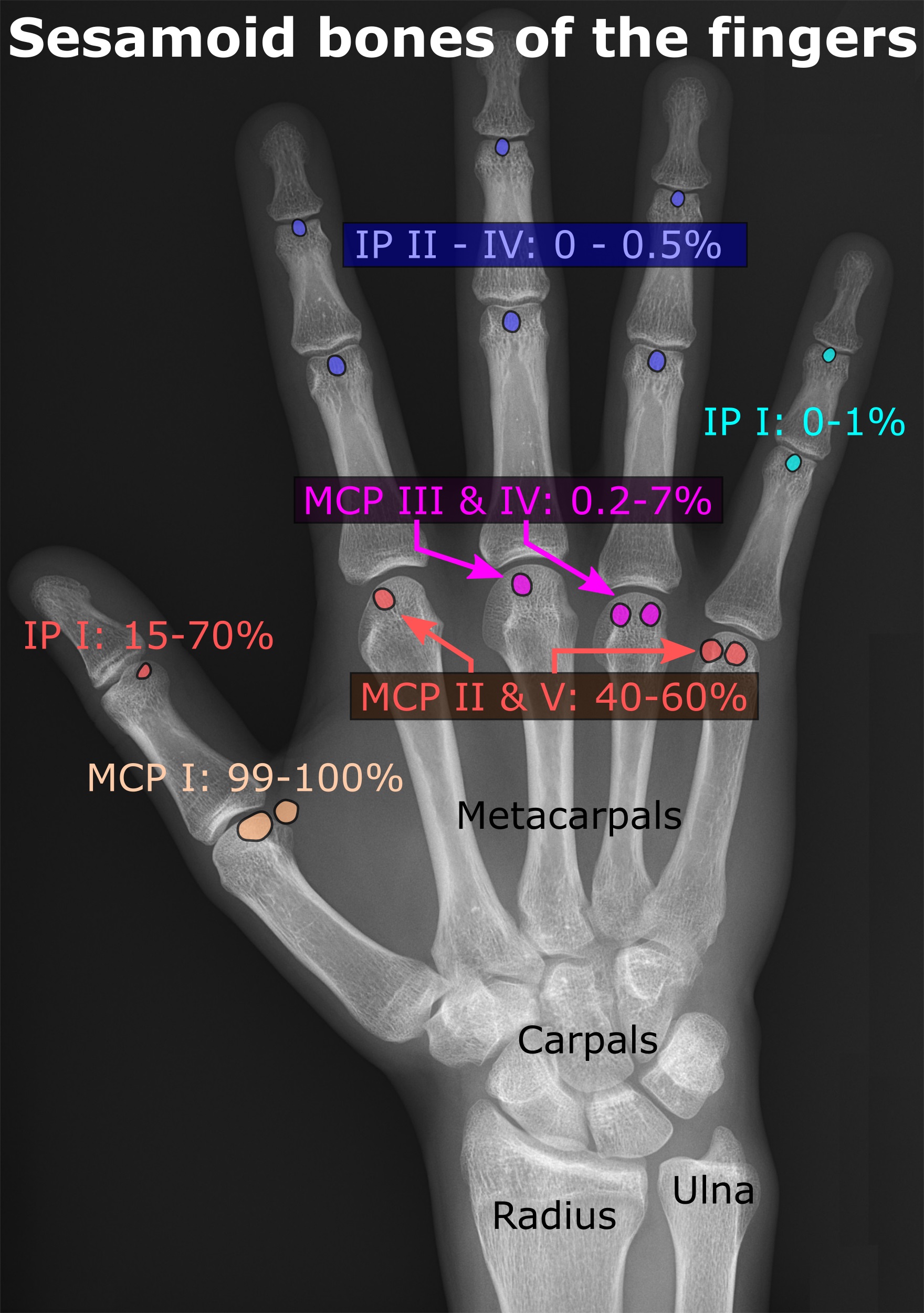
In anatomy, a sesamoid bone () is a bone embedded within a tendon or a muscle. Its name is derived from the Greek word for 'sesame seed', indicating the small size of most sesamoids. Often, these bones form in response to strain, or can be present as a anatomical variation, normal variant. The patella is the largest sesamoid bone in the body. Sesamoids act like pulleys, providing a smooth surface for tendons to slide over, increasing the tendon's ability to transmit Muscle#Function, muscular forces. Structure Sesamoid bones can be found on joints throughout the human body, including: * In the knee—the patella (within the quadriceps tendon). This is the largest sesamoid bone. * In the hand—two sesamoid bones are commonly found in the Anatomical terms of location#Arms, distal portions of the first metacarpal bone (within the tendons of adductor pollicis and flexor pollicis brevis). There is also commonly a sesamoid bone in distal portions of the second metacarpal bone and fif ... [...More Info...] [...Related Items...] OR: [Wikipedia] [Google] [Baidu] |
Tibia
The tibia (; : tibiae or tibias), also known as the shinbone or shankbone, is the larger, stronger, and anterior (frontal) of the two Leg bones, bones in the leg below the knee in vertebrates (the other being the fibula, behind and to the outside of the tibia); it connects the knee with the ankle bones, ankle. The tibia is found on the anatomical terms of location#Medial, medial side of the leg next to the fibula and closer to the median plane. The tibia is connected to the fibula by the interosseous membrane of leg, forming a type of fibrous joint called a syndesmosis with very little movement. The tibia is named for the flute ''aulos, tibia''. It is the second largest bone in the human body, after the femur. The leg bones are the strongest long bones as they support the rest of the body. Structure In human anatomy, the tibia is the second largest bone next to the femur. As in other vertebrates the tibia is one of two bones in the lower leg, the other being the fibula, and is a ... [...More Info...] [...Related Items...] OR: [Wikipedia] [Google] [Baidu] |
Plantaris Muscle
The plantaris is one of the superficial muscles of the superficial posterior compartment of the leg, one of the fascial compartments of the leg. It is composed of a thin muscle belly and a long thin tendon. While not as thick as the achilles tendon, the plantaris tendon (which tends to be between in length) is the longest tendon in the human body. Not including the tendon, the plantaris muscle is approximately long and is absent in 8-12% of the population. It is one of the plantar flexors in the posterior compartment of the leg, along with the gastrocnemius and soleus muscles. The plantaris is considered to have become an unimportant muscle when human ancestors switched from climbing trees to bipedalism and in anatomically modern humans it mainly acts with the gastrocnemius. Structure The plantaris muscle arises from the inferior part of the lateral supracondylar ridge of the femur at a position slightly superior to the origin of the lateral head of gastrocnemius. It pas ... [...More Info...] [...Related Items...] OR: [Wikipedia] [Google] [Baidu] |
Triceps Surae Muscle
The triceps surae consists of two muscles located at the calf – the two-headed gastrocnemius and the soleus. These muscles both insert into the calcaneus, the bone of the heel of the human foot, and form the major part of the muscle of the posterior leg, commonly known as the calf muscle. Structure The triceps surae is connected to the foot through the Achilles tendon, and has three heads deriving from the two major masses of muscle. * The superficial portion (the gastrocnemius) gives off two heads attaching to the base of the femur directly above the knee. * The deep (profundus) mass of muscle (the soleus) forms the remaining head which attaches to the superior posterior area of the tibia. The triceps surae is innervated by the tibial nerve, specifically, nerve roots L5–S2. Function Contraction of the triceps surae induce plantar flexion (sagittal plane) and stabilization of the ankle complex in the transverse plane. Functional activities include primarily movement ... [...More Info...] [...Related Items...] OR: [Wikipedia] [Google] [Baidu] |


