|
Fourth Metacarpal Bone
The fourth metacarpal bone (metacarpal bone of the ring finger) is shorter and smaller than the third. The base is small and quadrilateral; its superior surface presents two facets, a large one medially for articulation with the hamate, and a small one laterally for the capitate. On the radial side are two oval facets, for articulation with the third metacarpal; and on the ulnar side a single concave facet, for the fifth metacarpal. Clinical relevance A shortened fourth metacarpal bone can be a symptom of Kallmann syndrome, a genetic condition which results in the failure to commence or the non-completion of puberty. A short fourth metacarpal bone can also be found in Turner syndrome, a disorder involving sex chromosomes. A fracture of the fourth and/or fifth metacarpal bones transverse neck secondary due to axial loading is known as a boxer's fracture.Shultz, S. J., Houglum, P. A., Perrin, D. H. (2010). Examination of Musculoskeletal Injuries. Chicago: Human Kinetics Ossi ... [...More Info...] [...Related Items...] OR: [Wikipedia] [Google] [Baidu] |
Hamate
The hamate bone (from Latin language, Latin wiktionary:hamatus, hamatus, "hooked"), or unciform bone (from Latin language, Latin ''wikt:uncus, uncus'', "hook"), Latin os hamatum and occasionally abbreviated as just hamatum, is a bone in the human wrist readily distinguishable by its wedge shape and a hook-like process ("hamulus") projecting from its Radioulnar, palmar surface. Structure The hamate is an irregularly shaped carpal bone found within the hand. The hamate is found within the distal row of carpal bones, and abuts the metacarpals of the little finger and ring finger. Adjacent to the hamate on the ulnar side, and slightly proximal and ulnar to it, is the pisiform bone. Adjacent on the radial side is the capitate, and proximal is the lunate bone. Surfaces The hamate bone has six surfaces: * The ''superior'', the apex of the wedge, is narrow, convex, smooth, and articulates with the lunate bone, lunate. * The ''inferior'' articulates with the fourth and fifth metacarpal ... [...More Info...] [...Related Items...] OR: [Wikipedia] [Google] [Baidu] |
Fifth Metacarpal Bone
The fifth metacarpal bone (metacarpal bone of the little finger or pinky finger) is the most medial and second-shortest of the metacarpal bones. Surfaces It presents on its base one facet on its superior surface, which is concavo-convex and articulates with the hamate, and one on its radial side, which articulates with the fourth metacarpal. On its ulnar side is a prominent tubercle for the insertion of the tendon of the extensor carpi ulnaris muscle. The dorsal surface of the body is divided by an oblique ridge, which extends from near the ulnar side of the base to the radial side of the head. The lateral part of this surface serves for the attachment of the fourth interosseus dorsalis; the medial part is smooth, triangular, and covered by the extensor tendons of the little finger. The palmar surface is similarly divided: Its lateral side (facing the fourth metacarpal) provides the origin for the third palmar interosseus, its medial side contains the insertion of opponens ... [...More Info...] [...Related Items...] OR: [Wikipedia] [Google] [Baidu] |
Third Metacarpal Bone
The third metacarpal bone (metacarpal bone of the middle finger) is a little smaller than the second. The dorsal aspect of its base presents on its radial side a pyramidal eminence, the styloid process, which extends upward behind the capitate; immediately distal to this is a rough surface for the attachment of the extensor carpi radialis brevis muscle. The carpal articular facet is concave behind, flat in front, and articulates with the capitate. On the radial side is a smooth, concave facet for articulation with the second metacarpal, and on the ulnar side two small oval facets for the fourth metacarpal. Ossification The ossification process begins in the shaft during prenatal life, and in the head between the 11th and 27th months. Additional images File:Third metacarpal bone (left hand) - animation01.gif, Third metacarpal bone of the left hand (shown in red). Animation. File:Third metacarpal bone (left hand) - animation02.gif, Third metacarpal bone of the left hand. ... [...More Info...] [...Related Items...] OR: [Wikipedia] [Google] [Baidu] |
Second Metacarpal Bone
The second metacarpal bone (metacarpal bone of the index finger) is the longest, and its base the largest, of all the Metacarpus, metacarpal bones.''Gray's Anatomy'' (1918). See infobox. Human anatomy Its base is prolonged upward and medialward, forming a prominent ridge. It presents four articular facets, three on the upper surface and one on the ulnar side: * Of the facets on the upper surface: ** the ''intermediate'' is the largest and is concave from side to side, convex from before backward for articulation with the lesser multangular; ** the ''lateral'' is small, flat and oval for articulation with the greater multangular; ** the ''medial'', on the summit of the ridge, is long and narrow for articulation with the capitate. * The facet on the ulnar side articulates with the third metacarpal. The extensor carpi radialis longus muscle is inserted on the dorsal surface and the flexor carpi radialis muscle on the Anatomical terms of location#Hands and feet, volar surface of t ... [...More Info...] [...Related Items...] OR: [Wikipedia] [Google] [Baidu] |
First Metacarpal Bone
The first metacarpal bone or the metacarpal bone of the thumb is the first bone proximal to the thumb. It is connected to the trapezium of the carpus at the first carpometacarpal joint and to the proximal thumb phalanx at the first metacarpophalangeal joint. Characteristics The first metacarpal bone is short and thick with a shaft thicker and broader than those of the other metacarpal bones. Its narrow shaft connects its widened base and rounded head; the former consisting of a thick cortical bone surrounding the open medullary canal; the latter two consisting of cancellous bone surrounded by a thin cortical shell. Head The head is less rounded and less spherical than those of the other metacarpals, making it better suited for a hinge-like articulation. The distal articular surface is quadrilateral, wide, and flat; thicker and broader transversely and extends much further palmarly than dorsally. On the palmar aspect of the articular surface there is a pair of emin ... [...More Info...] [...Related Items...] OR: [Wikipedia] [Google] [Baidu] |
Metacarpus
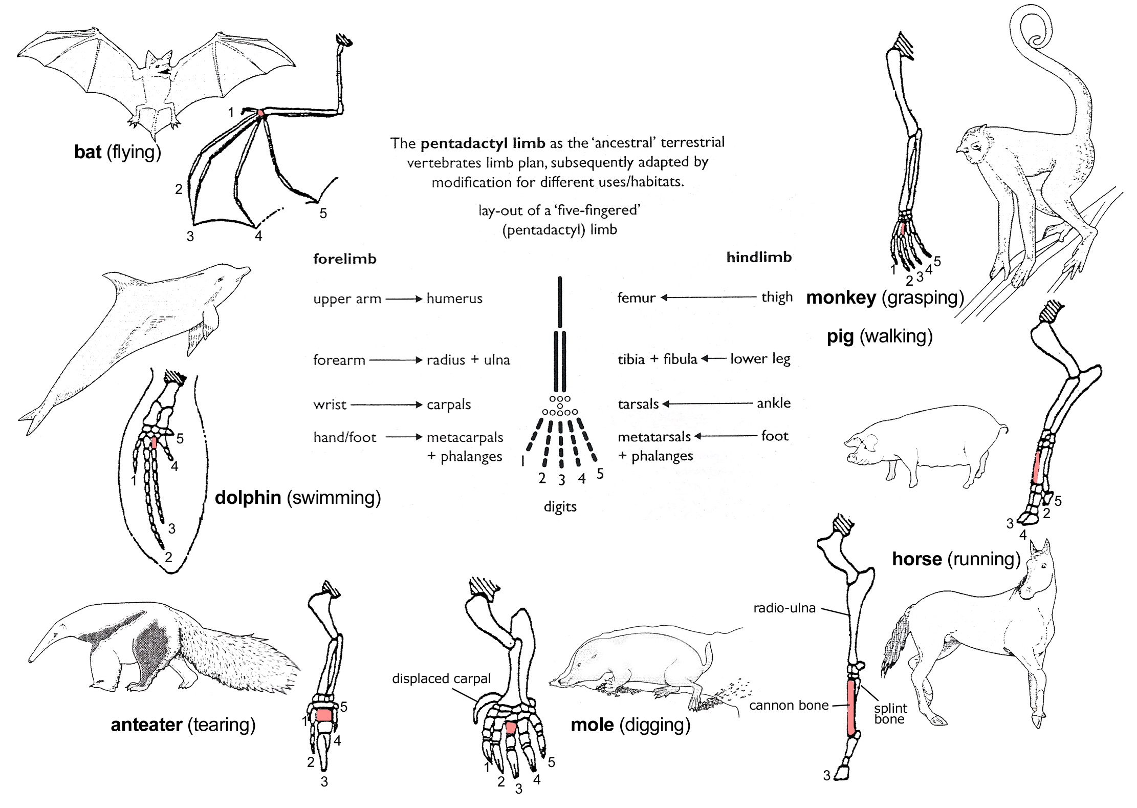
In human anatomy, the metacarpal bones or metacarpus, also known as the "palm bones", are the appendicular skeleton, appendicular bones that form the intermediate part of the hand between the phalanges (fingers) and the carpal bones (wrist, wrist bones), which joint, articulate with the forearm. The metacarpal bones are homologous to the metatarsal bones in the foot. Structure The metacarpals form a transverse arch to which the rigid row of distal carpal bones are fixed. The peripheral metacarpals (those of the thumb and little finger) form the sides of the cup of the palmar gutter and as they are brought together they deepen this concavity. The index metacarpal is the most firmly fixed, while the thumb metacarpal articulates with the trapezium and acts independently from the others. The middle metacarpals are tightly united to the carpus by intrinsic interlocking bone elements at their bases. The ring metacarpal is somewhat more mobile while the fifth metacarpal is semi-indepen ... [...More Info...] [...Related Items...] OR: [Wikipedia] [Google] [Baidu] |
Boxer's Fracture
A boxer's fracture is the break of the fifth metacarpal bone of the hand near the knuckle. Occasionally, it is used to refer to fractures of the fourth metacarpal as well. Symptoms include pain and a depressed knuckle. Classically, it occurs after a person hits an object with a closed fist. The knuckle is then bent towards the palm of the hand. Diagnosis is generally suspected based on symptoms and confirmed with X-rays. For most fractures with less than 70 degrees of angulation, buddy taping and a tensor bandage resulted in similar outcomes to reduction with splinting. In those with more than 70 degrees of angulation or in which the broken finger is rotated, reduction and splinting may be recommended. They represent about a fifth of hand fractures. They occur more commonly in males than females. Both short and long term outcomes are generally good. The knuckle, however, typically remains somewhat deformed. Signs and symptoms The symptoms are pain and tenderness in the ... [...More Info...] [...Related Items...] OR: [Wikipedia] [Google] [Baidu] |
Bone Fracture
A bone fracture (abbreviated FRX or Fx, Fx, or #) is a medical condition in which there is a partial or complete break in the continuity of any bone in the body. In more severe cases, the bone may be broken into several fragments, known as a ''comminuted fracture''. An open fracture (or compound fracture) is a bone fracture where the broken bone breaks through the skin. A bone fracture may be the result of high force Impact force, impact or Stress fracture, stress, or a minimal trauma injury as a result of certain medical conditions that weaken the bones, such as osteoporosis, osteopenia, bone cancer, or osteogenesis imperfecta, where the fracture is then properly termed a pathologic fracture. Most bone fractures require urgent medical attention to prevent further injury. Signs and symptoms Although bone tissue contains no nociceptors, pain receptors, a bone fracture is painful for several reasons: * Breaking in the continuity of the periosteum, with or without similar disconti ... [...More Info...] [...Related Items...] OR: [Wikipedia] [Google] [Baidu] |
Capitate
The capitate bone is a bone in the human wrist found in the center of the carpal bone region, located at the distal end of the radius and ulna bones. It articulates with the third metacarpal bone (the middle finger) and forms the third carpometacarpal joint. The capitate bone is the largest of the carpal bones in the human hand. It presents, above, a rounded portion or head, which is received into the concavity formed by the scaphoid and lunate bones; a constricted portion or neck; and below this, the body.''Gray's Anatomy'' (1918). See infobox. The bone is also found in many other mammals, and is homologous with the "third distal carpal" of reptiles and amphibians. Structure The capitate is the largest carpal bone found within the hand. The capitate is found within the distal row of carpal bones. The capitate lies directly adjacent to the metacarpal of the ring finger on its distal surface, has the hamate on its ulnar surface and trapezoid on its radial surface, and abuts the ... [...More Info...] [...Related Items...] OR: [Wikipedia] [Google] [Baidu] |
Turner Syndrome
Turner syndrome (TS), commonly known as 45,X, or 45,X0,Also written as 45,XO. is a chromosomal disorder in which cells of females have only one X chromosome instead of two, or are partially missing an X chromosome (sex chromosome monosomy) leading to the complete or partial deletion of the pseudoautosomal regions (PAR1, PAR2) in the affected X chromosome. Typically, people have two sex chromosomes (XX for females or XY for males). The chromosomal abnormality is often present in just some cells, in which case it is known as Turner syndrome with mosaicism. 45,X0 with mosaicism can occur in males or females, but Turner syndrome without mosaicism only occurs in females. Signs and symptoms vary among those affected but often include additional skin folds on the neck, arched palate, low-set ears, low hairline at the nape of the neck, short stature, and lymphedema of the hands and feet. Those affected do not normally develop menstrual periods or mammary glands without hormone trea ... [...More Info...] [...Related Items...] OR: [Wikipedia] [Google] [Baidu] |
Puberty
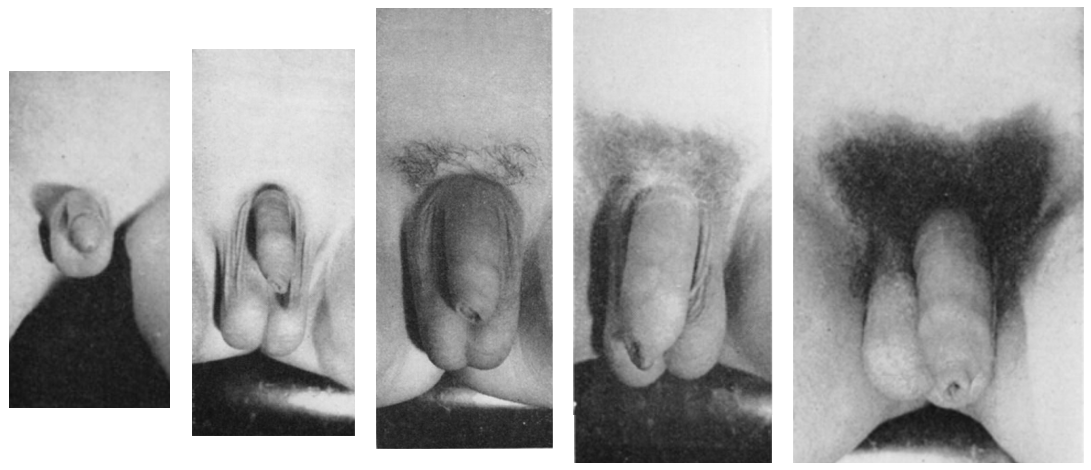
Puberty is the process of physical changes through which a child's body matures into an adult body capable of sexual reproduction. It is initiated by hormonal signals from the brain to the gonads: the ovaries in a female, the testicles in a male. In response to the signals, the gonads produce hormones that stimulate libido and the growth, function, and transformation of the brain, bones, muscle, blood, skin, hair, breasts, and sex organs. Physical growth—height and weight—accelerates in the first half of puberty and is completed when an adult body has been developed. Before puberty, the external sex organs, known as primary sexual characteristics, are sex characteristics that distinguish males and females. Puberty leads to sexual dimorphism through the development of the secondary sex characteristics, which further distinguish the sexes. On average, females begin puberty at age 10½ and complete puberty at ages 15-17; males begin at ages 11½-12 and complete pube ... [...More Info...] [...Related Items...] OR: [Wikipedia] [Google] [Baidu] |


