|
Foramen Ovale (heart)
In the fetal heart, the foramen ovale (), also foramen Botalli or the ostium secundum of Born, allows blood to enter the left atrium from the right atrium. It is one of two fetal cardiac shunts, the other being the ductus arteriosus (which allows blood that still escapes to the right ventricle to bypass the pulmonary circulation). Another similar adaptation in the fetus is the ductus venosus. In most individuals, the foramen ovale closes at birth. It later forms the fossa ovalis. Development The foramen ovale () forms in the late fourth week of gestation, as a small passageway between the septum secundum and the ostium secundum. Initially the atria are separated from one another by the septum primum except for a small opening below the septum, the ostium primum. As the septum primum grows, the ostium primum narrows and eventually closes. Before it does so, bloodflow from the inferior vena cava wears down a portion of the septum primum, forming the ostium secundum. Some e ... [...More Info...] [...Related Items...] OR: [Wikipedia] [Google] [Baidu] |
Pulmonary Vein
The pulmonary veins are the veins that transfer Blood#Oxygen transport, oxygenated blood from the lungs to the heart. The largest pulmonary veins are the four ''main pulmonary veins'', two from each lung that drain into the left atrium of the heart. The pulmonary veins are part of the pulmonary circulation. Structure There are four main pulmonary veins, two from each lung – an inferior and a superior main vein, emerging from each lung hilum, hilum. The main pulmonary veins receive blood from three or four feeding veins in each lung, and drain into the left atrium. The peripheral feeding veins do not follow the bronchial tree. They run between the pulmonary segments from which they drain the blood. At the root of the lung, the right superior pulmonary vein lies in front of and a little below the pulmonary artery; the inferior is situated at the lowest part of the lung hilum. Behind the pulmonary artery is the bronchus. The right main pulmonary veins (contains oxygenated bloo ... [...More Info...] [...Related Items...] OR: [Wikipedia] [Google] [Baidu] |
Programmed Cell Death
Programmed cell death (PCD) sometimes referred to as cell, or cellular suicide is the death of a cell (biology), cell as a result of events inside of a cell, such as apoptosis or autophagy. PCD is carried out in a biological process, which usually confers advantage during an organism's biological life cycle, lifecycle. For example, the Limb development, differentiation of fingers and toes in a developing human embryo occurs because cells between the fingers apoptose; the result is that the digits are separate. PCD serves fundamental functions during both plant and animal tissue development. Apoptosis and autophagy are both forms of programmed cell death. Necrosis is the death of a cell caused by external factors such as trauma or infection and occurs in several different forms. Necrosis was long seen as a non-physiological process that occurs as a result of infection or injury, but in the 2000s, a form of programmed necrosis, called necroptosis, was recognized as an alternative f ... [...More Info...] [...Related Items...] OR: [Wikipedia] [Google] [Baidu] |
Cardiology
Cardiology () is the study of the heart. Cardiology is a branch of medicine that deals with disorders of the heart and the cardiovascular system. The field includes medical diagnosis and treatment of congenital heart defects, coronary artery disease, heart failure, valvular heart disease, and electrophysiology. Physicians who specialize in this field of medicine are called cardiologists, a sub-specialty of internal medicine. Pediatric cardiologists are pediatricians who specialize in cardiology. Physicians who specialize in cardiac surgery are called cardiothoracic surgeons or cardiac surgeons, a specialty of general surgery. Specializations All cardiologists in the branch of medicine study the disorders of the heart, but the study of adult and child heart disorders each require different training pathways. Therefore, an adult cardiologist (often simply called "cardiologist") is inadequately trained to take care of children, and pediatric cardiologists are not trained to treat ... [...More Info...] [...Related Items...] OR: [Wikipedia] [Google] [Baidu] |
Coronary Arteries
The coronary arteries are the arteries, arterial blood vessels of coronary circulation, which transport oxygenated blood to the Cardiac muscle, heart muscle. The heart requires a continuous supply of oxygen to function and survive, much like any other tissue or organ of the body. The coronary arteries wrap around the entire heart. The two main branches are the left coronary artery and right coronary artery. The arteries can additionally be categorized based on the area of the heart for which they provide circulation. These categories are called ''epicardial'' (above the epicardium, or the outermost tissue of the heart) and ''microvascular'' (close to the endocardium, or the innermost tissue of the heart). Reduced function of the coronary arteries can lead to decreased flow of oxygen and nutrients to the heart. Not only does this affect supply to the heart muscle itself, but it also can affect the ability of the heart to pump blood throughout the body. Therefore, any disorder or d ... [...More Info...] [...Related Items...] OR: [Wikipedia] [Google] [Baidu] |
Transesophageal Echocardiogram
A transesophageal echocardiogram (TEE; also spelled transoesophageal echocardiogram; TOE in British English) is an alternative way to perform an echocardiogram. A specialized probe containing an ultrasound transducer at its tip is passed into the patient's esophagus. This allows image and Doppler evaluation which can be recorded. It is commonly used during cardiac surgery and is an excellent modality for assessing the aorta, although there are some limitations. It has several advantages and some disadvantages compared with a transthoracic echocardiogram (TTE). Details TEE is a semi-invasive procedure in that the probe must enter the body but does not require surgical (i.e., invasive) cutting for this procedure. Before inserting the probe, mild to moderate sedation is induced in the patient to ease the discomfort and to decrease the gag reflex. Usually a local anesthetic spray (e.g., lidocaine, benzocaine, xylocaine) is used for the back of the throat or as a je ... [...More Info...] [...Related Items...] OR: [Wikipedia] [Google] [Baidu] |
Stroke
Stroke is a medical condition in which poor cerebral circulation, blood flow to a part of the brain causes cell death. There are two main types of stroke: brain ischemia, ischemic, due to lack of blood flow, and intracranial hemorrhage, hemorrhagic, due to bleeding. Both cause parts of the brain to stop functioning properly. Signs and symptoms of stroke may include an hemiplegia, inability to move or feel on one side of the body, receptive aphasia, problems understanding or expressive aphasia, speaking, dizziness, or homonymous hemianopsia, loss of vision to one side. Signs and symptoms often appear soon after the stroke has occurred. If symptoms last less than 24 hours, the stroke is a transient ischemic attack (TIA), also called a mini-stroke. subarachnoid hemorrhage, Hemorrhagic stroke may also be associated with a thunderclap headache, severe headache. The symptoms of stroke can be permanent. Long-term complications may include pneumonia and Urinary incontinence, loss of b ... [...More Info...] [...Related Items...] OR: [Wikipedia] [Google] [Baidu] |
Embolism
An embolism is the lodging of an embolus, a blockage-causing piece of material, inside a blood vessel. The embolus may be a blood clot (thrombus), a fat globule (fat embolism), a bubble of air or other gas (air embolism, gas embolism), amniotic fluid (amniotic fluid embolism), or foreign body, foreign material. An embolism can cause partial or total blockage of blood flow in the affected vessel. Such a blockage (vascular occlusion) may affect a part of the body distant from the origin of the embolus. An embolism in which the embolus is a piece of thrombus is called a thromboembolism. An embolism is usually a pathological event, caused by illness or injury. Sometimes it is created intentionally for a therapeutic reason, such as to hemostasis, stop bleeding or to kill a cancerous tumor by stopping its blood supply. Such therapy is called embolization. Classification There are different types of embolism, some of which are listed below. Embolism can be classified based on wher ... [...More Info...] [...Related Items...] OR: [Wikipedia] [Google] [Baidu] |
Paradoxical Embolism
An embolus, is described as a free-floating mass, located inside blood vessels that can travel from one site in the blood stream to another. An embolus can be made up of solid (like a blood clot), liquid (like amniotic fluid), or gas (like air). Once these masses get "stuck" in a different blood vessel, it is then known as an "embolism." An embolism can cause ischemia—damage to an organ from lack of oxygen. A paradoxical embolism is a specific type of embolism in which the embolus travels from the right side of the heart (venous circulation) to the left side of the heart (arterial circulation) and lodges itself in a blood vessel known as an artery. It is termed "paradoxical" because venous emboli will usually be lodged in pulmonary artery in an event called pulmonary embolism, instead of systemic circulation. Pathophysiology An embolism may be made from any one of numerous materials that may find itself in a blood vessel, including a piece of a thrombus, known as a thromboembo ... [...More Info...] [...Related Items...] OR: [Wikipedia] [Google] [Baidu] |
Atrial Septal Defect
Atrial septal defect (ASD) is a congenital heart defect in which blood flows between the atrium (heart), atria (upper chambers) of the heart. Some flow is a normal condition both pre-birth and immediately post-birth via the Foramen ovale (heart), foramen ovale; however, when this does not naturally close after birth it is referred to as a patent (open) foramen ovale (PFO). It is common in patients with a congenital interatrial septum, atrial septal aneurysm (ASA). After PFO closure the atria normally are separated by a dividing wall, the interatrial septum. If this septum is defective or absent, then oxygen-rich blood can flow directly from the left side of the heart to mix with the oxygen-poor blood in the right side of the heart; or the opposite, depending on whether the left or right atrium has the higher blood pressure. In the absence of other heart defects, the left atrium has the higher pressure. This can lead to lower-than-normal oxygen levels in the arterial blood that su ... [...More Info...] [...Related Items...] OR: [Wikipedia] [Google] [Baidu] |
Placenta
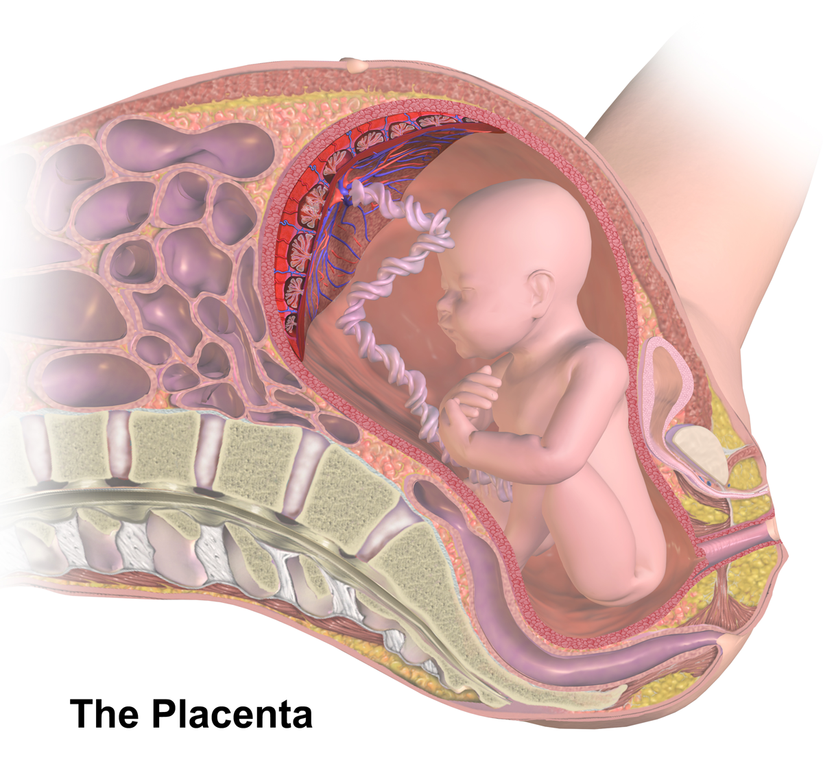
The placenta (: placentas or placentae) is a temporary embryonic and later fetal organ that begins developing from the blastocyst shortly after implantation. It plays critical roles in facilitating nutrient, gas, and waste exchange between the physically separate maternal and fetal circulations, and is an important endocrine organ, producing hormones that regulate both maternal and fetal physiology during pregnancy. The placenta connects to the fetus via the umbilical cord, and on the opposite aspect to the maternal uterus in a species-dependent manner. In humans, a thin layer of maternal decidual ( endometrial) tissue comes away with the placenta when it is expelled from the uterus following birth (sometimes incorrectly referred to as the 'maternal part' of the placenta). Placentas are a defining characteristic of placental mammals, but are also found in marsupials and some non-mammals with varying levels of development. Mammalian placentas probably first evolved abou ... [...More Info...] [...Related Items...] OR: [Wikipedia] [Google] [Baidu] |
Fetus
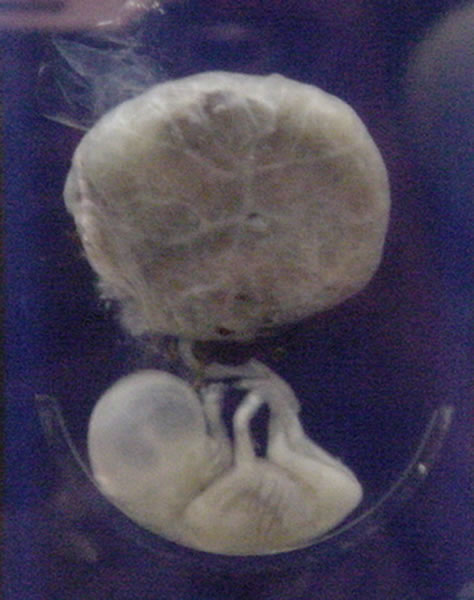
A fetus or foetus (; : fetuses, foetuses, rarely feti or foeti) is the unborn offspring of a viviparous animal that develops from an embryo. Following the embryonic development, embryonic stage, the fetal stage of development takes place. Prenatal development is a continuum, with no clear defining feature distinguishing an embryo from a fetus. However, in general a fetus is characterized by the presence of all the major body organs, though they will not yet be fully developed and functional, and some may not yet be situated in their final Anatomy, anatomical location. In human prenatal development, fetal development begins from the ninth week after Human fertilization, fertilization (which is the eleventh week of Gestational age (obstetrics), gestational age) and continues until the childbirth, birth of a newborn. Etymology The word ''wikt:fetus#English, fetus'' (plural ''wikt:fetuses#English, fetuses'' or rarely, the solecism ''wikt:feti#English, feti''''Oxford English Dict ... [...More Info...] [...Related Items...] OR: [Wikipedia] [Google] [Baidu] |


