|
Fetal Membranes
The fetal membranes are the four extraembryonic membranes, associated with the developing embryo, and fetus in humans and other mammals. They are the amnion, chorion, allantois, and yolk sac. The amnion and the chorion are the chorioamniotic membranes that make up the amniotic sac which surrounds and protects the embryo. The fetal membranes are four of six accessory organs developed by the conceptus that are not part of the embryo itself, the other two are the placenta, and the umbilical cord. Structure The fetal membranes surround the developing embryo and form the fetal-maternal interface. The fetal membranes are derived from the trophoblast layer (outer layer of cells) of the implanting blastocyst. The trophoblast layer differentiates into amnion and the chorion, which then comprise the fetal membranes. The amnion is the innermost layer and, therefore, contacts the amniotic fluid, the fetus and the umbilical cord. The internal pressure of the amniotic fluid cau ... [...More Info...] [...Related Items...] OR: [Wikipedia] [Google] [Baidu] |
Extraembryonic Membrane
The extraembryonic membranes are four membranes which assist in the development of an animal's embryo. Such membranes occur in a range of animals from humans to insects. They originate from the zygote, but are not considered part of the embryo. They typically perform roles in nutrition, gas exchange and waste removal. In amniotes There are four standard extraembryonic membranes in amniote, amniotes, i.e. reptiles (including birds) and mammals: #the yolk sac which surrounds the yolk #the amnion which surrounds and cushions the embryo #the allantois which among avians stores embryonic waste and assists with the exchange of carbon dioxide with oxygen as well as the resorption of calcium from the shell, and #the chorion which surrounds all of these and in avians successively merges with the allantois in the later stages of egg development to form a combined respiratory and excretory organ called the chorioallantois. In humans and other mammals they are more usually called fetal membran ... [...More Info...] [...Related Items...] OR: [Wikipedia] [Google] [Baidu] |
Amniotic Fluid
The amniotic fluid is the protective liquid contained by the amniotic sac of a gravid amniote. This fluid serves as a cushion for the growing fetus, but also serves to facilitate the exchange of nutrients, water, and biochemical products between mother and fetus. Colloquially, the amniotic fluid is commonly called water or waters (Latin liquor amnii). Development Amniotic fluid is present from the formation of the gestational sac. Amniotic fluid is in the amniotic sac. It is generated from maternal plasma, and passes through the fetal membranes by osmotic and hydrostatic forces. When fetal kidneys begin to function around week 16, fetal urine also contributes to the fluid. In earlier times, it was believed that the amniotic fluid was composed entirely of excreted fetal urine. The fluid is absorbed through the fetal tissue and skin. After 22 to 25 week of pregnancy, keratinization of an embryo's skin occurs. When this process completes around the 25th week, the fluid is pr ... [...More Info...] [...Related Items...] OR: [Wikipedia] [Google] [Baidu] |
Human
Humans (''Homo sapiens'') or modern humans are the most common and widespread species of primate, and the last surviving species of the genus ''Homo''. They are Hominidae, great apes characterized by their Prehistory of nakedness and clothing#Evolution of hairlessness, hairlessness, bipedality, bipedalism, and high Human intelligence, intelligence. Humans have large Human brain, brains, enabling more advanced cognitive skills that facilitate successful adaptation to varied environments, development of sophisticated tools, and formation of complex social structures and civilizations. Humans are Sociality, highly social, with individual humans tending to belong to a Level of analysis, multi-layered network of distinct social groups — from families and peer groups to corporations and State (polity), political states. As such, social interactions between humans have established a wide variety of Value theory, values, norm (sociology), social norms, languages, and traditions (co ... [...More Info...] [...Related Items...] OR: [Wikipedia] [Google] [Baidu] |
Bilaminar Embryonic Disc
The bilaminar embryonic disc, bilaminar blastoderm or embryonic disc is the distinct two-layered structure of cells formed in an embryo. In the development of the human embryo this takes place by day eight. It is formed when the inner cell mass, also known as the embryoblast, forms a bilaminar disc of two layers, an upper layer called the epiblast (primitive ectoderm) and a lower layer called the hypoblast (primitive endoderm), which will eventually form into fetus. These two layers of cells are stretched between two fluid-filled cavities at either end: the primitive yolk sac and the amniotic sac. The epiblast is adjacent to the trophoblast and made of columnar epithelial cells; the hypoblast is closest to the blastocoel (blastocystic cavity) and made of cuboidal cells. As the two layers become evident, a basement membrane forms between the layers. This distinction of layers of the bilaminar disc defines the primitive dorso ventral axis and polarity in embryogenesis. The epib ... [...More Info...] [...Related Items...] OR: [Wikipedia] [Google] [Baidu] |
Hypoblast
In amniote embryology, the hypoblast is one of two distinct layers arising from the inner cell mass in the mammalian blastocyst, or from the blastodisc in reptiles and birds. The hypoblast gives rise to the yolk sac. The hypoblast is a layer of cells in fish and amniote embryos. The hypoblast helps determine the embryo's body axes, and its migration determines the cell movements that accompany the formation of the primitive streak, and helps to orient the embryo, and create bilateral symmetry. The other layer of the inner cell mass, the epiblast, differentiates into the three primary germ layers, ectoderm, mesoderm, and endoderm. Structure The hypoblast lies beneath the epiblast and consists of small cuboidal cells. The hypoblast in fish (but not in birds and mammals) contains the precursors of both the endoderm and mesoderm. In birds and mammals, it contains precursors to the extraembryonic endoderm of the yolk sac. In chick embryos, early cleavage forms an area opaca ... [...More Info...] [...Related Items...] OR: [Wikipedia] [Google] [Baidu] |
Chorionic Villi
Chorionic villi are Wiktionary:villus, villi that sprout from the chorion to provide maximal contact area with maternal blood. They are an essential element in pregnancy from a histology, histomorphologic perspective, and are, by definition, a products of conception, product of conception. Branches of the umbilical arteries carry embryonic blood to the villi. After circulating through the capillaries of the villi, blood returns to the embryo through the umbilical vein. Thus, villi are part of the border between maternal and fetal blood during pregnancy. Structure Villi can also be classified by their relations: * Floating villi float freely in the intervillous space. They exhibit a bi-layered epithelium consisting of cytotrophoblasts with overlaying syncytium (syncytiotrophoblast). * Anchoring (stem) villi stabilize the mechanical integrity of the placental-maternal interface. Development The chorion undergoes rapid proliferation and forms numerous processes, the chorionic vil ... [...More Info...] [...Related Items...] OR: [Wikipedia] [Google] [Baidu] |
Connective Tissue
Connective tissue is one of the four primary types of animal tissue, a group of cells that are similar in structure, along with epithelial tissue, muscle tissue, and nervous tissue. It develops mostly from the mesenchyme, derived from the mesoderm, the middle embryonic germ layer. Connective tissue is found in between other tissues everywhere in the body, including the nervous system. The three meninges, membranes that envelop the brain and spinal cord, are composed of connective tissue. Most types of connective tissue consists of three main components: elastic and collagen fibers, ground substance, and cells. Blood and lymph are classed as specialized fluid connective tissues that do not contain fiber. All are immersed in the body water. The cells of connective tissue include fibroblasts, adipocytes, macrophages, mast cells and leukocytes. The term "connective tissue" (in German, ) was introduced in 1830 by Johannes Peter Müller. The tissue was already recognized as ... [...More Info...] [...Related Items...] OR: [Wikipedia] [Google] [Baidu] |
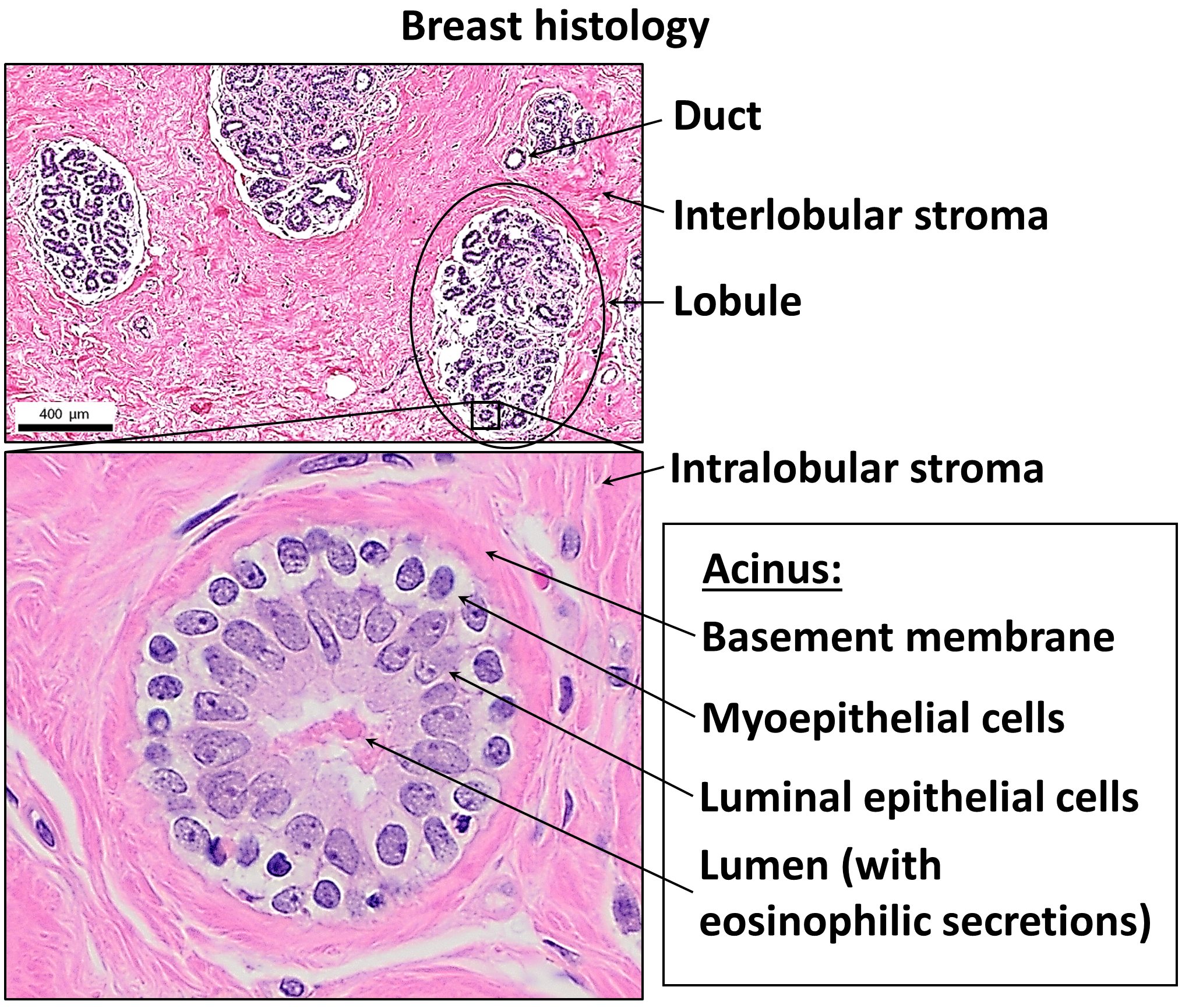
Basement Membrane
The basement membrane, also known as base membrane, is a thin, pliable sheet-like type of extracellular matrix that provides cell and tissue support and acts as a platform for complex signalling. The basement membrane sits between epithelial tissues including mesothelium and endothelium, and the underlying connective tissue. Structure As seen with the electron microscope, the basement membrane is composed of two layers, the basal lamina and the reticular lamina. The underlying connective tissue attaches to the basal lamina with collagen VII anchoring fibrils and fibrillin microfibrils. The basal lamina layer can further be subdivided into two layers based on their visual appearance in electron microscopy. The lighter-colored layer closer to the epithelium is called the lamina lucida, while the denser-colored layer closer to the connective tissue is called the lamina densa. The electron-dense lamina densa layer is about 30–70 nanometers thick and consists of an und ... [...More Info...] [...Related Items...] OR: [Wikipedia] [Google] [Baidu] |
Glycocalyx
The glycocalyx (: glycocalyces or glycocalyxes), also known as the pericellular matrix and cell coat, is a layer of glycoproteins and glycolipids which surround the cell membranes of bacteria, epithelial cells, and other cells. Animal epithelial cells have a fuzz-like coating on the external surface of their plasma membranes. This viscous coating is the glycocalyx that consists of several carbohydrate moieties of membrane glycolipids and glycoproteins, which serve as backbone molecules for support. Generally, the carbohydrate portion of the glycolipids found on the surface of plasma membranes helps these molecules contribute to cell–cell recognition, communication, and intercellular adhesion. The glycocalyx is a type of identifier that the body uses to distinguish between its own healthy cells and transplanted tissues, diseased cells, or invading organisms. Included in the glycocalyx are cell-adhesion molecules that enable cells to adhere to each other and guide the mo ... [...More Info...] [...Related Items...] OR: [Wikipedia] [Google] [Baidu] |
Microvillus
Microvilli (: microvillus) are microscopic cellular membrane protrusions that increase the surface area for diffusion and minimize any increase in volume, and are involved in a wide variety of functions, including absorption, secretion, cellular adhesion, and mechanotransduction. Structure Microvilli are covered in plasma membrane, which encloses cytoplasm and microfilaments. Though these are cellular extensions, there are little or no cellular organelles present in the microvilli. Each microvillus has a dense bundle of cross-linked actin filaments, which serves as its structural core. 20 to 30 tightly bundled actin filaments are cross-linked by bundling proteins fimbrin (or plastin-1), villin and espin to form the core of the microvilli. In the enterocyte microvillus, the structural core is attached to the plasma membrane along its length by lateral arms made of myosin 1a and Ca2+ binding protein calmodulin. Myosin 1a functions through a binding site for filamentous ac ... [...More Info...] [...Related Items...] OR: [Wikipedia] [Google] [Baidu] |
Fetal Surface Vessels
Chorionic (plate) vessels, also fetal surface vessels are blood vessels, including both arteries and veins, that carry blood through the chorion in the fetoplacental circulation. Chorionic arteries branch off the umbilical artery, and supply the capillaries of the chorionic villi. Increased vasocontractility of chorionic arteries may contribute to preeclampsia Pre-eclampsia is a multi-system disorder specific to pregnancy, characterized by the new onset of high blood pressure and often a significant amount of protein in the urine or by the new onset of high blood pressure along with significant end- .... References Embryology of cardiovascular system {{circulatory-stub ... [...More Info...] [...Related Items...] OR: [Wikipedia] [Google] [Baidu] |
Avascular
Blood vessels are the tubular structures of a circulatory system that transport blood throughout many animals’ bodies. Blood vessels transport blood cells, nutrients, and oxygen to most of the tissues of a body. They also take waste and carbon dioxide away from the tissues. Some tissues such as cartilage, epithelium, and the lens and cornea of the eye are not supplied with blood vessels and are termed ''avascular''. There are five types of blood vessels: the arteries, which carry the blood away from the heart; the arterioles; the capillaries, where the exchange of water and chemicals between the blood and the tissues occurs; the venules; and the veins, which carry blood from the capillaries back towards the heart. The word ''vascular'', is derived from the Latin ''vas'', meaning ''vessel'', and is mostly used in relation to blood vessels. Etymology * artery – late Middle English; from Latin ''arteria'', from Greek ''artēria'', probably from ''airein'' ("raise"). ... [...More Info...] [...Related Items...] OR: [Wikipedia] [Google] [Baidu] |


