|
Extensor Pollicis Et Indicis Communis Muscle
In human anatomy, the extensor pollicis et indicis communis is an aberrant muscle in the posterior compartment of forearm. It was first described in 1863. The muscle has a prevalence from 0.5% to 4%. Structure The structure of the extensor pollicis et indicis communis resembles both the characteristics of the extensor pollicis longus and the extensor indicis proprius. It originates from the distal end of ulna. Its tendon passes through the extensor retinaculum in the fourth extensor compartment, splits into two and inserts to both thumb and index finger. The presence of the extensor pollicis et indicis communis, on the other hand, may impair thumb adduction. It was reported as an unusual juncturae tendinum, a tendinous connection between tendon of the extensor pollicis longus and tendon of the extensor digitorum communis to the index finger. It was also identified as a slip of the extensor indicis proprius to the extensor pollicis longus in an Indian cadaver. In other anim ... [...More Info...] [...Related Items...] OR: [Wikipedia] [Google] [Baidu] |
Ulna
The ulna (''pl''. ulnae or ulnas) is a long bone found in the forearm that stretches from the elbow to the smallest finger, and when in anatomical position, is found on the medial side of the forearm. That is, the ulna is on the same side of the forearm as the little finger. It runs parallel to the radius, the other long bone in the forearm. The ulna is usually slightly longer than the radius, but the radius is thicker. Therefore, the radius is considered to be the larger of the two. Structure The ulna is a long bone found in the forearm that stretches from the elbow to the smallest finger, and when in anatomical position, is found on the medial side of the forearm. It is broader close to the elbow, and narrows as it approaches the wrist. Close to the elbow, the ulna has a bony process, the olecranon process, a hook-like structure that fits into the olecranon fossa of the humerus. This prevents hyperextension and forms a hinge joint with the trochlea of the humerus. There ... [...More Info...] [...Related Items...] OR: [Wikipedia] [Google] [Baidu] |
Tendon
A tendon or sinew is a tough, high-tensile-strength band of dense fibrous connective tissue that connects muscle to bone. It is able to transmit the mechanical forces of muscle contraction to the skeletal system without sacrificing its ability to withstand significant amounts of tension. Tendons are similar to ligaments; both are made of collagen. Ligaments connect one bone to another, while tendons connect muscle to bone. Structure Histologically, tendons consist of dense regular connective tissue. The main cellular component of tendons are specialized fibroblasts called tendon cells (tenocytes). Tenocytes synthesize the extracellular matrix of tendons, abundant in densely packed collagen fibers. The collagen fibers are parallel to each other and organized into tendon fascicles. Individual fascicles are bound by the endotendineum, which is a delicate loose connective tissue containing thin collagen fibrils and elastic fibres. Groups of fascicles are bounded by the epit ... [...More Info...] [...Related Items...] OR: [Wikipedia] [Google] [Baidu] |
Extensor Indicis Proprius
In human anatomy, the extensor indicis roprius'' is a narrow, elongated skeletal muscle in the deep layer of the dorsal forearm, placed medial to, and parallel with, the extensor pollicis longus. Its tendon goes to the index finger, which it extends. Structure It arises from the distal third of the dorsal part of the body of ulna and from the interosseous membrane. It runs through the fourth tendon compartment together with the extensor digitorum, from where it projects into the dorsal aponeurosis of the index finger. Opposite the head of the second metacarpal bone, it joins the ulnar side of the tendon of the extensor digitorum which belongs to the index finger. Like the extensor digiti minimi (i.e. the extensor of the little finger), the tendon of the extensor indicis runs and inserts on the ulnar side of the tendon of the common extensor digitorum. The extensor indicis lacks the juncturae tendinum interlinking the tendons of the extensor digitorum on the dorsal side of the ... [...More Info...] [...Related Items...] OR: [Wikipedia] [Google] [Baidu] |
Common Chimpanzee
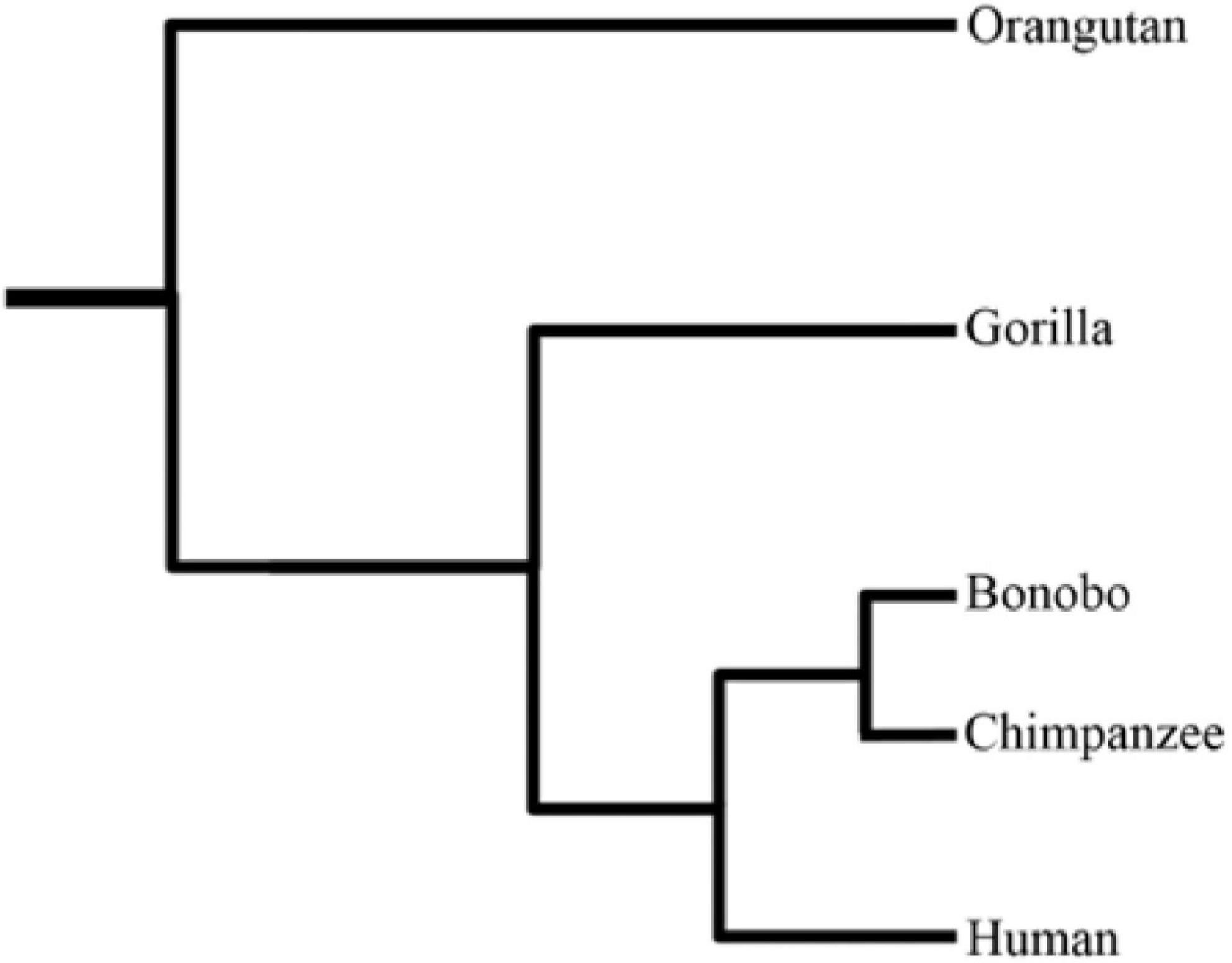
The chimpanzee (''Pan troglodytes''), also known as simply the chimp, is a species of great ape native to the forest and savannah of tropical Africa. It has four confirmed subspecies and a fifth proposed subspecies. When its close relative the bonobo was more commonly known as the pygmy chimpanzee, this species was often called the common chimpanzee or the robust chimpanzee. The chimpanzee and the bonobo are the only species in the genus ''Pan''. Evidence from fossils and DNA sequencing shows that ''Pan'' is a sister taxon to the human lineage and is humans' closest living relative. The chimpanzee is covered in coarse black hair, but has a bare face, fingers, toes, palms of the hands, and soles of the feet. It is larger and more robust than the bonobo, weighing for males and for females and standing . The chimpanzee lives in groups that range in size from 15 to 150 members, although individuals travel and forage in much smaller groups during the day. The species lives i ... [...More Info...] [...Related Items...] OR: [Wikipedia] [Google] [Baidu] |
List Of Anatomical Variations
This article lists anatomical variations that are not deemed inherently pathological. {{incomplete list, date=December 2013 Accessory features Bones * Cervical rib * Fabella * Foramen tympanicum * Supracondylar process of the humerus * Sternal foramen * Stafne bone cavity * Episternal ossicles * Fossa navicularis magna * Transverse basilar fissure - or ''Saucer's fissure'' * Canalis basilaris medianus * Craniopharyngeal canal * Intermediate condylar canal * Foramen arcuale * Os odontoideum * Os acromiale * Ossiculum terminale (of dens) * Scapular foramina and tunnels Muscles * Accessory soleus muscle * Axillary arch * Epitrochleoanconeus muscle - or ''anconeous epitrochlearis'' * Extensor medii proprius muscle * Extensor digitorum brevis manus muscle * Extensor indicis et medii communis muscle * Extensor pollicis et indicis communis muscle * Extensor carpi radialis tertius muscle - or ''extensor carpi radialis accessorius'' * Linburg-Comstock variation - o ... [...More Info...] [...Related Items...] OR: [Wikipedia] [Google] [Baidu] |
New World Monkey
New World monkeys are the five families of primates that are found in the tropical regions of Mexico, Central and South America: Callitrichidae, Cebidae, Aotidae, Pitheciidae, and Atelidae. The five families are ranked together as the Ceboidea (), the only extant superfamily in the parvorder Platyrrhini (). Platyrrhini is derived from the Greek for "broad nosed", and their noses are flatter than those of other simians, with sideways-facing nostrils. Monkeys in the family Atelidae, such as the spider monkey, are the only primates to have prehensile tails. New World monkeys' closest relatives are the other simians, the Catarrhini ("down-nosed"), comprising Old World monkeys and apes. New World monkeys descend from African simians that colonized South America, a line that split off about 40 million years ago. Evolutionary history About 40 million years ago, the Simiiformes infraorder split into the parvorders Platyrrhini (New World monkeys) and Catarrhini ( apes and O ... [...More Info...] [...Related Items...] OR: [Wikipedia] [Google] [Baidu] |
Extensor Digitorum Communis
The extensor digitorum muscle (also known as extensor digitorum communis) is a muscle of the posterior forearm present in humans and other animals. It extends the medial four digits of the hand. Extensor digitorum is innervated by the posterior interosseous nerve, which is a branch of the radial nerve. Structure The extensor digitorum muscle arises from the lateral epicondyle of the humerus, by the common tendon; from the intermuscular septa between it and the adjacent muscles, and from the antebrachial fascia. It divides below into four tendons, which pass, together with that of the extensor indicis proprius, through a separate compartment of the dorsal carpal ligament, within a mucous sheath. The tendons then diverge on the back of the hand, and are inserted into the middle and distal phalanges of the fingers in the following manner.'' Gray's anatomy'' (1918), see infobox Opposite the metacarpophalangeal articulation each tendon is bound by fasciculi to the collateral ligame ... [...More Info...] [...Related Items...] OR: [Wikipedia] [Google] [Baidu] |
Juncturae Tendinum
In human anatomy, juncturae tendinum or ''connexus intertendinei'' refers to the connective tissues that link the tendons of the extensor digitorum communis, and sometimes, to the tendon of the extensor digiti minimi. Juncturae tendinum are located on the dorsal aspect of the hand in the first, second and third inter-metacarpal spaces proximal to the metacarpophalangeal joint. Structure Juncturae tendinum are narrow bands of connective tissues that extend between the tendons of the extensor digitorum communis and the extensor digiti minimi. It is classified into three distinct types (Type 1, 2 and 3) depending on morphology. * Type 1: This is a thin and filamentous juncturae tendinum. Its shape can either be square, rhomboidal or triangular. * Type 2: This type is more tendinous and thicker than type 1 juncturae, and it is also located more distal than the type 1. * Type 3: Type 3 juncturae refers to the slips from the extensor digitorum communis. Type 3 juncture is further ... [...More Info...] [...Related Items...] OR: [Wikipedia] [Google] [Baidu] |
Anatomical Terms Of Motion
Motion, the process of movement, is described using specific anatomical terms. Motion includes movement of organs, joints, limbs, and specific sections of the body. The terminology used describes this motion according to its direction relative to the anatomical position of the body parts involved. Anatomists and others use a unified set of terms to describe most of the movements, although other, more specialized terms are necessary for describing unique movements such as those of the hands, feet, and eyes. In general, motion is classified according to the anatomical plane it occurs in. ''Flexion'' and ''extension'' are examples of ''angular'' motions, in which two axes of a joint are brought closer together or moved further apart. ''Rotational'' motion may occur at other joints, for example the shoulder, and are described as ''internal'' or ''external''. Other terms, such as ''elevation'' and ''depression'', describe movement above or below the horizontal plane. Many ana ... [...More Info...] [...Related Items...] OR: [Wikipedia] [Google] [Baidu] |
Extensor Tendon Compartments Of The Wrist
Extensor tendon compartments of the wrist are anatomical tunnels on the back of the wrist that contain tendons of muscles that extend (as opposed to flex) the wrist and the digits (fingers and thumb). The extensor tendons are held in place by the extensor retinaculum. As the tendons travel over the posterior (back) aspect of the wrist they are enclosed within synovial tendon sheaths. These sheaths reduce the friction to the extensor tendons as they traverse the compartments that are formed by the attachments of the extensor retinaculum to the distal (far end) of the radius and ulna. Structure The compartments are numbered with each compartment containing specific extensor tendons. Clinical significance Any of the dorsal compartments of the wrist can develop tenosynovial inflammation. The first compartment is the most frequently affected site, called De Quervain's disease (syndrome or tenosynovitis). The other two most commonly injured are the sixth (extensor carpi ulnaris ... [...More Info...] [...Related Items...] OR: [Wikipedia] [Google] [Baidu] |
Extensor Retinaculum Of The Hand
The extensor retinaculum (dorsal carpal ligament, or posterior annular ligament) is an anatomical term for the thickened part of the antebrachial fascia that holds the tendons of the extensor muscles in place. It is located on the back of the forearm, just proximal to the hand. It is continuous with the palmar carpal ligament, which is located on the anterior side of the forearm. Structure The extensor retinaculum is a strong, fibrous band, extending obliquely downward and medialward across the back of the wrist. It consists of part of the deep fascia of the back of the forearm, strengthened by the addition of some transverse fibers. The extensor retinaculum is attached laterally to the lateral margin of the radius. However, it is not attached to the ulna, as the distance between these two bones varies with supination and pronation of the forearm. Instead the medial attachment is to the pisiform bone and triquetral bone. Other authors may state the medial attachment of extenso ... [...More Info...] [...Related Items...] OR: [Wikipedia] [Google] [Baidu] |
Extensor Indicis Muscle
In human anatomy, the extensor indicis roprius'' is a narrow, elongated skeletal muscle in the deep layer of the dorsal forearm, placed medial to, and parallel with, the extensor pollicis longus In human anatomy, the extensor pollicis longus muscle (EPL) is a skeletal muscle located dorsally on the forearm. It is much larger than the extensor pollicis brevis, the origin of which it partly covers and acts to stretch the thumb together wi .... Its tendon goes to the index finger, which it extends. Structure It arises from the distal third of the dorsal part of the body of ulna and from the interosseous membrane. It runs through the fourth tendon compartment together with the extensor digitorum, from where it projects into the dorsal aponeurosis of the index finger. Opposite the head of the second metacarpal bone, it joins the ulnar side of the tendon of the extensor digitorum which belongs to the index finger. Like the extensor digiti minimi (i.e. the extensor of the ... [...More Info...] [...Related Items...] OR: [Wikipedia] [Google] [Baidu] |


