|
Epidural Venous Plexus
The epidural venous plexus is a venous plexus embedded within the epidural fat of the vertebral canal. It is situated within the anterior epidural space (the outermost part of the spinal canal). The plexus extends from the skull base to the sacrum. It is surrounded by sparse fat (although its levels increase inferiorly). It drains into the cavernous sinus of the cranial cavity The cranial cavity, also known as intracranial space, is the space within the skull that accommodates the brain. The skull is also known as the cranium. The cranial cavity is formed by eight cranial bones known as the neurocranium that in human ...; it also communicates with the radicular veins. References Spinal cord {{circulatory-stub ... [...More Info...] [...Related Items...] OR: [Wikipedia] [Google] [Baidu] |
Venous Plexus
In vertebrates, a venous plexus is a normal congregation anywhere in the body of multiple veins. A list of venous plexuses: * Basilar plexus * Batson venous plexus * Epidural venous plexus * External vertebral venous plexuses * Internal vertebral venous plexuses * Pampiniform venous plexus * Prostatic venous plexus * Pterygoid plexus * Rectal venous plexus * Soleal venous plexus * Submucosal venous plexus of the nose * Suboccipital venous plexus * Uterine venous plexus * Vaginal venous plexus * Venous plexus of hypoglossal canal * Vesical venous plexus The vesical venous plexus is a venous plexus situated at the fundus of the urinary bladder. It collects venous blood from the urinary bladder in both sexes, from the accessory sex glands in males, and from the corpora cavernosa of clitoris in fem ... References Veins {{Circulatory-stub ... [...More Info...] [...Related Items...] OR: [Wikipedia] [Google] [Baidu] |
Epidural Fat
Epidural administration (from Ancient Greek ἐπί, "upon" + '' dura mater'') is a method of medication administration in which a medicine is injected into the epidural space around the spinal cord. The epidural route is used by physicians and nurse anesthetists to administer local anesthetic agents, analgesics, diagnostic medicines such as radiocontrast agents, and other medicines such as glucocorticoids. Epidural administration involves the placement of a catheter into the epidural space, which may remain in place for the duration of the treatment. The technique of intentional epidural administration of medication was first described in 1921 by the Spanish Aragonese military surgeon Fidel Pagés. Epidural anaesthesia causes a loss of sensation, including pain, by blocking the transmission of signals through nerve fibres in or near the spinal cord. For this reason, epidurals are commonly used for pain control during childbirth and surgery, for which the technique is consid ... [...More Info...] [...Related Items...] OR: [Wikipedia] [Google] [Baidu] |
Epidural Space
In anatomy, the epidural space is the potential space between the dura mater and vertebrae ( spine). The anatomy term "epidural space" has its origin in the Ancient Greek language; , "on, upon" + dura mater also known as "epidural cavity", "extradural space" or "peridural space". In humans the epidural space contains lymphatics, spinal nerve roots, loose connective tissue, adipose tissue, small arteries, dural venous sinuses and a network of internal vertebral venous plexuses. Cranial epidural space In the skull, the periosteal layer of the dura mater adheres to the inner surface of the skull bones while the meningeal layer lays over the arachnoid mater. Between them is the epidural space. The two layers of the dura mater separate at several places, with the meningeal layer projecting deeper into the brain parenchyma forming fibrous septa that compartmentalize the brain tissue. At these sites, the epidural space is wide enough to house the epidural venous sinuses. There are ... [...More Info...] [...Related Items...] OR: [Wikipedia] [Google] [Baidu] |
Spinal Canal
In human anatomy, the spinal canal, vertebral canal or spinal cavity is an elongated body cavity enclosed within the dorsal bony arches of the vertebral column, which contains the spinal cord, spinal roots and dorsal root ganglia. It is a process of the dorsal body cavity formed by alignment of the vertebral foramina. Under the vertebral arches, the spinal canal is also covered anteriorly by the posterior longitudinal ligament and posteriorly by the ligamentum flavum. The potential space between these ligaments and the dura mater covering the spinal cord is known as the epidural space. Spinal nerves exit the spinal canal via the intervertebral foramina under the corresponding vertebral pedicles. In humans, the spinal cord gets outgrown by the vertebral column during development into adulthood, and the lower section of the spinal canal is occupied by the filum terminale and a bundle of spinal nerves known as the cauda equina instead of the actual spinal cord, which fi ... [...More Info...] [...Related Items...] OR: [Wikipedia] [Google] [Baidu] |
Skull Base
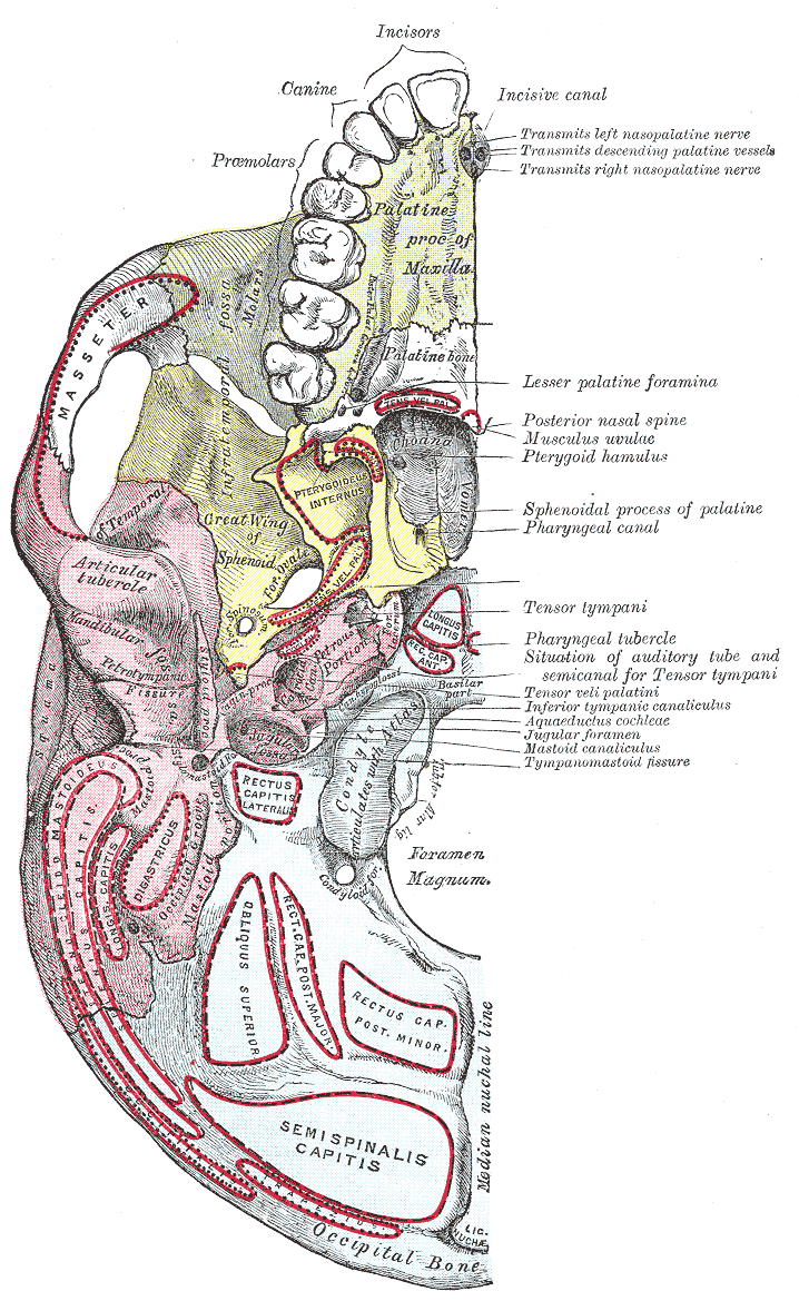
The base of skull, also known as the cranial base or the cranial floor, is the most Anatomical terms of location#Superior and inferior, inferior area of the human skull, skull. It is composed of the endocranium and the lower parts of the Calvaria (skull), calvaria. Structure Structures found at the base of the skull are for example: Bones There are five bones that make up the base of the skull: *Ethmoid bone *Sphenoid bone *Occipital bone *Frontal bone *Temporal bone Sinuses *Occipital sinus *Superior sagittal sinus *Superior petrosal sinus Foramina of the skull *Foramen cecum (frontal bone), Foramen cecum *Optic foramen *Foramen lacerum *Foramen rotundum *Foramen magnum *Foramen ovale (skull), Foramen ovale *Jugular foramen *Internal auditory meatus *Mastoid foramen *Sphenoidal emissary foramen *Foramen spinosum Sutures *Frontoethmoidal suture *Sphenofrontal suture *Sphenopetrosal suture *Sphenoethmoidal suture *Petrosquamous suture *Sphenosquamosal suture Other *Sph ... [...More Info...] [...Related Items...] OR: [Wikipedia] [Google] [Baidu] |
Sacrum
The sacrum (: sacra or sacrums), in human anatomy, is a triangular bone at the base of the spine that forms by the fusing of the sacral vertebrae (S1S5) between ages 18 and 30. The sacrum situates at the upper, back part of the pelvic cavity, between the two wings of the pelvis. It forms joints with four other bones. The two projections at the sides of the sacrum are called the alae (wings), and articulate with the ilium at the L-shaped sacroiliac joints. The upper part of the sacrum connects with the last lumbar vertebra (L5), and its lower part with the coccyx (tailbone) via the sacral and coccygeal cornua. The sacrum has three different surfaces which are shaped to accommodate surrounding pelvic structures. Overall, it is concave (curved upon itself). The base of the sacrum, the broadest and uppermost part, is tilted forward as the sacral promontory internally. The central part is curved outward toward the posterior, allowing greater room for the pelvic cavity. In a ... [...More Info...] [...Related Items...] OR: [Wikipedia] [Google] [Baidu] |
University Of Washington
The University of Washington (UW and informally U-Dub or U Dub) is a public research university in Seattle, Washington, United States. Founded in 1861, the University of Washington is one of the oldest universities on the West Coast of the United States. The university has a main campus located in the city's University District. It also has satellite campuses in nearby cities of Tacoma and Bothell. Overall, UW encompasses more than 500 buildings and over 20 million gross square footage of space, including one of the largest library systems in the world with more than 26 university libraries, art centers, museums, laboratories, lecture halls, and stadiums. Washington is the flagship institution of the six public universities in Washington State. It is known for its medical, engineering, and scientific research. Washington is a member of the Association of American Universities. According to the National Science Foundation, UW spent $1.73 billion on research and develo ... [...More Info...] [...Related Items...] OR: [Wikipedia] [Google] [Baidu] |
Cavernous Sinus
The cavernous sinus within the human head is one of the dural venous sinuses creating a cavity called the lateral sellar compartment bordered by the temporal bone of the skull and the sphenoid bone, lateral to the sella turcica. Structure The cavernous sinus is one of the dural venous sinuses of the head. It is a network of veins that sit in a cavity. It sits on both sides of the sphenoidal bone and pituitary gland, approximately 1 × 2 cm in size in an adult. The carotid siphon of the internal carotid artery, and cranial nerves III, IV, V (branches V1 and V2) and VI all pass through this blood filled space. Both sides of cavernous sinus are connected to each other via intercavernous sinuses. The cavernous sinus lies in between the inner and outer layers of dura mater. Nearby structures * Above: optic tract, optic chiasma, internal carotid artery. * Inferiorly: foramen lacerum, and the junction of the body and greater wing of sphenoid bone. * Medially: p ... [...More Info...] [...Related Items...] OR: [Wikipedia] [Google] [Baidu] |
Cranial Cavity
The cranial cavity, also known as intracranial space, is the space within the skull that accommodates the brain. The skull is also known as the cranium. The cranial cavity is formed by eight cranial bones known as the neurocranium that in humans includes the skull cap and forms the protective case around the brain. The remainder of the skull is the facial skeleton. The meninges are three protective membranes that surround the brain to minimize damage to the brain in the case of head trauma. Meningitis is the inflammation of meninges caused by bacterial or viral infections. Structure The capacity of an adult human cranial cavity is 1,200–1,700 cm3. The spaces between meninges and the brain are filled with a clear cerebrospinal fluid, increasing the protection of the brain. Facial bones of the skull are not included in the cranial cavity. There are only eight cranial bones: The occipital, sphenoid, frontal, ethmoid, two parietal, and two temporal bones are fused to ... [...More Info...] [...Related Items...] OR: [Wikipedia] [Google] [Baidu] |
Radicular Veins
Radicular veins (or segmental medullary veins) are segmental veins providing venous drainage of the spinal cord and canal. They communicate with anterior and posterior spinal veins as well as epidural venous plexus. They exit the spinal canal through the intervertebral foramina The intervertebral foramen (also neural foramen) (often abbreviated as IV foramen or IVF) is an opening between (the intervertebral notches of) two pedicles (one above and one below) of adjacent vertebra in the articulated spine. Each interve ..., accompanying the corresponding of radicular arteries. References Veins {{Circulatory-stub ... [...More Info...] [...Related Items...] OR: [Wikipedia] [Google] [Baidu] |


