|
Eosinophilic Gastroenteritis
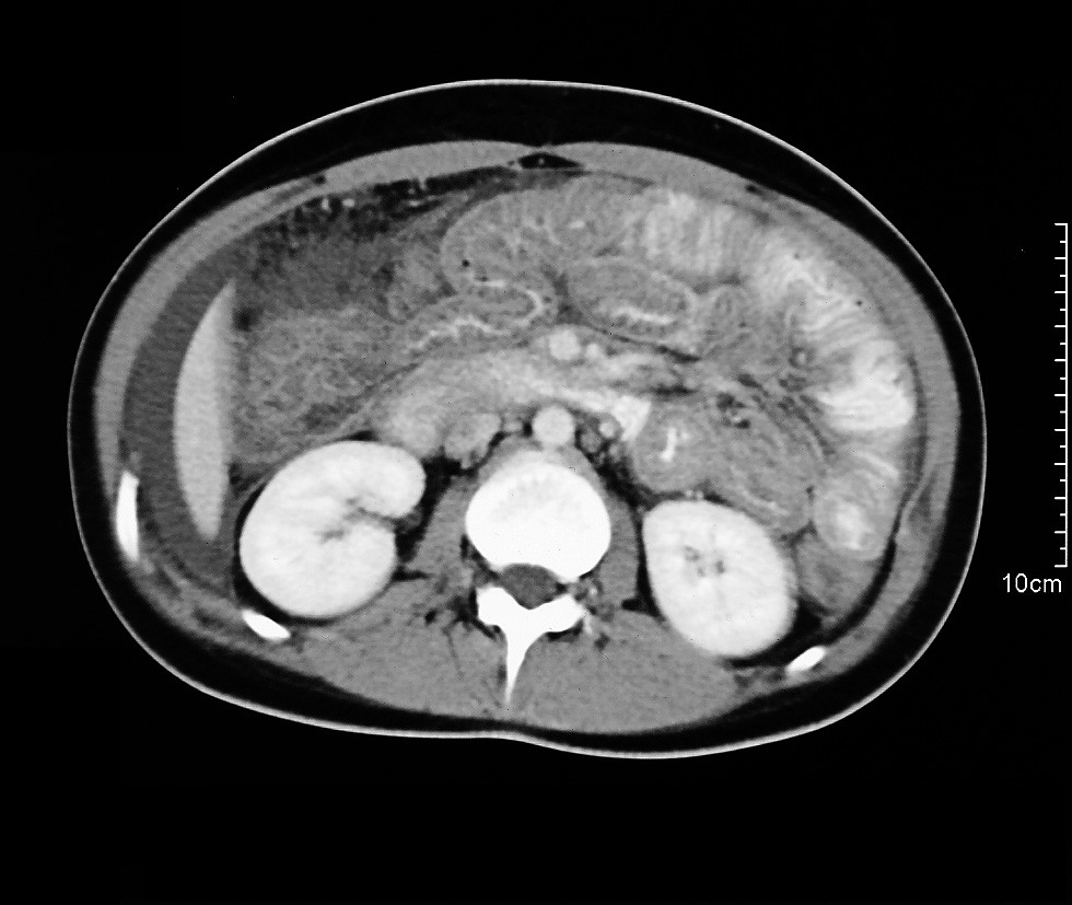
Eosinophilic gastroenteritis (EG or EGE), also known as eosinophilic enteritis, is a rare and heterogeneous condition characterized by patchy or diffuse eosinophilic infiltration of gastrointestinal (GI) tissue, first described by Kaijser in 1937.Kaijser R. Zur Kenntnis der allergischen Affektionen des Verdauugskanals vom Standpunkt des Chirurgen aus. Arch Klin Chir 1937; 188:36–64. Presentation may vary depending on location as well as depth and extent of bowel wall involvement and usually runs a chronic relapsing course. It can be classified into mucosal, muscular and serosal types based on the depth of involvement. Any part of the GI tract can be affected, and isolated biliary tract involvement has also been reported. The stomach is the organ most commonly affected, followed by the small intestine and the colon. Signs and symptoms EG typically presents with a combination of chronic nonspecific GI symptoms which include abdominal pain, diarrhea, occasional nausea and vomitin ... [...More Info...] [...Related Items...] OR: [Wikipedia] [Google] [Baidu] |
Eosinophilic
Eosinophilic (Greek suffix '' -phil'', meaning ''eosin-loving'') describes the staining of tissues, cells, or organelles after they have been washed with eosin, a dye commonly used in histological staining. Eosin is an acidic dye for staining cell cytoplasm, collagen, and muscle fibers. ''Eosinophilic'' describes the appearance of cells and structures seen in histological sections that take up the staining dye eosin. Such eosinophilic structures are, in general, composed of protein. Eosin is usually combined with a stain called hematoxylin to produce a hematoxylin- and eosin-stained section (also called an H&E stain, HE or H+E section). It is the most widely used histological stain for a medical diagnosis. When a pathologist examines a biopsy of a suspected cancer, they will stain the biopsy with H&E. Some structures seen inside cells are described as being eosinophilic; for example, Lewy and Mallory bodies. [...More Info...] [...Related Items...] OR: [Wikipedia] [Google] [Baidu] |
Mucosal
A mucous membrane or mucosa is a membrane that lines various cavities in the body of an organism and covers the surface of internal organs. It consists of one or more layers of epithelial cells overlying a layer of loose connective tissue. It is mostly of endodermal origin and is continuous with the skin at body openings such as the eyes, eyelids, ears, inside the nose, inside the mouth, lips, the genital areas, the urethral opening and the anus. Some mucous membranes secrete mucus, a thick protective fluid. The function of the membrane is to stop pathogens and dirt from entering the body and to prevent bodily tissues from becoming dehydrated. Structure The mucosa is composed of one or more layers of epithelial cells that secrete mucus, and an underlying lamina propria of loose connective tissue. The type of cells and type of mucus secreted vary from organ to organ and each can differ along a given tract. Mucous membranes line the digestive, respiratory and reproductive trac ... [...More Info...] [...Related Items...] OR: [Wikipedia] [Google] [Baidu] |
Duodenal Ulcer
Peptic ulcer disease is when the inner part of the stomach's gastric mucosa (lining of the stomach), the first part of the small intestine, or sometimes the lower esophagus, gets damaged. An ulcer in the stomach is called a gastric ulcer, while one in the first part of the intestines is a duodenal ulcer. The most common symptoms of a duodenal ulcer are waking at night with epigastrium, upper abdominal pain, and upper abdominal pain that improves with eating. With a gastric ulcer, the pain may worsen with eating. The pain is often described as a dyspepsia, burning or dull ache. Other symptoms include belching, vomiting, weight loss, or Anorexia (symptom), poor appetite. About a third of older people with peptic ulcers have no symptoms. Complications may include gastrointestinal bleeding, bleeding, gastrointestinal perforation, perforation, and gastric outlet obstruction, blockage of the stomach. Bleeding occurs in as many as 15% of cases. Common causes include infection with ''Hel ... [...More Info...] [...Related Items...] OR: [Wikipedia] [Google] [Baidu] |
Appendicitis
Appendicitis is inflammation of the Appendix (anatomy), appendix. Symptoms commonly include right lower abdominal pain, nausea, vomiting, fever and anorexia (symptom), decreased appetite. However, approximately 40% of people do not have these typical symptoms. Severe complications of a ruptured appendix include widespread, agonising and awful peritonitis, inflammation of the inner lining of the abdominal wall and sepsis. Appendicitis is primarily caused by a blockage of the Lumen (anatomy), hollow portion in the appendix. This blockage typically results from a Fecalith, faecolith, a calcified "stone" made of feces. Some studies show a correlation between appendicoliths and disease severity. Other factors such as inflamed Mucosa-associated lymphoid tissue, lymphoid tissue from a viral infection, Human parasite, intestinal parasites, gallstone, or Neoplasm, tumors may also lead to this blockage. When the appendix becomes blocked, it experiences increased pressure, reduced blood f ... [...More Info...] [...Related Items...] OR: [Wikipedia] [Google] [Baidu] |
Splenitis
Splenomegaly is an enlargement of the spleen. The spleen usually lies in the left upper quadrant (LUQ) of the human abdomen. Splenomegaly is one of the four cardinal signs of ''hypersplenism'' which include: some reduction in number of circulating blood cells affecting granulocytes, erythrocytes or platelets in any combination; a compensatory proliferative response in the bone marrow; and the potential for correction of these abnormalities by splenectomy. Splenomegaly is usually associated with increased workload (such as in hemolytic anemias), which suggests that it is a response to hyperfunction. It is therefore not surprising that splenomegaly is associated with any disease process that involves abnormal red blood cells being destroyed in the spleen. Other common causes include congestion due to portal hypertension and infiltration by leukemias and lymphomas. Thus, the finding of an enlarged spleen, along with caput medusae, is an important sign of portal hypertension. Definiti ... [...More Info...] [...Related Items...] OR: [Wikipedia] [Google] [Baidu] |
Pancreatitis
Pancreatitis is a condition characterized by inflammation of the pancreas. The pancreas is a large organ behind the stomach that produces digestive enzymes and a number of hormone A hormone (from the Ancient Greek, Greek participle , "setting in motion") is a class of cell signaling, signaling molecules in multicellular organisms that are sent to distant organs or tissues by complex biological processes to regulate physio ...s. There are two main types, acute pancreatitis and chronic pancreatitis. Signs and symptoms of pancreatitis include epigastrium, pain in the upper abdomen, nausea, and vomiting. The pain often goes into the back and is usually severe. In acute pancreatitis, a fever may occur; symptoms typically resolve in a few days. In chronic pancreatitis, weight loss, steatorrhea, fatty stool, and diarrhea may occur. Complications may include infection, bleeding, diabetes mellitus, or problems with other organs. The two most common causes of acute pancreatitis ar ... [...More Info...] [...Related Items...] OR: [Wikipedia] [Google] [Baidu] |
Corticosteroids
Corticosteroids are a class of steroid hormones that are produced in the adrenal cortex of vertebrates, as well as the synthetic analogues of these hormones. Two main classes of corticosteroids, glucocorticoids and mineralocorticoids, are involved in a wide range of physiological processes, including stress response, immune response, and regulation of inflammation, carbohydrate metabolism, protein catabolism, blood electrolyte levels, and behavior. Some common naturally occurring steroid hormones are cortisol (), corticosterone (), cortisone () and aldosterone () (cortisone and aldosterone are isomers). The main corticosteroids produced by the adrenal cortex are cortisol and aldosterone. The etymology of the '' cortico-'' part of the name refers to the adrenal cortex, which makes these steroid hormones. Thus a corticosteroid is a "cortex steroid". Classes * Glucocorticoids such as cortisol affect carbohydrate, fat, and protein metabolism, and have anti-inflam ... [...More Info...] [...Related Items...] OR: [Wikipedia] [Google] [Baidu] |
Serous Membrane
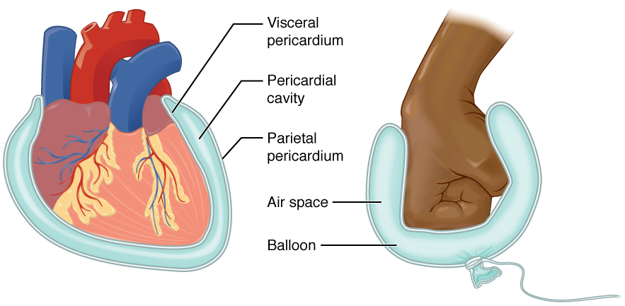
The serous membrane (or serosa) is a smooth epithelial membrane of mesothelium lining the contents and inner walls of body cavity, body cavities, which secrete serous fluid to allow lubricated sliding (motion), sliding movements between opposing surfaces. The serous membrane that covers Viscera, internal organs (viscera) is called ''visceral'', while the one that covers the cavity wall is called ''parietal''. For instance the peritoneum, parietal peritoneum is attached to the abdominal wall and the pelvic walls. The peritoneum, visceral peritoneum is wrapped around the visceral organs. For the heart, the layers of the serous membrane are called parietal and visceral pericardium. For the lungs they are called parietal and visceral pleura. The visceral serosa of the uterus is called the perimetrium. The potential space between two opposing serosal surfaces is mostly empty except for the small amount of serous fluid. The Latin anatomical name is Tunica (biology)#Anatomy usages, tu ... [...More Info...] [...Related Items...] OR: [Wikipedia] [Google] [Baidu] |
Intussusception (medical Disorder)
Intussusception is a medical condition in which a part of the intestine folds into the section immediately ahead of it. It typically involves the small intestine and less commonly the large intestine. Symptoms include abdominal pain which may come and go, vomiting, abdominal bloating, and bloody stool. It often results in a small bowel obstruction. Other complications may include peritonitis or bowel perforation. The cause in children is typically unknown; in adults a ''lead point'' is sometimes present. Risk factors in children include certain infections, diseases like cystic fibrosis, and intestinal polyps. Risk factors in adults include endometriosis, bowel adhesions, and intestinal tumors. Diagnosis is often supported by medical imaging. In children, ultrasound is preferred while in adults a CT scan is preferred. Intussusception is an emergency requiring rapid treatment. Treatment in children is typically by an enema with surgery used if this is not successful. Dexa ... [...More Info...] [...Related Items...] OR: [Wikipedia] [Google] [Baidu] |
Cecum
The cecum ( caecum, ; plural ceca or caeca, ) is a pouch within the peritoneum that is considered to be the beginning of the large intestine. It is typically located on the right side of the body (the same side of the body as the appendix (anatomy), appendix, to which it is joined). The term stems from the Latin ''wikt:caecus, caecus'', meaning "blindness, blind". It receives chyme from the ileum, and connects to the ascending colon of the large intestine. It is separated from the ileum by the ileocecal valve (ICV), also called Bauhin's valve. It is also separated from the Large intestine#Structure, colon by the cecocolic junction. While the cecum is usually intraperitoneal, the ascending colon is Retroperitoneal space, retroperitoneal. In herbivores, the cecum stores food material where bacteria are able to break down the cellulose. In humans, the cecum is involved in absorption of Salt (chemistry), salts and Electrolyte, electrolytes and lubricates the solid waste that pas ... [...More Info...] [...Related Items...] OR: [Wikipedia] [Google] [Baidu] |
Lower Gastrointestinal Bleeding
Lower gastrointestinal bleeding (LGIB) is any form of gastrointestinal bleeding in the lower gastrointestinal tract. LGIB is a common reason for seeking medical attention at a hospital's emergency department. LGIB accounts for 30–40% of all gastrointestinal bleeding and is less common than upper gastrointestinal bleeding (UGIB). It is estimated that UGIB accounts for 100–200 per 100,000 cases versus 20–27 per 100,000 cases for LGIB. Approximately 85% of lower gastrointestinal bleeding involves the large intestine, 10% are from bleeds that are actually upper gastrointestinal bleeds, and 3–5% involve the small intestine. Signs and symptoms A lower gastrointestinal bleed is defined as bleeding originating distal to the ileocecal valve, which includes the colon, rectum, and anus. LGIB was previously defined as any bleed that occurs distal to the ligament of Treitz, which included the aforementioned parts of the intestine and also included the last 1/4 of the duodenum and t ... [...More Info...] [...Related Items...] OR: [Wikipedia] [Google] [Baidu] |


