|
Enamel Pearl
Enamel pearls are developmental variations of teeth that present as beads or nodules of enamel in places where they are not normally observed. Appearance and Location Enamel pearls most commonly present as spheroid in shape, but can also be conical, cylindrical, oval, teardrop, and irregularly shaped. They vary in size with an average diameter of 1.7mm. Enamel pearls are most frequently found in the root furcation of molars, and are not commonly seen in teeth with a single root, such as incisors and canines. Mechanisms of Formation Several theories of enamel pearl development exist, although no definitive mechanism has been established. Enamel pearls are composed primarily of enamel, but most also have a core of dentin within them. The most widely accepted theory suggests enamel pearls are formed from remnants of Hertwig’s epithelial root sheath (HERS), which adhere to the tooth surface after root development. During normal tooth development, after dentin formation is ... [...More Info...] [...Related Items...] OR: [Wikipedia] [Google] [Baidu] |
Dentistry
Dentistry, also known as dental medicine and oral medicine, is the branch of medicine focused on the teeth, gums, and mouth. It consists of the study, diagnosis, prevention, management, and treatment of diseases, disorders, and conditions of the mouth, most commonly focused on dentition (the development and arrangement of teeth) as well as the oral mucosa. Dentistry may also encompass other aspects of the craniofacial complex including the temporomandibular joint. The practitioner is called a dentist. The history of dentistry is almost as ancient as the history of humanity and civilization with the earliest evidence dating from 7000 BC to 5500 BC. Dentistry is thought to have been the first specialization in medicine which have gone on to develop its own accredited degree with its own specializations. Dentistry is often also understood to subsume the now largely defunct medical specialty of stomatology (the study of the mouth and its disorders and diseases) for which ... [...More Info...] [...Related Items...] OR: [Wikipedia] [Google] [Baidu] |
Tooth Enamel
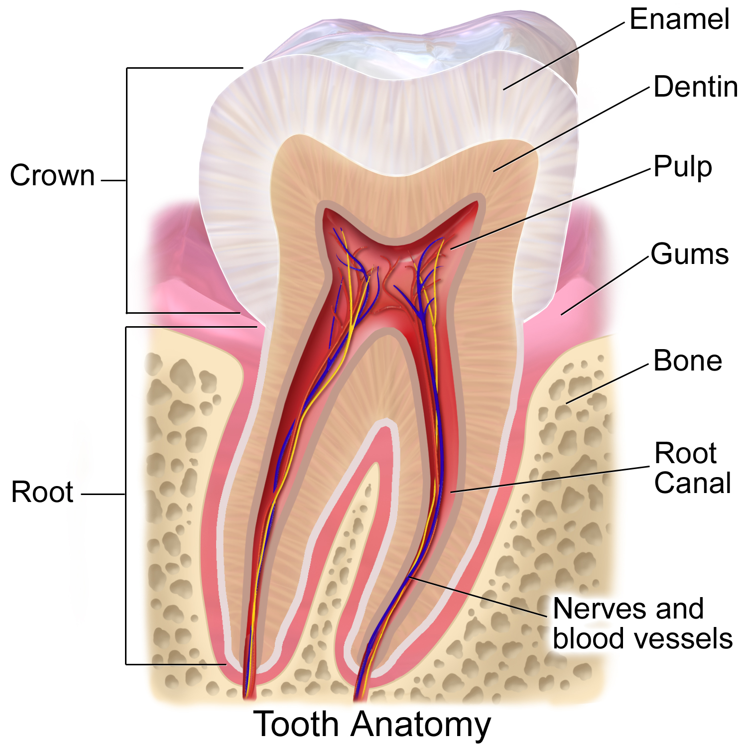
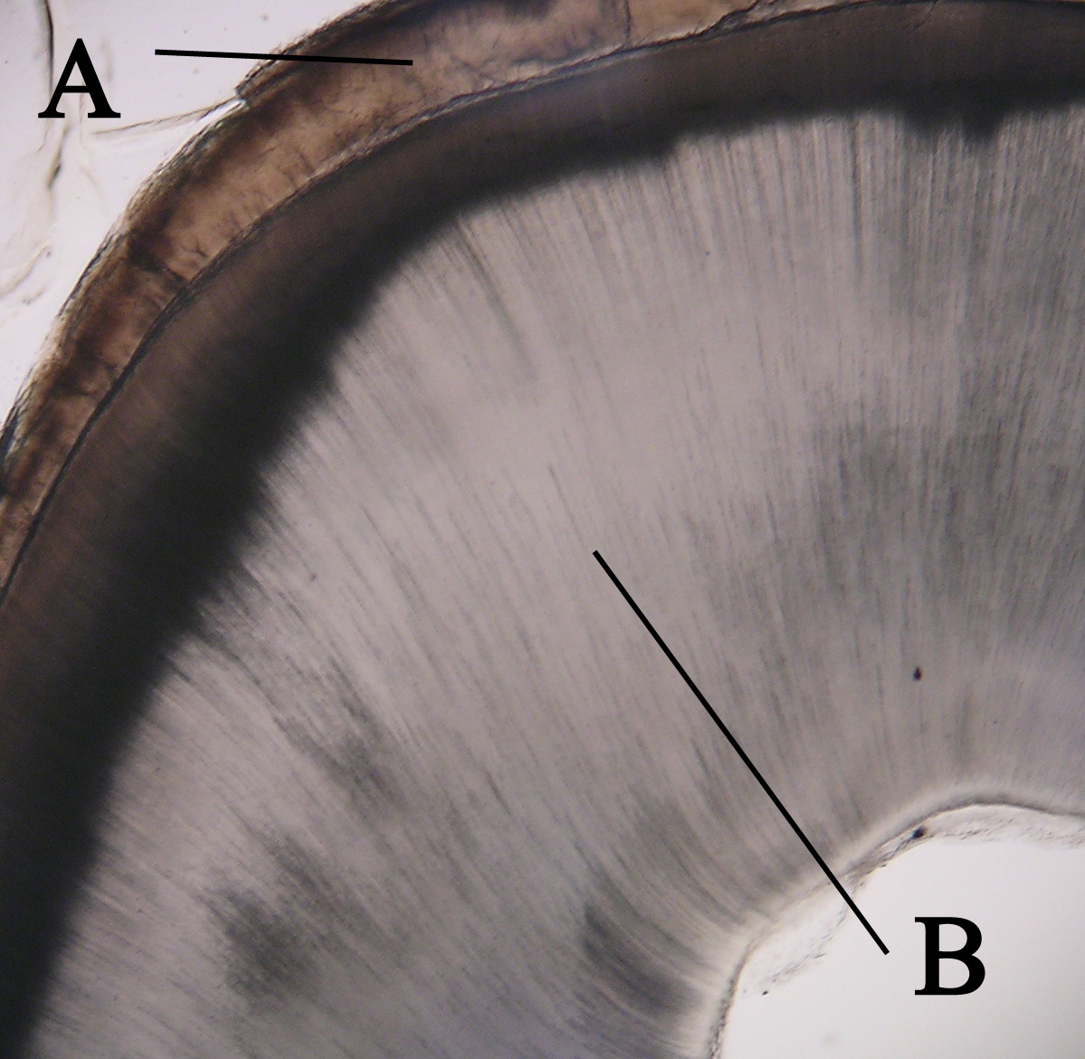
Tooth enamel is one of the four major tissues that make up the tooth in humans and many other animals, including some species of fish. It makes up the normally visible part of the tooth, covering the crown. The other major tissues are dentin, cementum, and dental pulp. It is a very hard, white to off-white, highly mineralised substance that acts as a barrier to protect the tooth but can become susceptible to degradation, especially by acids from food and drink. Calcium hardens the tooth enamel. In rare circumstances enamel fails to form, leaving the underlying dentin exposed on the surface. Features Enamel is the hardest substance in the human body and contains the highest percentage of minerals (at 96%),Ross ''et al.'', p. 485 with water and organic material composing the rest.Ten Cate's Oral Histology, Nancy, Elsevier, pp. 70–94 The primary mineral is hydroxyapatite, which is a crystalline calcium phosphate. Enamel is formed on the tooth while the tooth develops wit ... [...More Info...] [...Related Items...] OR: [Wikipedia] [Google] [Baidu] |
Molar (tooth)
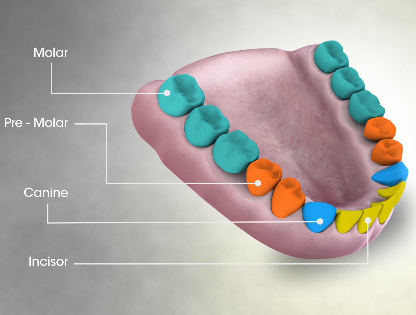
The molars or molar teeth are large, flat teeth A tooth ( : teeth) is a hard, calcified structure found in the jaws (or mouths) of many vertebrates and used to break down food. Some animals, particularly carnivores and omnivores, also use teeth to help with capturing or wounding prey, te ... at the back of the mouth. They are more developed in mammal, mammals. They are used primarily to comminution, grind food during mastication, chewing. The name ''molar'' derives from Latin, ''molaris dens'', meaning "millstone tooth", from ''mola'', millstone and ''dens'', tooth. Molars show a great deal of diversity in size and shape across mammal groups. The third molar of humans is sometimes vestigial. Human anatomy In humans, the molar teeth have either four or five cusp (dentistry), cusps. Adult humans have 12 molars, in four groups of three at the back of the mouth. The third, rearmost molar in each group is called a Third molar, wisdom tooth. It is the last tooth to appear, ... [...More Info...] [...Related Items...] OR: [Wikipedia] [Google] [Baidu] |
Incisor
Incisors (from Latin ''incidere'', "to cut") are the front teeth present in most mammals. They are located in the premaxilla above and on the mandible below. Humans have a total of eight (two on each side, top and bottom). Opossums have 18, whereas armadillos have none. Structure Adult humans normally have eight incisors, two of each type. The types of incisor are: * maxillary central incisor (upper jaw, closest to the center of the lips) * maxillary lateral incisor (upper jaw, beside the maxillary central incisor) * mandibular central incisor (lower jaw, closest to the center of the lips) * mandibular lateral incisor (lower jaw, beside the mandibular central incisor) Children with a full set of deciduous teeth (primary teeth) also have eight incisors, named the same way as in permanent teeth. Young children may have from zero to eight incisors depending on the stage of their tooth eruption and tooth development. Typically, the mandibular central incisors erupt first, ... [...More Info...] [...Related Items...] OR: [Wikipedia] [Google] [Baidu] |
Canine Tooth
In mammalian oral anatomy, the canine teeth, also called cuspids, dog teeth, or (in the context of the upper jaw) fangs, eye teeth, vampire teeth, or vampire fangs, are the relatively long, pointed teeth. They can appear more flattened however, causing them to resemble incisors and leading them to be called ''incisiform''. They developed and are used primarily for firmly holding food in order to tear it apart, and occasionally as weapons. They are often the largest teeth in a mammal's mouth. Individuals of most species that develop them normally have four, two in the upper jaw and two in the lower, separated within each jaw by incisors; humans and dogs are examples. In most species, canines are the anterior-most teeth in the maxillary bone. The four canines in humans are the two maxillary canines and the two mandibular canines. Details There are generally four canine teeth: two in the upper (maxillary) and two in the lower (mandibular) arch. A canine is placed laterall ... [...More Info...] [...Related Items...] OR: [Wikipedia] [Google] [Baidu] |
Dentin
Dentin () (American English) or dentine ( or ) (British English) ( la, substantia eburnea) is a calcified tissue of the body and, along with enamel, cementum, and pulp, is one of the four major components of teeth. It is usually covered by enamel on the crown and cementum on the root and surrounds the entire pulp. By volume, 45% of dentin consists of the mineral hydroxyapatite, 33% is organic material, and 22% is water. Yellow in appearance, it greatly affects the color of a tooth due to the translucency of enamel. Dentin, which is less mineralized and less brittle than enamel, is necessary for the support of enamel. Dentin rates approximately 3 on the Mohs scale of mineral hardness. There are two main characteristics which distinguish dentin from enamel: firstly, dentin forms throughout life; secondly, dentin is sensitive and can become hypersensitive to changes in temperature due to the sensory function of odontoblasts, especially when enamel recedes and dentin channels b ... [...More Info...] [...Related Items...] OR: [Wikipedia] [Google] [Baidu] |
Hertwig's Epithelial Root Sheath
The Hertwig epithelial root sheath (HERS) or epithelial root sheath is a proliferation of epithelial cells located at the cervical loop of the enamel organ in a developing tooth. Hertwig epithelial root sheath initiates the formation of dentin in the root of a tooth by causing the differentiation of odontoblasts from the dental papilla. The root sheath eventually disintegrates with the periodontal ligament, but residual pieces that do not completely disappear are seen as epithelial cell rests of Malassez (ERM). These rests can become cystic, presenting future periodontal infections. Structure Hertwig epithelial root sheath is derived from the inner and outer enamel epithelium of the enamel organ.Illustrated Dental Embryology, Histology, and Anatomy, Bath-Balogh and Fehrenbach, Elsevier, 2011, p. 66 Function The sheath is also responsible for multiple or accessory roots (medial growth) and lateral or accessory canals in the root (break in epithelium).Ten Cate's Oral Histolo ... [...More Info...] [...Related Items...] OR: [Wikipedia] [Google] [Baidu] |
Cementum
Cementum is a specialized calcified substance covering the root of a tooth. The cementum is the part of the periodontium that attaches the teeth to the alveolar bone by anchoring the periodontal ligament.Illustrated Dental Embryology, Histology, and Anatomy, Bath-Balogh and Fehrenbach, Elsevier, 2011, page 170. Structure The cells of cementum are the entrapped cementoblasts, the cementocytes. Each cementocyte lies in its lacuna, similar to the pattern noted in bone. These lacunae also have canaliculi or canals. Unlike those in bone, however, these canals in cementum do not contain nerves, nor do they radiate outward. Instead, the canals are oriented toward the periodontal ligament and contain cementocytic processes that exist to diffuse nutrients from the ligament because it is vascularized. After the apposition of cementum in layers, the cementoblasts that do not become entrapped in cementum line up along the cemental surface along the length of the outer covering of the per ... [...More Info...] [...Related Items...] OR: [Wikipedia] [Google] [Baidu] |
Ameloblast
Ameloblasts are cells present only during tooth development that deposit tooth enamel, which is the hard outermost layer of the tooth forming the surface of the crown. Structure Each ameloblast is a columnar cell approximately 4 micrometers in diameter, 40 micrometers in length and is hexagonal in cross section. The secretory end of the ameloblast ends in a six-sided pyramid-like projection known as the Tomes' process. The angulation of the Tomes' process is significant in the orientation of enamel rods, the basic unit of tooth enamel. Distal terminal bars are junctional complexes that separate the Tomes' processes from ameloblast proper. Development Ameloblasts are derived from oral epithelium tissue of ectodermal origin. Their differentiation from preameloblasts (whose origin is from inner enamel epithelium) is a result of signaling from the ectomesenchymal cells of the dental papilla. Initially the preameloblasts will differentiate into presecretory ameloblasts and then ... [...More Info...] [...Related Items...] OR: [Wikipedia] [Google] [Baidu] |
Pulp (tooth)
The pulp is the connective tissue, nerves, blood vessels, and odontoblasts that comprise the innermost layer of a tooth. The pulp's activity and signalling processes regulate its behaviour. Anatomy The pulp is the neurovascular bundle central to each tooth, permanent or primary. It is composed of a central pulp chamber, pulp horns, and radicular canals. The large mass of the pulp is contained within the pulp chamber, which is contained in and mimics the overall shape of the crown of the tooth.Illustrated Dental Embryology, Histology, and Anatomy, Bath-Balogh and Fehrenbach, Elsevier, 2011, page 164. Because of the continuous deposition of the dentine, the pulp chamber becomes smaller with the age. This is not uniform throughout the coronal pulp but progresses faster on the floor than on the roof or sidewalls. Radicular pulp canals extend down from the cervical region of the crown to the root apex. They are not always straight but vary in shape, size, and number. They a ... [...More Info...] [...Related Items...] OR: [Wikipedia] [Google] [Baidu] |
Developmental Tooth Disorders
Development of the human body is the process of Cell growth, growth to Adulthood, maturity. The process begins with Human fertilization, fertilization, where an egg released from the ovary of a female is penetrated by a spermatozoan, sperm cell from a male. The resulting zygote develops through mitosis and cell differentiation, and the resulting embryo then Implantation (human embryo), implants in the uterus, where the embryo continues development through a fetus, fetal stage until Childbirth, birth. Further growth and development continues after birth, and includes both physical development, physical and psychological development, influenced by Genetics, genetic, hormonal, environmental and other factors. This continues throughout life: through childhood and adolescence into adulthood. Before birth Development before birth, or prenatal development () is the process in which a zygote, and later an embryo and then a fetus develops during gestation. Prenatal development starts wi ... [...More Info...] [...Related Items...] OR: [Wikipedia] [Google] [Baidu] |


