|
Eloquent Cortex
Eloquent cortex is a name used by neurologists for areas of cortex that—if removed—will result in loss of sensory processing or linguistic ability, or paralysis. The most common areas of eloquent cortex are in the left temporal and frontal lobes for speech and language, bilateral occipital lobes for vision, bilateral parietal lobes for sensation, and bilateral motor cortex for movement. Neuroimaging techniques such as magnetic resonance imaging, electroencephalography, or magnetoencephalography are especially useful non-invasive tools to locate eloquent cortices.{{cite journal, last2=Jeong, first2=JW, last3=Brown, first3=EC, last4=Rothermel, first4=R, last5=Kojima, first5=K, last6=Kambara, first6=T, last7=Shah, first7=A, last8=Mittal, first8=S, last9=Sood, first9=S, last10=Asano, first10=E, year=2017, title=Three- and four-dimensional mapping of speech and language in patients with epilepsy, journal=Brain, volume=140, pages=1351–1370, doi=10.1093/brain/awx051, pmid=28334963 ... [...More Info...] [...Related Items...] OR: [Wikipedia] [Google] [Baidu] |
|
 |
Neurology
Neurology (from , "string, nerve" and the suffix wikt:-logia, -logia, "study of") is the branch of specialty (medicine) , medicine dealing with the diagnosis and treatment of all categories of conditions and disease involving the nervous system, which comprises the Human brain, brain, the spinal cord and the peripheral nervous system , peripheral nerves. Neurological practice relies heavily on the field of neuroscience, the scientific study of the nervous system, using various techniques of neurotherapy. IEEE Brain (2019). "Neurotherapy: Treating Disorders by Retraining the Brain". ''The Future Neural Therapeutics White Paper''. Retrieved 23.01.2025 from: https://brain.ieee.org/topics/neurotherapy-treating-disorders-by-retraining-the-brain/#:~:text=Neurotherapy%20trains%20a%20patient's%20brain,wave%20activity%20through%20positive%20reinforcement International Neuromodulation Society, Retrieved 23 January 2025 from: https://www.neuromodulation.com/ Val Danilov I (2023). "The O ... [...More Info...] [...Related Items...] OR: [Wikipedia] [Google] [Baidu] |
 |
Touch
The somatosensory system, or somatic sensory system is a subset of the sensory nervous system. The main functions of the somatosensory system are the perception of external stimuli, the perception of internal stimuli, and the regulation of body position and balance ( proprioception). It is believed to act as a pathway between the different sensory modalities within the body. As of 2024 debate continued on the underlying mechanisms, correctness and validity of the somatosensory system model, and whether it impacts emotions in the body. The somatosensory system has been thought of as having two subdivisions; *one for the detection of mechanosensory information related to touch. Mechanosensory information includes that of light touch, vibration, pressure and tension in the skin. Much of this information belongs to the sense of touch which is a general somatic sense in contrast to the special senses of sight, smell, taste, hearing, and balance. * one for the nociception ... [...More Info...] [...Related Items...] OR: [Wikipedia] [Google] [Baidu] |
|
Subdural Space
The subdural space (or subdural cavity) is a potential space that can be opened by the separation of the arachnoid mater from the dura mater as the result of trauma, pathologic process, or the absence of cerebrospinal fluid as seen in a cadaver. In the cadaver, due to the absence of cerebrospinal fluid in the subarachnoid space, the arachnoid mater falls away from the dura mater. It may also be the site of trauma, such as a subdural hematoma, causing abnormal separation of dura and arachnoid mater. Hence, the subdural space is referred to as " potential" or "artificial" space. See also * Epidural space * Subarachnoid space * Meninges * Subdural hematoma A subdural hematoma (SDH) is a type of bleeding in which a collection of blood—usually but not always associated with a traumatic brain injury—gathers between the inner layer of the dura mater and the arachnoid mater of the meninges surrou ... References External links * * Meninges {{Neuroanatomy-stub ... [...More Info...] [...Related Items...] OR: [Wikipedia] [Google] [Baidu] |
|
|
Electrocorticography
Electrocorticography (ECoG), a type of intracranial electroencephalography (iEEG), is a type of electrophysiological monitoring that uses electrodes placed directly on the exposed surface of the brain to record electrical activity from the cerebral cortex. In contrast, conventional electroencephalography (EEG) electrodes monitor this activity from outside the skull. ECoG may be performed either in the operating room during surgery (intraoperative ECoG) or outside of surgery (extraoperative ECoG). Because a craniotomy (a surgical incision into the skull) is required to implant the electrode grid, ECoG is an invasive procedure. History ECoG was pioneered in the early 1950s by Wilder Penfield and Herbert Jasper, neurosurgeons at the Montreal Neurological Institute. The two developed ECoG as part of their groundbreakinMontreal procedure a surgical protocol used to treat patients with severe epilepsy. The cortical potentials recorded by ECoG were used to identify epileptogen ... [...More Info...] [...Related Items...] OR: [Wikipedia] [Google] [Baidu] |
|
 |
Magnetoencephalography
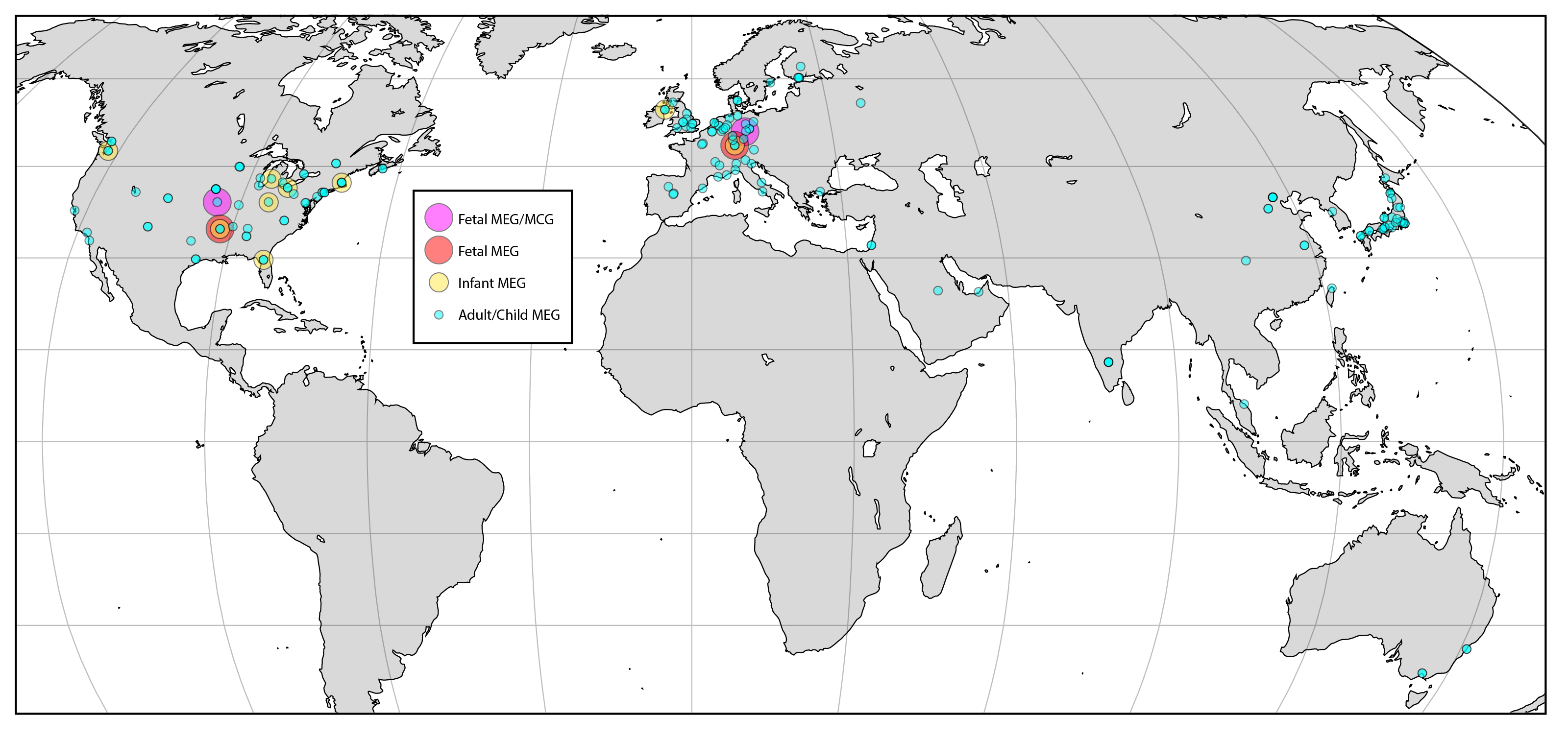
Magnetoencephalography (MEG) is a functional neuroimaging technique for mapping brain activity by recording magnetic fields produced by electric current, electrical currents occurring naturally in the human brain, brain, using very sensitive magnetometers. Arrays of SQUIDs (superconducting quantum interference devices) are currently the most common magnetometer, while the SERF (spin exchange relaxation-free) magnetometer is being investigated for future machines. Applications of MEG include basic research into perceptual and cognitive brain processes, localizing regions affected by pathology before surgical removal, determining the function of various parts of the brain, and neurofeedback. This can be applied in a clinical setting to find locations of abnormalities as well as in an experimental setting to simply measure brain activity. History MEG signals were first measured by University of Illinois physicist David Cohen (physicist), David Cohen in 1968, before the availabili ... [...More Info...] [...Related Items...] OR: [Wikipedia] [Google] [Baidu] |
 |
Electroencephalography
Electroencephalography (EEG) is a method to record an electrogram of the spontaneous electrical activity of the brain. The biosignal, bio signals detected by EEG have been shown to represent the postsynaptic potentials of pyramidal neurons in the neocortex and allocortex. It is typically non-invasive, with the EEG electrodes placed along the scalp (commonly called "scalp EEG") using the 10–20 system (EEG), International 10–20 system, or variations of it. Electrocorticography, involving surgical placement of electrodes, is sometimes called Electrocorticography, "intracranial EEG". Clinical interpretation of EEG recordings is most often performed by visual inspection of the tracing or quantitative EEG, quantitative EEG analysis. Voltage fluctuations measured by the EEG bioamplifier, bio amplifier and electrodes allow the evaluation of normal Brain activity and meditation, brain activity. As the electrical activity monitored by EEG originates in neurons in the underlying Huma ... [...More Info...] [...Related Items...] OR: [Wikipedia] [Google] [Baidu] |
|
Magnetic Resonance Imaging
Magnetic resonance imaging (MRI) is a medical imaging technique used in radiology to generate pictures of the anatomy and the physiological processes inside the body. MRI scanners use strong magnetic fields, magnetic field gradients, and radio waves to form images of the organs in the body. MRI does not involve X-rays or the use of ionizing radiation, which distinguishes it from computed tomography (CT) and positron emission tomography (PET) scans. MRI is a medical application of nuclear magnetic resonance (NMR) which can also be used for imaging in other NMR applications, such as NMR spectroscopy. MRI is widely used in hospitals and clinics for medical diagnosis, staging and follow-up of disease. Compared to CT, MRI provides better contrast in images of soft tissues, e.g. in the brain or abdomen. However, it may be perceived as less comfortable by patients, due to the usually longer and louder measurements with the subject in a long, confining tube, although ... [...More Info...] [...Related Items...] OR: [Wikipedia] [Google] [Baidu] |
|
 |
Neuroimaging
Neuroimaging is the use of quantitative (computational) techniques to study the neuroanatomy, structure and function of the central nervous system, developed as an objective way of scientifically studying the healthy human brain in a non-invasive manner. Increasingly it is also being used for quantitative research studies of brain disease and psychiatric illness. Neuroimaging is highly multidisciplinary involving neuroscience, computer science, psychology and statistics, and is not a medical specialty. Neuroimaging is sometimes confused with neuroradiology. Neuroradiology is a medical specialty that uses non-statistical brain imaging in a clinical setting, practiced by radiologists who are medical practitioners. Neuroradiology primarily focuses on recognizing brain lesions, such as vascular diseases, strokes, tumors, and inflammatory diseases. In contrast to neuroimaging, neuroradiology is qualitative (based on subjective impressions and extensive clinical training) but sometime ... [...More Info...] [...Related Items...] OR: [Wikipedia] [Google] [Baidu] |
 |
Motor Cortex
The motor cortex is the region of the cerebral cortex involved in the planning, motor control, control, and execution of voluntary movements. The motor cortex is an area of the frontal lobe located in the posterior precentral gyrus immediately anterior to the central sulcus. Components The motor cortex can be divided into three areas: 1. The primary motor cortex is the main contributor to generating neural impulses that pass down to the spinal cord and control the execution of movement. However, some of the other motor areas in the brain also play a role in this function. It is located on the anterior paracentral lobule on the medial surface. 2. The premotor cortex is responsible for some aspects of motor control, possibly including the preparation for movement, the sensory guidance of movement, the spatial guidance of reaching, or the direct control of some movements with an emphasis on control of proximal and trunk muscles of the body. Located anterior to the primary mo ... [...More Info...] [...Related Items...] OR: [Wikipedia] [Google] [Baidu] |
 |
Parietal Lobe
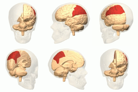
The parietal lobe is one of the four Lobes of the brain, major lobes of the cerebral cortex in the brain of mammals. The parietal lobe is positioned above the temporal lobe and behind the frontal lobe and central sulcus. The parietal lobe integrates sensory information among various sensory modality, modalities, including spatial sense and navigation (proprioception), the main sensory receptive area for the sense of touch in the somatosensory cortex which is just posterior to the central sulcus in the postcentral gyrus, and the two-streams hypothesis#Dorsal stream, dorsal stream of the visual system. The major sensory inputs from the skin (mechanoreceptor, touch, thermoreceptor, temperature, and nociceptor, pain receptors), relay through the thalamus to the parietal lobe. Several areas of the parietal lobe are important in language processing in the brain, language processing. The somatosensory cortex can be illustrated as a distorted figure – the cortical homunculus (Latin: "li ... [...More Info...] [...Related Items...] OR: [Wikipedia] [Google] [Baidu] |
 |
Cerebral Cortex
The cerebral cortex, also known as the cerebral mantle, is the outer layer of neural tissue of the cerebrum of the brain in humans and other mammals. It is the largest site of Neuron, neural integration in the central nervous system, and plays a key role in attention, perception, awareness, thought, memory, language, and consciousness. The six-layered neocortex makes up approximately 90% of the Cortex (anatomy), cortex, with the allocortex making up the remainder. The cortex is divided into left and right parts by the longitudinal fissure, which separates the two cerebral hemispheres that are joined beneath the cortex by the corpus callosum and other commissural fibers. In most mammals, apart from small mammals that have small brains, the cerebral cortex is folded, providing a greater surface area in the confined volume of the neurocranium, cranium. Apart from minimising brain and cranial volume, gyrification, cortical folding is crucial for the Neural circuit, brain circuitry ... [...More Info...] [...Related Items...] OR: [Wikipedia] [Google] [Baidu] |
|
Visual Perception
Visual perception is the ability to detect light and use it to form an image of the surrounding Biophysical environment, environment. Photodetection without image formation is classified as ''light sensing''. In most vertebrates, visual perception can be enabled by photopic vision (daytime vision) or scotopic vision (night vision), with most vertebrates having both. Visual perception detects light (photons) in the visible spectrum reflected by objects in the environment or emitted by light sources. The light, visible range of light is defined by what is readily perceptible to humans, though the visual perception of non-humans often extends beyond the visual spectrum. The resulting perception is also known as vision, sight, or eyesight (adjectives ''visual'', ''optical'', and ''ocular'', respectively). The various physiological components involved in vision are referred to collectively as the visual system, and are the focus of much research in linguistics, psychology, cognitive s ... [...More Info...] [...Related Items...] OR: [Wikipedia] [Google] [Baidu] |