|
Early Pregnancy Bleeding
Early pregnancy bleeding (also called first trimester bleeding) is vaginal bleeding before 13 weeks of gestational age. Early pregnancy bleeding is common and can occur in up to 25% of pregnancies. Many individuals with first trimester bleeding experience no additional complications. However, 50% of pregnancies with first trimester bleeding end in miscarriage. Common causes of early pregnancy bleeding include miscarriage, ectopic pregnancy, and subchorionic hematomas. Other causes include implantation bleeding, gestational trophoblastic disease, cervical changes, or infections. Assessment of first trimester bleeding includes history and physical exam (including speculum examination), imaging using ultrasound, and lab work such as beta-hCG and ABO/Rh blood tests. Treatment depends on the underlying cause. Emergent management is indicated for patients with significant blood loss or hemodynamic instability. Anti-D immune globulin is usually recommended in those who are Rh-n ... [...More Info...] [...Related Items...] OR: [Wikipedia] [Google] [Baidu] |
Obstetrics
Obstetrics is the field of study concentrated on pregnancy, childbirth and the postpartum period. As a medical specialty, obstetrics is combined with gynecology under the discipline known as obstetrics and gynecology (OB/GYN), which is a surgical field. Main areas Prenatal care Prenatal care is important in screening for various complications of pregnancy. This includes routine office visits with physical exams and routine lab tests along with telehealth care for women with low-risk pregnancies: Image:Ultrasound_image_of_a_fetus.jpg, 3D ultrasound of fetus (about 14 weeks gestational age) Image:Sucking his thumb and waving.jpg, Fetus at 17 weeks Image:3dultrasound 20 weeks.jpg, Fetus at 20 weeks First trimester Routine tests in the first trimester of pregnancy generally include: * Complete blood count * Blood type ** Rh-negative antenatal patients should receive RhoGAM at 28 weeks to prevent Rh disease. * Indirect Coombs test (AGT) to assess risk of hem ... [...More Info...] [...Related Items...] OR: [Wikipedia] [Google] [Baidu] |
Rh-negative
The Rh blood group system is a human blood group system. It contains proteins on the surface of red blood cells. After the ABO blood group system, it is most likely to be involved in transfusion reactions. The Rh blood group system consisted of 49 defined blood group antigens in 2005. there are over 50 antigens, of which the five antigens D, C, c, E, and e are among the most prominent. There is no d antigen. Rh(D) status of an individual is normally described with a ''positive'' (+) or ''negative'' (−) suffix after the ABO type (e.g., someone who is A+ has the A antigen and Rh(D) antigen, whereas someone who is A− has the A antigen but lacks the Rh(D) antigen). The terms ''Rh factor'', ''Rh positive'', and ''Rh negative'' refer to the Rh(D) antigen only. Antibodies to Rh antigens can be involved in hemolytic transfusion reactions and antibodies to the Rh(D) and Rh antigens confer significant risk of hemolytic disease of the newborn. Nomenclature The Rh blood group syst ... [...More Info...] [...Related Items...] OR: [Wikipedia] [Google] [Baidu] |
Uterine Rupture
Uterine rupture is when the muscular wall of the uterus tears during pregnancy or childbirth. Symptoms, while classically including increased pain, vaginal bleeding, or a change in contractions, are not always present. Disability or death of the mother or baby may result. Risk factors include vaginal birth after cesarean section (VBAC), other uterine scars, obstructed labor, induction of labor, trauma, and cocaine use. While typically rupture occurs during labor it may occasionally happen earlier in pregnancy. Diagnosis may be suspected based on a rapid drop in the baby's heart rate during labor. Uterine dehiscence is a less severe condition in which there is only incomplete separation of the old scar. Treatment involves rapid surgery to control bleeding and delivery of the baby. A hysterectomy may be required to control the bleeding. Blood transfusions may be given to replace blood loss. Women who have had a prior rupture are generally recommended to have C-sections in sub ... [...More Info...] [...Related Items...] OR: [Wikipedia] [Google] [Baidu] |
Placental Abruption
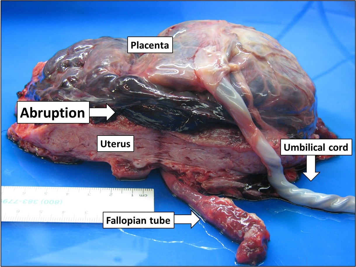
Placental abruption is when the placenta separates early from the uterus, in other words separates before childbirth. It occurs most commonly around 25 weeks of pregnancy. Symptoms may include vaginal bleeding, lower abdominal pain, and dangerously low blood pressure. Complications for the mother can include disseminated intravascular coagulopathy and kidney failure. Complications for the baby can include fetal distress, low birthweight, preterm delivery, and stillbirth. The cause of placental abruption is not entirely clear. Risk factors include smoking, pre-eclampsia, prior abruption (most important and predictive risk factor), trauma during pregnancy, cocaine use, and previous cesarean section. Diagnosis is based on symptoms and supported by ultrasound. It is classified as a complication of pregnancy. For small abruption, bed rest may be recommended, while for more significant abruptions or those that occur near term, delivery may be recommended. If everything is sta ... [...More Info...] [...Related Items...] OR: [Wikipedia] [Google] [Baidu] |
Vasa Praevia
Vasa praevia or vasa previa is a complication of obstetrics in which fetal blood vessels cross or run near the internal opening of the uterus. Since these vessels are not protected by the umbilical cord or placental tissue, the rupture of the fetal membranes during birth causes them also to rupture, leading rapidly to death of the fetus. The term is derived from the Latin; ''vasa'' means "vessels" and ''praevia'' comes from ''pre'' meaning "before" and ''via'' meaning "way". In other words, vessels lie before the fetus in the birth canal and in the way. Risk factors include low-lying placenta and in vitro fertilization. Vasa praevia occurs in about 0.6 per 1,000 pregnancies. Cause In vasa praevia, blood vessels from the fetoplacental circulation lie unprotected on the fetal membranes across or near (within 2 cm) the internal cervical os, either from a velamentous insertion of the umbilical cord or connecting an accessory ( succenturiate) lobe of the placenta to its main ... [...More Info...] [...Related Items...] OR: [Wikipedia] [Google] [Baidu] |
Placenta Praevia
Placenta praevia or placenta previa is when the placenta attaches inside the uterus but in a position near or over the cervical opening. Symptoms include vaginal bleeding in the second half of pregnancy. The bleeding is bright red and tends not to be associated with pain. Complications may include placenta accreta, dangerously low blood pressure, or bleeding after delivery. Complications for the baby may include fetal growth restriction. Risk factors include pregnancy at an older age and smoking as well as prior cesarean section, labor induction, or termination of pregnancy. Diagnosis is by ultrasound. It is classified as a complication of pregnancy. For those who are less than 36 weeks pregnant with only a small amount of bleeding recommendations may include bed rest and avoiding sexual intercourse. For those after 36 weeks of pregnancy or with a significant amount of bleeding, cesarean section is generally recommended. In those less than 36 weeks pregnant, corticostero ... [...More Info...] [...Related Items...] OR: [Wikipedia] [Google] [Baidu] |
Cervix
The cervix (: cervices) or cervix uteri is a dynamic fibromuscular sexual organ of the female reproductive system that connects the vagina with the uterine cavity. The human female cervix has been documented anatomically since at least the time of Hippocrates, over 2,000 years ago. The cervix is approximately 4 cm long with a diameter of approximately 3 cm and tends to be described as a cylindrical shape, although the front and back walls of the cervix are contiguous. The size of the cervix changes throughout a woman's life cycle. For example, women in the fertile years of their reproductive cycle tend to have larger cervixes than postmenopausal women; likewise, women who have produced offspring have a larger cervix than those who have not. In relation to the vagina, the part of the cervix that opens to the uterus is called the ''internal os'' and the opening of the cervix in the vagina is called the ''external os''. Between them is a conduit commonly called the cervic ... [...More Info...] [...Related Items...] OR: [Wikipedia] [Google] [Baidu] |
Gestational Trophoblastic Neoplasia
Gestational trophoblastic disease (GTD) is a term used for a group of pregnancy-related tumours. These tumours are rare, and they appear when cells in the womb start to proliferate uncontrollably. The cells that form gestational trophoblastic tumours are called trophoblasts and come from tissue that grows to form the placenta during pregnancy. There are several different types of GTD. A hydatidiform mole also known as a ''molar pregnancy'', is the most common and is usually benign. Sometimes it may develop into an invasive mole, or, more rarely into a choriocarcinoma. A choriocarcinoma is likely to spread quickly, but is very sensitive to chemotherapy, and has a very good prognosis. Trophoblasts are of particular interest to cell biologists because, like cancer, they can invade tissue (the uterus), but unlike cancer, they usually "know" when to stop. GTD can simulate pregnancy, because the uterus may contain fetal tissue, albeit abnormal. This tissue may grow at the same rate ... [...More Info...] [...Related Items...] OR: [Wikipedia] [Google] [Baidu] |
Uterus
The uterus (from Latin ''uterus'', : uteri or uteruses) or womb () is the hollow organ, organ in the reproductive system of most female mammals, including humans, that accommodates the embryonic development, embryonic and prenatal development, fetal development of one or more Fertilized egg, fertilized eggs until birth. The uterus is a hormone-responsive sex organ that contains uterine gland, glands in its endometrium, lining that secrete uterine milk for embryonic nourishment. (The term ''uterus'' is also applied to analogous structures in some non-mammalian animals.) In humans, the lower end of the uterus is a narrow part known as the Uterine isthmus, isthmus that connects to the cervix, the anterior gateway leading to the vagina. The upper end, the body of the uterus, is connected to the fallopian tubes at the uterine horns; the rounded part, the fundus, is above the openings to the fallopian tubes. The connection of the uterine cavity with a fallopian tube is called the utero ... [...More Info...] [...Related Items...] OR: [Wikipedia] [Google] [Baidu] |
Chorion
The chorion is the outermost fetal membrane around the embryo in mammals, birds and reptiles (amniotes). It is also present around the embryo of other animals, like insects and molluscs. Structure In humans and other therian mammals, the chorion is one of the fetal membranes that exist during pregnancy between the developing fetus and mother. The chorion and the amnion together form the amniotic sac. In humans it is formed by extraembryonic mesoderm and the two layers of trophoblast that surround the embryo and other membranes; the chorionic villi emerge from the chorion, invade the endometrium, and allow the transfer of nutrients from maternal blood to fetal blood. Layers The chorion consists of two layers: an outer formed by the trophoblast, and an inner formed by the extra-embryonic mesoderm. The trophoblast is made up of an internal layer of cubical or prismatic cells, the cytotrophoblast or layer of Langhans, and an external multinucleated layer, the syncytiotro ... [...More Info...] [...Related Items...] OR: [Wikipedia] [Google] [Baidu] |
Hematoma
A hematoma, also spelled haematoma, or blood suffusion is a localized bleeding outside of blood vessels, due to either disease or trauma including injury or surgery and may involve blood continuing to seep from broken capillaries. A hematoma is benign and is initially in liquid form spread among the tissues including in sacs between tissues where it may coagulate and solidify before blood is reabsorbed into blood vessels. An ecchymosis is a hematoma of the skin larger than 10 mm. They may occur among and or within many areas such as skin and other organs, connective tissues, bone, joints and muscle. A collection of blood (or even a hemorrhage) may be aggravated by anticoagulant medication (blood thinner). Blood seepage and collection of blood may occur if heparin is given via an intramuscular route; to avoid this, heparin must be given intravenously or subcutaneously. Signs and symptoms Some hematomas are visible under the surface of the skin (commonly called bruise ... [...More Info...] [...Related Items...] OR: [Wikipedia] [Google] [Baidu] |
Implantation Bleeding
Implantation, also known as nidation, is the stage in the mammalian embryonic development in which the blastocyst hatches, attaches, adheres, and penetrates into the endometrium of the female's uterus. Implantation is the first stage of gestation, and, when successful, the female is considered to be pregnant. An implanted embryo is detected by the presence of increased levels of human chorionic gonadotropin (hCG) in a pregnancy test. The implanted embryo will receive oxygen and nutrients in order to grow. For implantation to take place the uterus must become receptive. Uterine receptivity involves much cross-talk between the embryo and the uterus, initiating changes to the endometrium. This stage gives a synchrony that opens a window of implantation that enables successful implantation of a viable embryo. The endocannabinoid system plays a vital role in this synchrony in the uterus, influencing uterine receptivity, and embryo implantation. The embryo expresses cannabinoid recep ... [...More Info...] [...Related Items...] OR: [Wikipedia] [Google] [Baidu] |


