|
Dyssynergic Defecation
Anismus or dyssynergic defecation is the failure of normal relaxation of pelvic floor muscles during attempted defecation. It can occur in both children and adults, and in both men and women (although it is more common in women). It can be caused by physical defects or it can occur for other reasons or unknown reasons. Anismus that has a behavioral cause could be viewed as having similarities with parcopresis, or psychogenic fecal retention. Symptoms include tenesmus (the sensation of incomplete emptying of the rectum after defecation has occurred) and constipation. Retention of stool may result in fecal loading (retention of a mass of stool of any consistency) or fecal impaction (retention of a mass of hard stool). This mass may stretch the walls of the rectum and colon, causing megarectum and/or megacolon, respectively. Liquid stool may leak around a fecal impaction, possibly causing degrees of liquid fecal incontinence. This is usually termed encopresis or soiling in children ... [...More Info...] [...Related Items...] OR: [Wikipedia] [Google] [Baidu] |
Defecating Proctogram
Defecography (also known as proctography, defecating/defecation proctography, evacuating/evacuation proctography or dynamic rectal examination) is a type of medical radiology, radiological imaging in which the mechanics of a patient's defecation are visualized in real time using a fluoroscope. The anatomy and function of the anorectum and pelvic floor can be dynamically studied at various stages during defecation. History Defecating proctography was pioneered in 1945, during World War II. The procedure gained popularity at this time in the midst of an outbreak of whipworm, which is known to cause rectal prolapse. It has since become used for diagnosis of various anorectal disorders, including anismus and other causes of obstructed defecation. It has fallen out of favor due to inadequate training in the technique. It is now only performed at a few institutions. Many radiology residents refer to the procedure as the "Def Proc", "Defogram", or "Stool Finale". Indications Defecograp ... [...More Info...] [...Related Items...] OR: [Wikipedia] [Google] [Baidu] |
Digital Rectal Examination
Digital rectal examination (DRE), also known as a prostate exam (), is an internal examination of the rectum performed by a healthcare provider. Prior to a 2018 report from the United States Preventive Services Task Force, a digital exam was a common component of annual medical examination for older men, as it was thought to be a reliable screening test for prostate cancer. Usage This examination may be used: * for the diagnosis of prostatic disorders, benign prostatic hyperplasia and the four types of prostatitis. Chronic prostatitis/chronic pelvic pain syndrome, chronic bacterial prostatitis, acute (sudden) bacterial prostatitis, and asymptomatic inflammatory prostatitis. The DRE has a 50% specificity for benign prostatic hyperplasia. Vigorous examination of the prostate in suspected acute prostatitis can lead to seeding of septic emboli and should never be done. Its utility as a screening method for prostate cancer however is not supported by the evidence; * for the eva ... [...More Info...] [...Related Items...] OR: [Wikipedia] [Google] [Baidu] |
Peristalsis
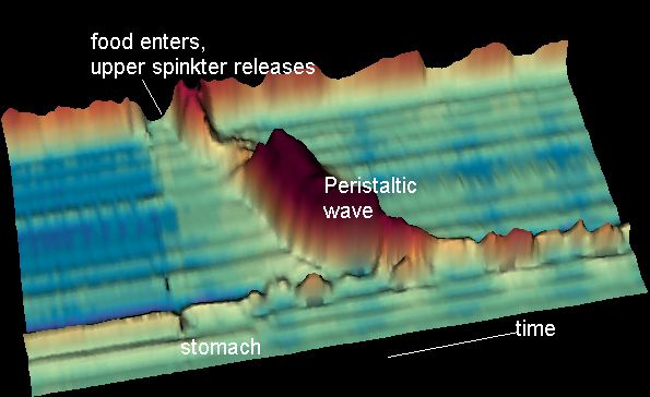
Peristalsis ( , ) is a type of intestinal motility, characterized by symmetry in biology#Radial symmetry, radially symmetrical contraction and relaxation of muscles that propagate in a wave down a tube, in an wikt:anterograde, anterograde direction. Peristalsis is progression of coordinated contraction of involuntary circular muscles, which is preceded by a simultaneous contraction of the longitudinal muscle and relaxation of the circular muscle in the lining of the gut. In much of a digestive tract, such as the human gastrointestinal tract, smooth muscle tissue contracts in sequence to produce a peristaltic wave, which propels a ball of food (called a bolus (digestion), bolus before being transformed into chyme in the stomach) along the tract. The peristaltic movement comprises relaxation of circular smooth muscles, then their contraction behind the chewed material to keep it from moving backward, then longitudinal contraction to push it forward. Earthworms use a similar mec ... [...More Info...] [...Related Items...] OR: [Wikipedia] [Google] [Baidu] |
Human Defecation Posture
Humans mostly use one of two types of defecation postures to defecate: squatting and sitting. People use the squatting postures when using squat toilets or when defecating in the open in the absence of toilets. The sitting posture on the other hand is used in toilets that have a pedestal or "throne", where users generally lean forward or sit at 90 degrees to a toilet seat. Sitting The sitting defecation posture involves sitting with hips and knees at approximately right angles, as on a chair. So-called "Western-style" flush toilets and also many types of dry toilets are designed to be used in a sitting posture. In Europe, America and other western countries most people are accustomed to sitting toilets, although this fashion has only been present for around 100 years. Sitting toilets only came into widespread use in Europe in the nineteenth century. Sitting toilets requires users to strain in an unnatural position. In the sitting position, the puborectalis muscle chokes the ... [...More Info...] [...Related Items...] OR: [Wikipedia] [Google] [Baidu] |
Pubic Bone
In vertebrates, the pubis or pubic bone () forms the lower and anterior part of each side of the hip bone. The pubis is the most forward-facing (ventral and anterior) of the three bones that make up the hip bone. The left and right pubic bones are each made up of three sections; a superior ramus, an inferior ramus, and a body. Structure The pubic bone is made up of a ''body'', ''superior ramus'', and ''inferior ramus'' (). The left and right coxal bones join at the pubic symphysis. It is covered by a layer of fat – the mons pubis. The pubis is the lower limit of the suprapubic region. In the female, the pubis is anterior to the urethral sponge. Body The body of pubis has: * a superior border or the pubic crest * a pubic tubercle at the lateral end of the pubic crest * three surfaces (anterior, posterior and medial). The body forms the wide, strong, middle and flat part of the pubic bone. The bodies of the left and right pubic bones join at the pubic symphysis. The rough u ... [...More Info...] [...Related Items...] OR: [Wikipedia] [Google] [Baidu] |
Skeletal Muscle
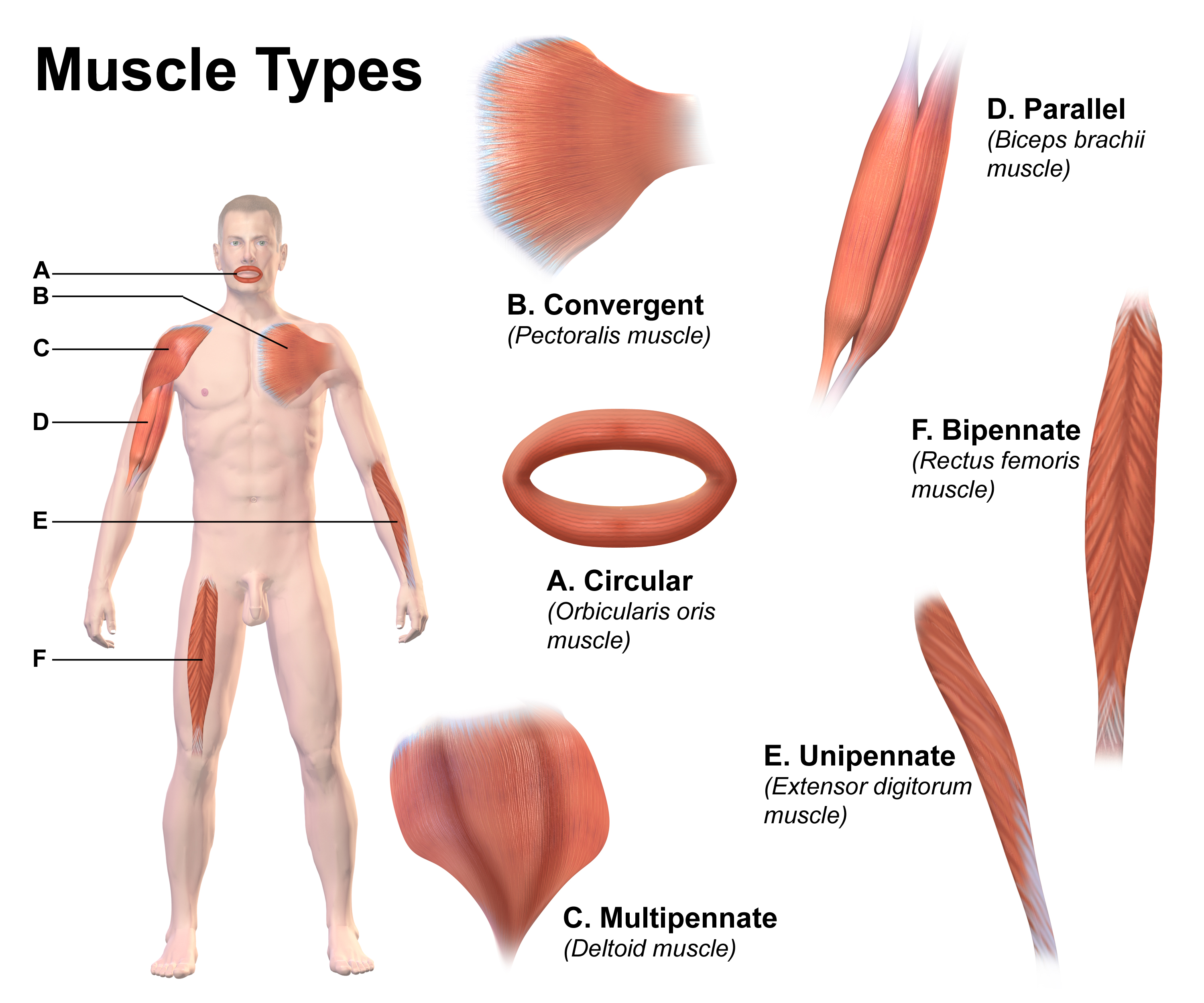
Skeletal muscle (commonly referred to as muscle) is one of the three types of vertebrate muscle tissue, the others being cardiac muscle and smooth muscle. They are part of the somatic nervous system, voluntary muscular system and typically are attached by tendons to bones of a skeleton. The skeletal muscle cells are much longer than in the other types of muscle tissue, and are also known as ''muscle fibers''. The tissue of a skeletal muscle is striated muscle tissue, striated – having a striped appearance due to the arrangement of the sarcomeres. A skeletal muscle contains multiple muscle fascicle, fascicles – bundles of muscle fibers. Each individual fiber and each muscle is surrounded by a type of connective tissue layer of fascia. Muscle fibers are formed from the cell fusion, fusion of developmental myoblasts in a process known as myogenesis resulting in long multinucleated cells. In these cells, the cell nucleus, nuclei, termed ''myonuclei'', are located along the inside ... [...More Info...] [...Related Items...] OR: [Wikipedia] [Google] [Baidu] |
Sphincter Ani Internus Muscle
The internal anal sphincter, IAS, or sphincter ani internus is a ring of smooth muscle that surrounds about 2.5–4.0 cm of the anal canal. It is about 5 mm thick, and is formed by an aggregation of the smooth (involuntary) circular muscle fibers of the rectum. The internal anal sphincter aids the sphincter ani externus to occlude the anal aperture and aids in the expulsion of the feces. Its action is entirely involuntary. It is normally in a state of continuous maximal contraction to prevent leakage of faeces or gases. Sympathetic stimulation stimulates and maintains the sphincter's contraction, and parasympathetic stimulation inhibits it. It becomes relaxed in response to distention of the rectal ampulla, requiring voluntary contraction of the puborectalis and external anal sphincter to maintain continence, and also contracts during the bulbospongiosus reflex. Structure The internal anal sphincter is the specialised thickened terminal portion of the inner circular ... [...More Info...] [...Related Items...] OR: [Wikipedia] [Google] [Baidu] |
Human Anus
In humans, the anus (: anuses or ani; from Latin ''ānus'', "ring", "circle") is the external opening of the rectum located inside the intergluteal cleft. Two sphincters control the exit of Human feces, feces from the body during an act of defecation, which is the primary function of the anus. These are the internal anal sphincter and the external anal sphincter, which are circular muscles that normally maintain constriction of the orifice and which relax as required by normal physiological functioning. The inner sphincter is involuntary and the outer is voluntary. Above the anus is the perineum, which is also located beneath the vulva or scrotum. In part owing to its exposure to feces, a number of medical conditions may affect the anus, such as hemorrhoids. The anus is the site of potential infections and other conditions, including cancer (see anal cancer). With anal sex, the anus can play a role in Human sexuality, sexuality. Attitudes toward anal sex vary, and it is illeg ... [...More Info...] [...Related Items...] OR: [Wikipedia] [Google] [Baidu] |
Large Intestine
The large intestine, also known as the large bowel, is the last part of the gastrointestinal tract and of the Digestion, digestive system in tetrapods. Water is absorbed here and the remaining waste material is stored in the rectum as feces before being removed by defecation. The Colon (anatomy), colon (progressing from the ascending colon to the transverse colon, transverse, the descending colon, descending and finally the sigmoid colon) is the longest portion of the large intestine, and the terms "large intestine" and "colon" are often used interchangeably, but most sources define the large intestine as the combination of the cecum, colon, rectum, and anal canal. Some other sources exclude the anal canal. In humans, the large intestine begins in the right iliac region of the pelvis, just at or below the waist, where it is joined to the end of the small intestine at the cecum, via the ileocecal valve. It then continues as the colon ascending colon, ascending the abdomen, across t ... [...More Info...] [...Related Items...] OR: [Wikipedia] [Google] [Baidu] |
Sigmoid Colon
The sigmoid colon (or pelvic colon) is the part of the large intestine that is closest to the rectum and anus. It forms a loop that averages about in length. The loop is typically shaped like a Greek letter sigma (ς) or Latin letter S (thus ''sigma'' + '' -oid''). This part of the colon normally lies within the pelvis, but due to its freedom of movement it is liable to be displaced into the abdominal cavity. Structure The sigmoid colon begins at the superior aperture of the lesser pelvis, where it is continuous with the iliac colon, and passes transversely across the front of the sacrum to the right side of the pelvis. It then curves on itself and turns toward the left to reach the middle line at the level of the third piece of the sacrum, where it bends downward and ends in the rectum. Its function is to expel solid and gaseous waste from the gastrointestinal tract. The curving path it takes toward the anus allows it to store gas in the superior arched portion, enabling ... [...More Info...] [...Related Items...] OR: [Wikipedia] [Google] [Baidu] |
Sphincter Ani Externus Muscle
The external anal sphincter (or sphincter ani externus) is an oval tube of Skeletal muscle#Skeletal muscle cells, skeletal muscle fibers. Distally, it is adherent to the integument, skin surrounding the margin of the anus. It exhibits a resting state of tonical contraction and also contracts during the bulbospongiosus reflex. Anatomy The external anal sphincter is far more substantial than the internal anal sphincter. The proximal portion of external anal sphincter overlaps the internal anal sphincter (which terminates distally a little distance proximal to the anal orifice) superficially; where the two overlap, they are separated by the intervening conjoint longitudinal muscle. Structure Historically, the sphincter was described as consisting of three parts (deep, superficial, and subcutaneous). This is not supported by current anatomical knowledge. Some sources still describe it in two layers, deep (or proximal) and superficial (or distal or subcutaneous). Some of the muscles ... [...More Info...] [...Related Items...] OR: [Wikipedia] [Google] [Baidu] |
Puborectalis
The levator ani is a broad, thin muscle group, situated on either side of the pelvis. It is formed from three muscle components: the pubococcygeus, the iliococcygeus, and the puborectalis. It is attached to the inner surface of each side of the lesser pelvis, and these unite to form the greater part of the pelvic floor. The coccygeus muscle completes the pelvic floor, which is also called the ''pelvic diaphragm''. It supports the viscera in the pelvic cavity, and surrounds the various structures that pass through it. The levator ani is the main pelvic floor muscle and contracts rhythmically during female orgasm, and painfully during vaginismus. Structure The levator ani is made up of 3 parts: * Iliococcygeus muscle * Pubococcygeus muscle * Puborectalis muscle The iliococcygeus arises from the inner side of the ischium (the lower and back part of the hip bone) and from the posterior part of the tendinous arch of the obturator fascia, and is attached to the coccyx and anoco ... [...More Info...] [...Related Items...] OR: [Wikipedia] [Google] [Baidu] |


