|
Dok-7
Dok-7 is a non-catalytic cytoplasmic adaptor protein that is expressed specifically in muscle and is essential for the formation of neuromuscular synapses. Further, Dok-7 contains pleckstrin homology (PH) and phosphotyrosine-binding (PTB) domains that are critical for Dok-7 function. Finally, mutations in Dok-7 are commonly found in patients with limb-girdle congenital myasthenia. Dok-7 regulates neuromuscular synapse formation by activating MuSK The formation of neuromuscular synapses requires the muscle-specific receptor tyrosine kinase (MuSK). In mice genetically mutant for MuSK, acetylcholine receptors (AChRs) fail to cluster and motor neurons fail to differentiate. Because Dok-7 mutant mice are indistinguishable from MuSK mutant mice, these observations suggest Dok-7 might regulate MuSK activation. Indeed, Dok-7 binds phosphorylated MuSK and activates MuSK in purified protein preparations and in muscle in-vivo by transgenic overexpression. Furthermore, the nerve-derived orga ... [...More Info...] [...Related Items...] OR: [Wikipedia] [Google] [Baidu] |
MuSK Protein
MuSK (for Muscle-Specific Kinase) is a receptor tyrosine kinase required for the formation and maintenance of the neuromuscular junction. It is activated by a nerve-derived proteoglycan called agrin, which is similarly also required for neuromuscular junction formation. MuSK signaling Upon activation by its ligand agrin, MuSK signals via the proteins called casein kinase 2 (CK2), Dok-7 and rapsyn, to induce "clustering" of acetylcholine receptors ( AChR). Both CK2 and Dok-7 are required for MuSK-induced formation of the neuromuscular junction, since mice lacking Dok-7 failed to form AChR clusters or neuromuscular synapses, and since downregulation of CK2 also impedes recruitment of AChR to the primary MuSK scaffold. In addition to the proteins mentioned, other proteins are then gathered, to form the endplate to the neuromuscular junction. The nerve terminates onto the endplate, forming the neuromuscular junction - a structure required to transmit nerve impulses to the muscle, a ... [...More Info...] [...Related Items...] OR: [Wikipedia] [Google] [Baidu] |
Neuromuscular Synapse
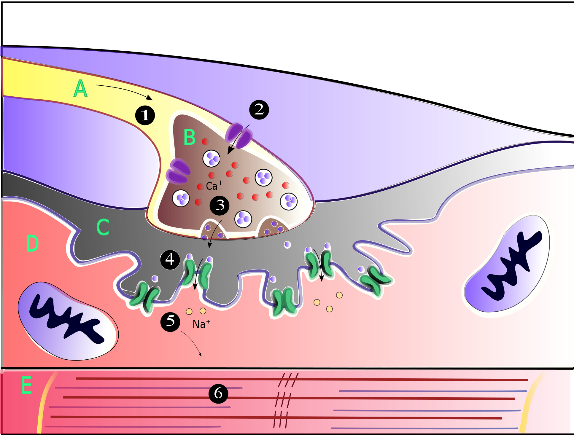
A neuromuscular junction (or myoneural junction) is a chemical synapse between a motor neuron and a muscle fiber. It allows the motor neuron to transmit a signal to the muscle fiber, causing muscle contraction. Muscles require innervation to function—and even just to maintain muscle tone, avoiding atrophy. In the neuromuscular system, nerves from the central nervous system and the peripheral nervous system are linked and work together with muscles. Synaptic transmission at the neuromuscular junction begins when an action potential reaches the presynaptic terminal of a motor neuron, which activates voltage-gated calcium channels to allow calcium ions to enter the neuron. Calcium ions bind to sensor proteins (synaptotagmins) on synaptic vesicles, triggering vesicle fusion with the cell membrane and subsequent neurotransmitter release from the motor neuron into the synaptic cleft. In vertebrates, motor neurons release acetylcholine (ACh), a small molecule neurotransmitter, which ... [...More Info...] [...Related Items...] OR: [Wikipedia] [Google] [Baidu] |
Salbutamol
Salbutamol, also known as albuterol and sold under the brand name Ventolin among others, is a medication that opens up the medium and large airways in the lungs. It is a short-acting β2 adrenergic receptor agonist that causes relaxation of airway smooth muscle. It is used to treat asthma, including asthma attacks and exercise-induced bronchoconstriction, as well as chronic obstructive pulmonary disease (COPD). It may also be used to treat high blood potassium levels. Salbutamol is usually used with an inhaler or nebulizer, but it is also available in a pill, liquid, and intravenous solution. Onset of action of the inhaled version is typically within 15 minutes and lasts for two to six hours. Common side effects include shakiness, headache, fast heart rate, dizziness, and feeling anxious. Serious side effects may include worsening bronchospasm, irregular heartbeat, and low blood potassium levels. It can be used during pregnancy and breastfeeding, but safety is no ... [...More Info...] [...Related Items...] OR: [Wikipedia] [Google] [Baidu] |
Congenital Myasthenic Syndrome
Congenital myasthenic syndrome (CMS) is an inherited neuromuscular disorder caused by defects of several types at the neuromuscular junction. The effects of the disease are similar to Lambert-Eaton Syndrome and myasthenia gravis, the difference being that CMS is not an autoimmune disorder. There are only 600 known family cases of this disorder and it is estimated that its overall frequency in the human population is 1 in 200,000. Types The types of CMS are classified into three categories: presynaptic, postsynaptic, and synaptic. * ''Presynaptic'' symptoms include brief stops in breathing, weakness of the eye, mouth, and throat muscles. These symptoms often result in double vision and difficulty chewing and swallowing. * ''Postsynaptic'' symptoms in infants include severe muscle weakness, feeding and respiratory problems, and delays in the ability to sit, crawl, and walk. * ''Synaptic'' symptoms include early childhood feeding and respiratory problems, reduced mobility, curvat ... [...More Info...] [...Related Items...] OR: [Wikipedia] [Google] [Baidu] |
Phosphotyrosine-binding Domain
In molecular biology, phosphotyrosine-binding domains are protein domains which bind to phosphotyrosine. The phosphotyrosine-binding domain (PTB, also phosphotyrosine-interaction or PI domain) in the protein tensin tends to be found at the C-terminus. Tensin is a multi-domain protein that binds to actin filaments and functions as a focal-adhesion molecule (focal adhesions are regions of plasma membrane through which cells attach to the extracellular matrix). Human tensin has actin-binding sites, an SH2 () domain and a region similar to the tumour suppressor PTEN. The PTB domain interacts with the cytoplasmic tails of beta integrin by binding to an NPXY motif. The phosphotyrosine-binding domain of insulin receptor substrate-1 is not related to the phosphotyrosine-binding domain of tensin. Insulin receptor substrate-1 proteins contain both a pleckstrin homology domain and a phosphotyrosine binding (PTB) domain. The PTB domains facilitate interaction with the activated t ... [...More Info...] [...Related Items...] OR: [Wikipedia] [Google] [Baidu] |
Agrin
Agrin is a large proteoglycan whose best-characterised role is in the development of the neuromuscular junction during embryogenesis. Agrin is named based on its involvement in the aggregation of acetylcholine receptors during synaptogenesis. In humans, this protein is encoded by the ''AGRN'' gene. This protein has nine domains homologous to protease inhibitors. It may also have functions in other tissues and during other stages of development. It is a major proteoglycan component in the glomerular basement membrane and may play a role in the renal filtration and cell-matrix interactions. Agrin functions by activating the MuSK protein (for Muscle-Specific Kinase), which is a receptor tyrosine kinase required for the formation and maintenance of the neuromuscular junction. Agrin is required to activate MuSK. Agrin is also required for neuromuscular junction formation. Discovery Agrin was first identified by the U.J. McMahan laboratory, Stanford University. Mechani ... [...More Info...] [...Related Items...] OR: [Wikipedia] [Google] [Baidu] |
PTB Domain
In molecular biology, phosphotyrosine-binding domains are protein domains which bind to phosphotyrosine. The phosphotyrosine-binding domain (PTB, also phosphotyrosine-interaction or PI domain) in the protein tensin tends to be found at the C-terminus. Tensin is a multi-domain protein that binds to actin filaments and functions as a focal-adhesion molecule (focal adhesions are regions of plasma membrane through which cells attach to the extracellular matrix). Human tensin has actin-binding sites, an SH2 () domain and a region similar to the tumour suppressor PTEN. The PTB domain interacts with the cytoplasmic tails of beta integrin by binding to an NPXY motif. The phosphotyrosine-binding domain of insulin receptor substrate-1 is not related to the phosphotyrosine-binding domain of tensin. Insulin receptor substrate-1 proteins contain both a pleckstrin homology domain and a phosphotyrosine binding (PTB) domain. The PTB domains facilitate interaction with the activated tyro ... [...More Info...] [...Related Items...] OR: [Wikipedia] [Google] [Baidu] |
Signal Transducing Adaptor Protein
Signal transducing adaptor proteins (STAPs) are proteins that are accessory to main proteins in a signal transduction pathway. Adaptor proteins contain a variety of protein-binding modules that link protein-binding partners together and facilitate the creation of larger signaling complexes. These proteins tend to lack any intrinsic enzymatic activity themselves, instead mediating specific protein–protein interactions that drive the formation of Multiprotein complex, protein complexes. Examples of adaptor proteins include MYD88, Grb2 and SHC1. Signaling components Much of the specificity of signal transduction depends on the recruitment of several signalling components such as protein kinases and G-protein GTPases into short-lived active complexes in response to an activating signal such as a growth factor binding to its receptor (biochemistry), receptor. Domains Adaptor proteins usually contain several domains within their structure (e.g., SH2 domain, Src homology 2 (SH2) and S ... [...More Info...] [...Related Items...] OR: [Wikipedia] [Google] [Baidu] |
Cell Signaling
In biology, cell signaling (cell signalling in British English) is the Biological process, process by which a Cell (biology), cell interacts with itself, other cells, and the environment. Cell signaling is a fundamental property of all Cell (biology), cellular life in both prokaryotes and eukaryotes. Typically, the signaling process involves three components: the signal, the receptor, and the effector. In biology, signals are mostly chemical in nature, but can also be physical cues such as pressure, Membrane potential, voltage, temperature, or light. Chemical signals are molecules with the ability to bind and activate a specific Receptor (biochemistry), receptor. These molecules, also referred to as Ligand (biochemistry), ligands, are chemically diverse, including ions (e.g. Na+, K+, Ca2+, etc.), lipids (e.g. steroid, prostaglandin), peptides (e.g. insulin, ACTH), carbohydrates, glycosylated proteins (proteoglycans), nucleic acids, etc. Peptide and lipid ligands are particularly ... [...More Info...] [...Related Items...] OR: [Wikipedia] [Google] [Baidu] |
Muscle Contraction
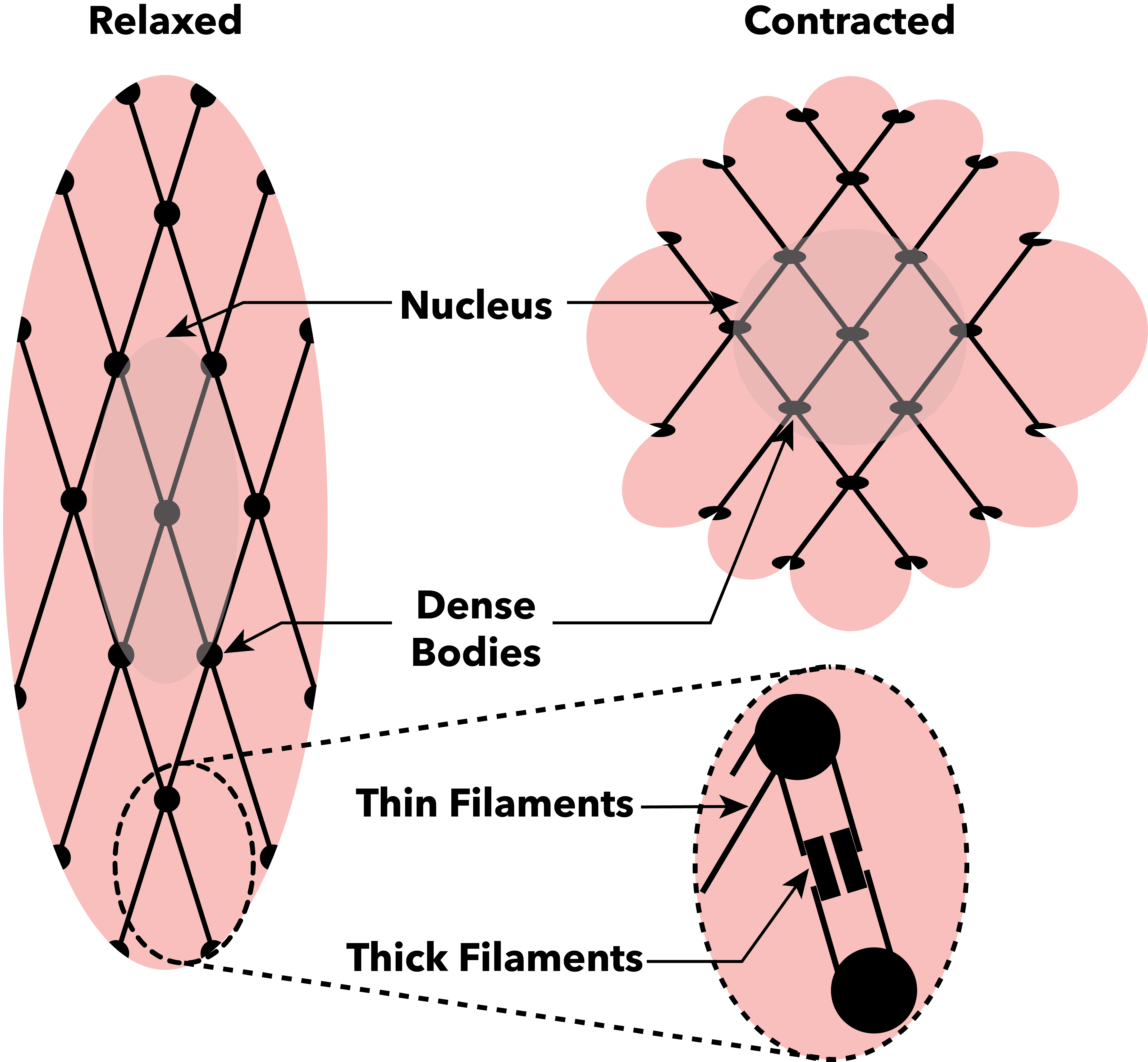
Muscle contraction is the activation of Tension (physics), tension-generating sites within muscle cells. In physiology, muscle contraction does not necessarily mean muscle shortening because muscle tension can be produced without changes in muscle length, such as when holding something heavy in the same position. The termination of muscle contraction is followed by muscle relaxation, which is a return of the muscle fibers to their low tension-generating state. For the contractions to happen, the muscle cells must rely on the change in action of two types of Myofilament, filaments: thin and thick filaments. The major constituent of thin filaments is a chain formed by helical coiling of two strands of actin, and thick filaments dominantly consist of chains of the Motor protein, motor-protein myosin. Together, these two filaments form myofibrils - the basic functional organelles in the skeletal muscle system. In vertebrates, Muscle cell#Muscle contraction in striated muscle, skele ... [...More Info...] [...Related Items...] OR: [Wikipedia] [Google] [Baidu] |


