|
Dislocation Of Hip
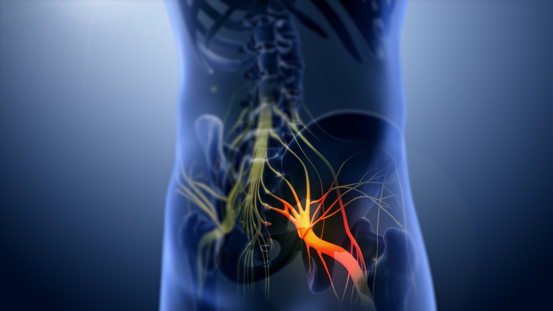
A hip dislocation is when the thighbone (femur) separates from the hip bone (pelvis). Specifically it is when the ball–shaped head of the femur (femoral head) separates from its cup–shaped socket in the hip bone, known as the acetabulum. The joint of the femur and pelvis (hip joint) is very stable, secured by both bony and soft-tissue constraints. With that, dislocation would require significant force which typically results from significant trauma such as from a motor vehicle collision or from a fall from elevation. Hip dislocations can also occur following a hip replacement or from a developmental abnormality known as hip dysplasia. Hip dislocations are classified by fracture association and by the positioning of the dislocated femoral head. A posteriorly positioned head is the most common dislocation type. Hip dislocations are a medical emergency, requiring prompt placement of the femoral head back into the acetabulum ( reduction). This reduction of the femoral head bac ... [...More Info...] [...Related Items...] OR: [Wikipedia] [Google] [Baidu] |
Orthopedics
Orthopedic surgery or orthopedics (American and British English spelling differences, alternative spelling orthopaedics) is the branch of surgery concerned with conditions involving the musculoskeletal system. Orthopedic surgeons use both surgical and nonsurgical means to treat musculoskeletal Physical trauma, trauma, Spinal disease, spine diseases, Sports injury, sports injuries, degenerative diseases, infections, tumors and congenital disorders. Etymology Nicholas Andry coined the word in French as ', derived from the Ancient Greek words ("correct", "straight") and ("child"), and published ''Orthopedie'' (translated as ''Orthopædia: Or the Art of Correcting and Preventing Deformities in Children'') in 1741. The word was Assimilation (linguistics), assimilated into English as ''orthopædics''; the Typographic ligature, ligature ''æ'' was common in that era for ''ae'' in Greek- and Latin-based words. As the name implies, the discipline was initially developed with atte ... [...More Info...] [...Related Items...] OR: [Wikipedia] [Google] [Baidu] |
Traction (orthopedics)
Traction is a set of mechanisms for straightening broken bones or relieving pressure on the spine and skeletal system. There are two types of traction: skin traction and skeletal traction. They are used in orthopedic medicine. Techniques Traction procedures have largely been replaced by more modern techniques, but certain approaches are still used today: * Milwaukee brace * Bryant's traction *Buck's traction, involving skin traction. It is widely used for femoral fractures, low back pain, acetabular fractures and hip fractures.Skin Traction - Lower Extremity from University of Stellenbosch, Department of Orthopaedic Surgery. Retrieved May 2, 2013 Skin traction rarely causes fracture reduction ... [...More Info...] [...Related Items...] OR: [Wikipedia] [Google] [Baidu] |
Ischiofemoral Ligament
The ischiofemoral ligament (ischiocapsular ligament or ischiocapsular band) consists of a triangular band of strong fibers on the posterior side of the hip joint. It is one of the four ligaments that reinforce the hip joint. It attaches to the posterior surface of the acetabular rim and acetabular labrum, and extends around the circumference of the joint to insert on the anterior aspect of the femur. The ischiofemoral ligament limits the internal rotation and adduction of the hip when it is in a flexed position. Some deeper fibres of the ligament are continuous with the fibres of the zona orbicularis The zona orbicularis or annular ligament is a ligament on the neck of the femur formed by the circular fibers of the articular capsule of the hip joint In vertebrate anatomy, the hip, or coxaLatin ''coxa'' was used by Celsus in the sense "hi ... of the capsule. This ligament is less well-defined than the other two capsular ligaments of the hip joint. Function Studies o ... [...More Info...] [...Related Items...] OR: [Wikipedia] [Google] [Baidu] |
Iliofemoral Ligament
The iliofemoral ligament is a thick and very tough triangular capsular ligament of the hip joint situated anterior to this joint. It attaches superiorly at the inferior portion of the anterior inferior iliac spine and adjacent portion of the margin of the acetabulum; it attaches inferiorly at the intertrochanteric line. It is also referred to as the Y-ligament (see below). the ligament of Bigelow, the ligament of Bertin and any combinations of these names. With a force strength exceeding 350 kg (772 lbs), the iliofemoral ligament is not only stronger than the two other ligaments of the hip joint, the ischiofemoral and the pubofemoral, but also the strongest ligament in the human body and as such is an important constraint to the hip joint. Structure The ligament is triangular in shape, with its apex represented by its pelvic attachment. The ligament has two though outer bands; it is thinner and weaker centrally. As the lateral portion is twisted like a screw, the t ... [...More Info...] [...Related Items...] OR: [Wikipedia] [Google] [Baidu] |
Degrees Of Freedom (mechanics)
In classical mechanics, physics, the number of degrees of freedom (DOF) of a mechanical system is the number of independent parameters required to completely specify its configuration or state. That number is an important property in the analysis of systems of bodies in mechanical engineering, structural engineering, aerospace engineering, robotics, and other fields. As an example, the position of a single railcar (engine) moving along a track has one degree of freedom because the position of the car can be completely specified by a single number expressing its distance along the track from some chosen origin. A train of rigid cars connected by hinges to an engine still has only one degree of freedom because the positions of the cars behind the engine are constrained by the shape of the track. For a second example, an automobile with a very stiff suspension can be considered to be a rigid body traveling on a plane (a flat, two-dimensional space). This body has three independe ... [...More Info...] [...Related Items...] OR: [Wikipedia] [Google] [Baidu] |
Ligament
A ligament is a type of fibrous connective tissue in the body that connects bones to other bones. It also connects flight feathers to bones, in dinosaurs and birds. All 30,000 species of amniotes (land animals with internal bones) have ligaments. It is also known as ''articular ligament'', ''articular larua'', ''fibrous ligament'', or ''true ligament''. Comparative anatomy Ligaments are similar to tendons and fasciae as they are all made of connective tissue. The differences among them are in the connections that they make: ligaments connect one bone to another bone, tendons connect muscle to bone, and fasciae connect muscles to other muscles. These are all found in the skeletal system of the human body. Ligaments cannot usually be regenerated naturally; however, there are periodontal ligament stem cells located near the periodontal ligament which are involved in the adult regeneration of periodontist ligament. The study of ligaments is known as . Humans Other ligame ... [...More Info...] [...Related Items...] OR: [Wikipedia] [Google] [Baidu] |
Tendon
A tendon or sinew is a tough band of fibrous connective tissue, dense fibrous connective tissue that connects skeletal muscle, muscle to bone. It sends the mechanical forces of muscle contraction to the skeletal system, while withstanding tension (physics), tension. Tendons, like ligaments, are made of collagen. The difference is that ligaments connect bone to bone, while tendons connect muscle to bone. There are about 4,000 tendons in the adult human body. Structure A tendon is made of dense regular connective tissue, whose main cellular components are special fibroblasts called tendon cells (tenocytes). Tendon cells synthesize the tendon's extracellular matrix, which abounds with densely-packed collagen fibers. The collagen fibers run parallel to each other and are grouped into fascicles. Each fascicle is bound by an endotendineum, which is a delicate loose connective tissue containing thin collagen fibrils and elastic fibers. A set of fascicles is bound by an epitenon, whi ... [...More Info...] [...Related Items...] OR: [Wikipedia] [Google] [Baidu] |
Ball-and-socket Joint
The ball-and-socket joint (or spheroid joint) is a type of synovial joint in which the ball-shaped surface of one rounded bone fits into the cup-like depression of another bone. The distal bone is capable of motion around an indefinite number of axes, which have one common center. This enables the joint to move in many directions. An enarthrosis is a special kind of spheroidal joint in which the socket covers the sphere beyond its equator.Platzer, Werner (2008) ''Color Atlas of Human Anatomy'', Volume 1p.28/ref> Examples of joints Examples of this form of articulation are found in the hip, where the round head of the femur (ball) rests in the cup-like acetabulum (socket) of the pelvis The pelvis (: pelves or pelvises) is the lower part of an Anatomy, anatomical Trunk (anatomy), trunk, between the human abdomen, abdomen and the thighs (sometimes also called pelvic region), together with its embedded skeleton (sometimes also c ...; and in the shoulder joint, where the roun ... [...More Info...] [...Related Items...] OR: [Wikipedia] [Google] [Baidu] |
Articulation (anatomy)
A joint or articulation (or articular surface) is the connection made between bones, ossicles, or other hard structures in the body which link an animal's skeletal system into a functional whole.Saladin, Ken. Anatomy & Physiology. 7th ed. McGraw-Hill Connect. Webp.274/ref> They are constructed to allow for different degrees and types of movement. Some joints, such as the knee, elbow, and shoulder, are self-lubricating, almost frictionless, and are able to withstand compression and maintain heavy loads while still executing smooth and precise movements. Other joints such as sutures between the bones of the skull permit very little movement (only during birth) in order to protect the brain and the sense organs. The connection between a tooth and the jawbone is also called a joint, and is described as a fibrous joint known as a gomphosis. Joints are classified both structurally and functionally. Joints play a vital role in the human body, contributing to movement, stability ... [...More Info...] [...Related Items...] OR: [Wikipedia] [Google] [Baidu] |
Sciatic Nerve
The sciatic nerve, also called the ischiadic nerve, is a large nerve in humans and other vertebrate animals. It is the largest branch of the sacral plexus and runs alongside the hip joint and down the right lower limb. It is the longest and widest single nerve in the human body, going from the top of the leg to the foot on the posterior aspect. The sciatic nerve has no cutaneous branches for the thigh. This nerve provides the connection to the nervous system for the skin of the lateral leg and the whole foot, the muscles of the back of the thigh, and those of the leg and foot. It is derived from Spinal nerve, spinal nerves Lumbar spinal nerve 4, L4 to Sacral spinal nerve 3, S3. It contains Axon, fibres from both the anterior and posterior divisions of the lumbosacral plexus. Structure In humans, the sciatic nerve is formed from the L4 to S3 segments of the sacral plexus, a collection of nerve fibres that emerge from the Sacrum, sacral part of the spinal cord. The lumbosacral trunk ... [...More Info...] [...Related Items...] OR: [Wikipedia] [Google] [Baidu] |
Teratology
Teratology is the study of abnormalities of physiological development in organisms during their life span. It is a sub-discipline in medical genetics which focuses on the classification of congenital abnormalities in dysmorphology caused by teratogens and also in pharmacology and toxicology. Teratogens are substances that may cause non-heritable birth defects via a toxic effect on an embryo or fetus. Defects include malformations, disruptions, deformations, and dysplasia that may cause stunted growth, delayed mental development, or other congenital disorders that lack structural malformations. These defects can be recognized prior to or at birth as well as later during early childhood. The related term developmental toxicity includes all manifestations of abnormal development that are caused by environmental insult. The extent to which teratogens will impact an embryo is dependent on several factors, such as how long the embryo has been exposed, the stage of development the ... [...More Info...] [...Related Items...] OR: [Wikipedia] [Google] [Baidu] |
Pipkin Classification
Femoral head fractures are very rare fractures of the upper end (femoral head) of the thigh bone (femur). They are a very rare kind of hip fracture that may be the result of a fall like most hip fractures but are more commonly caused by more violent incidents such as traffic accidents They are categorized according to the Pipkin classification based on the following bone fracture patterns: See also *Femoral neck The femoral neck (also femur neck or neck of the femur) is a flattened pyramidal process of bone, connecting the femoral head with the femoral shaft, and forming with the latter a wide angle opening medialward. Structure The neck is flattene ... References Orthobullets Hip fracture classifications {{Orthopedics-stub ... [...More Info...] [...Related Items...] OR: [Wikipedia] [Google] [Baidu] |


