|
Desmoplastic Small Round Cell Tumor
Desmoplastic small-round-cell tumor (DSRCT) is an aggressive and rare cancer that primarily occurs as masses in the abdomen. Other areas affected may include the lymph nodes, the lining of the abdomen, diaphragm, spleen, liver, chest wall, skull, spinal cord, large intestine, small intestine, bladder, brain, lungs, testicles, ovaries, and the pelvis. Reported sites of metastatic spread include the liver, lungs, lymph nodes A lymph node, or lymph gland, is a kidney-shaped Organ (anatomy), organ of the lymphatic system and the adaptive immune system. A large number of lymph nodes are linked throughout the body by the lymphatic vessels. They are major sites of lymphoc ..., brain, skull, and bones. It is characterized by the EWS-WT1 fusion protein. The tumor is classified as a soft tissue sarcoma and a small round blue cell tumor. It most often occurs in male children. The disease rarely occurs in females, but when it does the tumors can be mistaken for ovarian cancer. Signs and ... [...More Info...] [...Related Items...] OR: [Wikipedia] [Google] [Baidu] |
Micrograph
A micrograph is an image, captured photographically or digitally, taken through a microscope or similar device to show a magnify, magnified image of an object. This is opposed to a macrograph or photomacrograph, an image which is also taken on a microscope but is only slightly magnified, usually less than 10 times. Micrography is the practice or art of using microscopes to make photographs. A photographic micrograph is a photomicrograph, and one taken with an electron microscope is an electron micrograph. A micrograph contains extensive details of microstructure. A wealth of information can be obtained from a simple micrograph like behavior of the material under different conditions, the phases found in the system, failure analysis, grain size estimation, elemental analysis and so on. Micrographs are widely used in all fields of microscopy. Types Photomicrograph A light micrograph or photomicrograph is a micrograph prepared using an optical microscope, a process referred to ... [...More Info...] [...Related Items...] OR: [Wikipedia] [Google] [Baidu] |
Ovarian Cancer
Ovarian cancer is a cancerous tumor of an ovary. It may originate from the ovary itself or more commonly from communicating nearby structures such as fallopian tubes or the inner lining of the abdomen. The ovary is made up of three different cell types including epithelial cells, germ cells, and stromal cells. When these cells become abnormal, they have the ability to divide and form tumors. These cells can also invade or spread to other parts of the body. When this process begins, there may be no or only vague symptoms. Symptoms become more noticeable as the cancer progresses. These symptoms may include bloating, vaginal bleeding, pelvic pain, abdominal swelling, constipation, and loss of appetite, among others. Common areas to which the cancer may spread include the lining of the abdomen, lymph nodes, lungs, and liver. The risk of ovarian cancer increases with age. Most cases of ovarian cancer develop after menopause. It is also more common in women who have ovulated ... [...More Info...] [...Related Items...] OR: [Wikipedia] [Google] [Baidu] |
Chromosome 22
Chromosome 22 is one of the 23 pairs of chromosomes in human cells. Humans normally have two copies of chromosome 22 in each cell. Chromosome 22 is the second smallest human chromosome, spanning about 51 million DNA base pairs and representing between 1.5 and 2% of the total DNA in cells. In 1999, researchers working on the Human Genome Project announced they had determined the sequence of base pairs that make up this chromosome. Chromosome 22 was the first human chromosome to be fully sequenced. Human chromosomes are numbered by their apparent size in the karyotype, with chromosome 1 being the largest and chromosome 22 having originally been identified as the smallest. However, genome sequencing has revealed that chromosome 21 is actually smaller than chromosome 22. Genes Number of genes The following are some of the gene count estimates of human chromosome 22. Because researchers use different approaches to genome annotation, their predictions of the number of genes on e ... [...More Info...] [...Related Items...] OR: [Wikipedia] [Google] [Baidu] |
Locus (genetics)
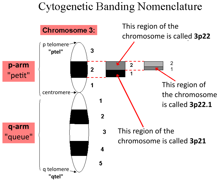
In genetics, a locus (: loci) is a specific, fixed position on a chromosome where a particular gene or genetic marker is located. Each chromosome carries many genes, with each gene occupying a different position or locus; in humans, the total number of Human genome#Coding sequences (protein-coding genes), protein-coding genes in a complete haploid set of 23 chromosomes is estimated at 19,000–20,000. Genes may possess multiple variants known as alleles, and an allele may also be said to reside at a particular locus. Diploid and polyploid cells whose chromosomes have the same allele at a given locus are called homozygote, homozygous with respect to that locus, while those that have different alleles at a given locus are called heterozygote, heterozygous. The ordered list of loci known for a particular genome is called a gene map. Gene mapping is the process of determining the specific locus or loci responsible for producing a particular phenotype or biological trait. Association ma ... [...More Info...] [...Related Items...] OR: [Wikipedia] [Google] [Baidu] |
Karyotype
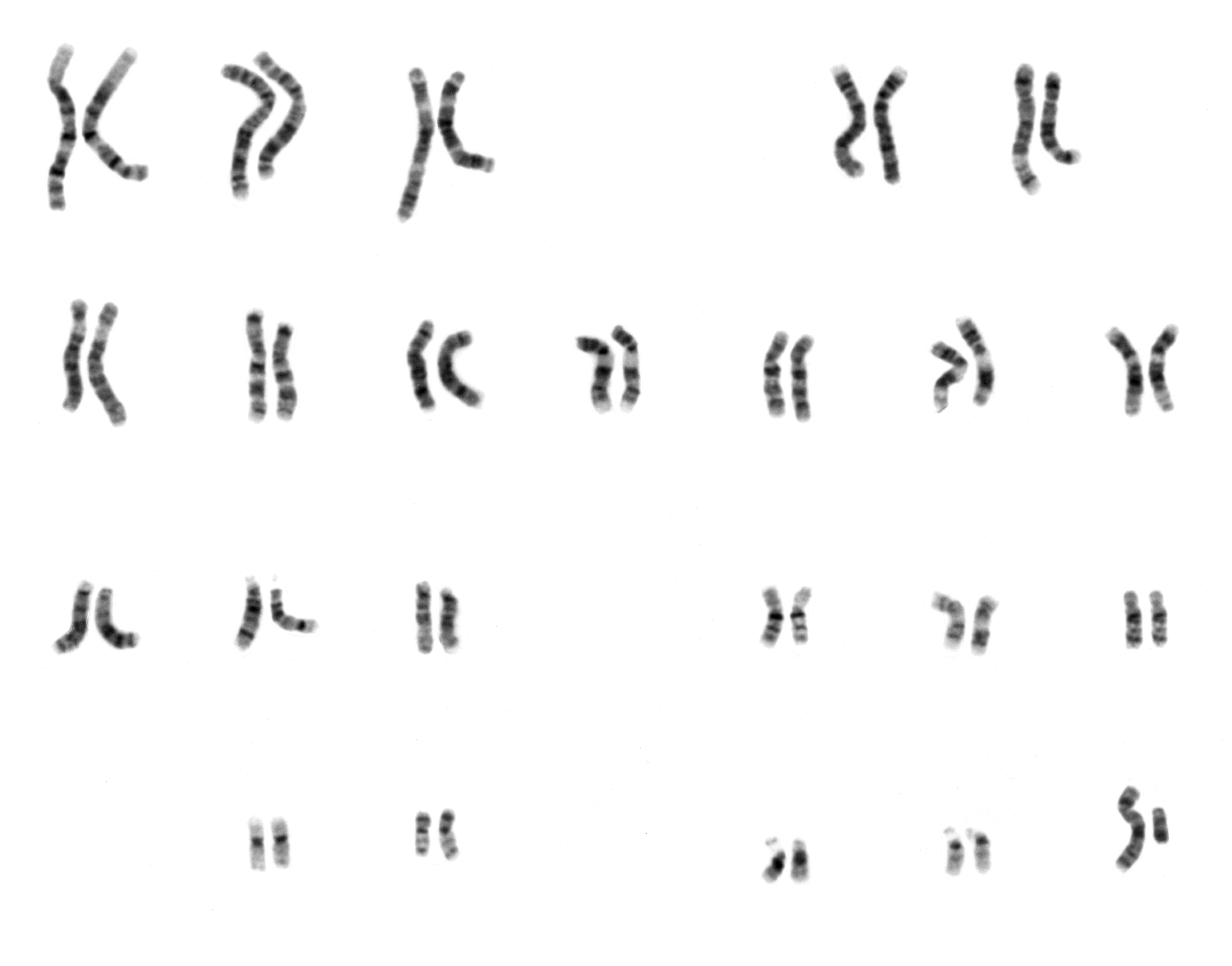
A karyotype is the general appearance of the complete set of chromosomes in the cells of a species or in an individual organism, mainly including their sizes, numbers, and shapes. Karyotyping is the process by which a karyotype is discerned by determining the chromosome complement of an individual, including the number of chromosomes and any abnormalities. A karyogram or idiogram is a graphical depiction of a karyotype, wherein chromosomes are generally organized in pairs, ordered by size and position of centromere for chromosomes of the same size. Karyotyping generally combines light microscopy and photography in the metaphase of the cell cycle, and results in a photomicrographic (or simply micrographic) karyogram. In contrast, a schematic karyogram is a designed graphic representation of a karyotype. In schematic karyograms, just one of the sister chromatids of each chromosome is generally shown for brevity, and in reality they are generally so close together that they look as ... [...More Info...] [...Related Items...] OR: [Wikipedia] [Google] [Baidu] |
FET Protein Family
The FET protein family (also known as the TET protein family) consists of three similarly structured and functioning proteins. They and the genes in the FET gene family which encode them (i.e. form the pre-messenger RNAs that are converted to the messenger RNAs responsible for their production) are: 1) the EWSR1 protein encoded by the ''EWSR1'' gene (also termed the ''Ewing sarcoma RNA binding protein, EWS RNA binding protein 1,'' or ''bK984G1.4'' gene) located at band 12.2 of the long (i.e. "q") arm of chromosome 22; 2) the FUS (i.e. fused in sarcoma) protein encoded by the ''FUS'' gene (also termed the ''FUS RNA binding protein, TLS, asTLS, ALS6, ETM4, FUS1, POMP75, altFUS'', or ''HNRNPP2'' gene) located at band 16 on the short arm of chromosome 16; and 3) the TAF15 protein encoded by the ''TAF15'' gene (also termed the ''TATA-box binding protein associated factor 15, Npl3, RBP56, TAF2N'', or ''TAFII68'' gene) located at band 12 on the long arm of chromosome 7 The FET in this ... [...More Info...] [...Related Items...] OR: [Wikipedia] [Google] [Baidu] |
EWSR1
RNA-binding protein EWS is a protein that in humans is encoded by the ''EWSR1'' gene on human chromosome 22, specifically 22q12.2. It is one of 3 proteins in the FET protein family. Clinical significance The q22.2 region of chromosome 22 encodes the N-terminal transactivation domain of the EWS protein and that region may become joined to one of several other chromosomes which encode various transcription factors; see EWS/FLI and OMIM-133450. The expression of a chimeric protein with the EWS transactivation domain fused to the DNA binding region of a transcription factor generates a powerful oncogenic protein causing Ewing sarcoma and other members of the Ewing family of tumors. These translocations can occur due to chromoplexy, a burst of complex chromosomal rearrangements seen in cancer cells. The normal EWS gene encodes an RNA binding protein closely related to FUS (gene) and TAF15, all of which have been associated to amyotrophic lateral sclerosis. Interactions The EWS ... [...More Info...] [...Related Items...] OR: [Wikipedia] [Google] [Baidu] |
Chromosomal Translocation
In genetics, chromosome translocation is a phenomenon that results in unusual rearrangement of chromosomes. This includes "balanced" and "unbalanced" translocation, with three main types: "reciprocal", "nonreciprocal" and "Robertsonian" translocation. Reciprocal translocation is a chromosome abnormality caused by exchange of parts between non-homologous chromosomes. Two detached fragments of two different chromosomes are switched. Robertsonian translocation occurs when two non-homologous chromosomes get attached, meaning that given two healthy pairs of chromosomes, one of each pair "sticks" and blends together homogeneously. Each type of chromosomal translocation can result in disorders for growth, function and the development of an individuals body, often resulting from a change in their genome. A gene fusion may be created when the translocation joins two otherwise-separated genes. It is detected on cytogenetics or a karyotype of affected cells. Translocations can be bala ... [...More Info...] [...Related Items...] OR: [Wikipedia] [Google] [Baidu] |
Wilms' Tumor
Wilms' tumor or Wilms tumor, also known as nephroblastoma, is a cancer of the kidneys that typically occurs in children (rarely in adults), and occurs most commonly as a renal tumor in child patients. It is named after Max Wilms, the German surgeon (1867–1918) who first described it. Approximately 650 cases are diagnosed in the U.S. annually. The majority of cases occur in children with no associated genetic syndromes; however, a minority of children with Wilms' tumor have a congenital abnormality. It is highly responsive to treatment, with about 90 percent of children being cured. Signs and symptoms Typical signs and symptoms of Wilms' tumor include the following: * a painless, palpable abdominal mass * loss of appetite * abdominal pain * fever * nausea and vomiting * blood in the urine (in about 20% of cases) * high blood pressure in some cases (especially if synchronous or metachronous bilateral kidney involvement) * Rarely as varicoceleErginel B, Vural S, Akın M ... [...More Info...] [...Related Items...] OR: [Wikipedia] [Google] [Baidu] |
Cachexia
Cachexia () is a syndrome that happens when people have certain illnesses, causing muscle loss that cannot be fully reversed with improved nutrition. It is most common in diseases like cancer, Heart failure, congestive heart failure, chronic obstructive pulmonary disease, chronic kidney disease, and AIDS. These conditions change how the body handles inflammation, metabolism, and brain signaling, leading to muscle loss and other harmful changes to body composition over time. Unlike weight loss from not eating enough, cachexia mainly affects muscle and can happen with or without fat loss. Diagnosis of cachexia is difficult because there are no clear guidelines, and its occurrence varies from one affected person to the next. Like malnutrition, cachexia can lead to worse health outcomes and lower quality of life. Definition Cachexia is hard to define because it often happens alongside malnutrition and sarcopenia. Since there are no clear rules separating these conditions, experts ... [...More Info...] [...Related Items...] OR: [Wikipedia] [Google] [Baidu] |


