|
Crux Cordis
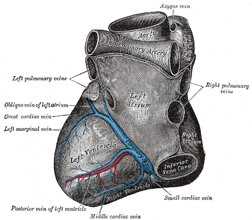
The crux cordis or crux of the heart (from Latin "crux" meaning "cross") is the area on the lower back side of the heart where the coronary sulcus (the groove separating the atria from the ventricles) and the posterior interventricular sulcus The posterior interventricular sulcus or posterior longitudinal sulcus is one of the two grooves separating the ventricles of the heart (the other being the anterior interventricular sulcus). They can be known as subsinosal interventricular groo ... (the groove separating the left from the right ventricle) meet. It is important surgically because the atrioventricular nodal artery, a small but vital vessel, passes in proximity to the crux of the heart. It is the anastomotic point of right and left coronary artery. References Cardiac anatomy {{circulatory-stub ... [...More Info...] [...Related Items...] OR: [Wikipedia] [Google] [Baidu] |
Human Heart
The heart is a muscular organ found in humans and other animals. This organ pumps blood through the blood vessels. The heart and blood vessels together make the circulatory system. The pumped blood carries oxygen and nutrients to the tissue, while carrying metabolic waste such as carbon dioxide to the lungs. In humans, the heart is approximately the size of a closed fist and is located between the lungs, in the middle compartment of the chest, called the mediastinum. In humans, the heart is divided into four chambers: upper left and right atria and lower left and right ventricles. Commonly, the right atrium and ventricle are referred together as the right heart and their left counterparts as the left heart. In a healthy heart, blood flows one way through the heart due to heart valves, which prevent backflow. The heart is enclosed in a protective sac, the pericardium, which also contains a small amount of fluid. The wall of the heart is made up of three layers: ep ... [...More Info...] [...Related Items...] OR: [Wikipedia] [Google] [Baidu] |
Coronary Sulcus
The coronary sulcus (also called coronary groove, auriculoventricular groove, atrioventricular groove, AV groove) is a Sulcus (morphology), groove on the surface of the heart at the base of right auricle that separates the Atrium (heart), atria from the Ventricle (heart), ventricles. The structure contains the trunks of the Coronary arteries, nutrient vessels of the heart, and is deficient in front, where it is crossed by the root of the pulmonary trunk. On the posterior surface of the heart, the coronary sulcus contains the coronary sinus. The right coronary artery, circumflex branch of left coronary artery, and small cardiac vein all travel along parts of the coronary sulcus. Structure In relation to the rib cage, the coronary sulcus spans from the medial side of the 3rd left costal cartilage, to the middle of the right 6th costal cartilage. Pericardium, Epicardial Adipose tissue, fat tends to be concentrated along the coronary sulcus. There are two coronary sulci in the heart ... [...More Info...] [...Related Items...] OR: [Wikipedia] [Google] [Baidu] |
Atrium (heart)
The atrium (; : atria) is one of the two Heart#Chambers, upper chambers in the heart that receives blood from the circulatory system. The blood in the atria is pumped into the Ventricle (heart), heart ventricles through the atrioventricular valve, atrioventricular mitral valve, mitral and tricuspid valve, tricuspid heart valves. There are two atria in the human heart – the left atrium receives blood from the pulmonary circulation, and the right atrium receives blood from the venae cavae of the systemic circulation. During the cardiac cycle, the atria receive blood while relaxed in diastole, then contract in systole to move blood to the ventricles. Each atrium is roughly cube-shaped except for an ear-shaped projection called an atrial appendage, previously known as an auricle. All animals with a closed circulatory system have at least one atrium. The atrium was formerly called the 'auricle'. That term is still used to describe this chamber in some other animals, such as the ''Mo ... [...More Info...] [...Related Items...] OR: [Wikipedia] [Google] [Baidu] |
Ventricle (heart)
A ventricle is one of two large chambers located toward the bottom of the heart that collect and expel blood towards the peripheral beds within the body and lungs. The blood pumped by a ventricle is supplied by an atrium, an adjacent chamber in the upper heart that is smaller than a ventricle. Interventricular means between the ventricles (for example the interventricular septum), while intraventricular means within one ventricle (for example an intraventricular block). In a four-chambered heart, such as that in humans, there are two ventricles that operate in a double circulatory system: the right ventricle pumps blood into the pulmonary circulation to the lungs, and the left ventricle pumps blood into the systemic circulation through the aorta. Structure Ventricles have thicker walls than atria and generate higher blood pressures. The physiological load on the ventricles requiring pumping of blood throughout the body and lungs is much greater than the pressure generated by ... [...More Info...] [...Related Items...] OR: [Wikipedia] [Google] [Baidu] |
Posterior Interventricular Sulcus
The posterior interventricular sulcus or posterior longitudinal sulcus is one of the two grooves separating the ventricles of the heart (the other being the anterior interventricular sulcus). They can be known as subsinosal interventricular groove or paraconal interventricular groove respectively. It is located on the diaphragmatic surface of the heart near the right margin. It extends between the coronary sulcus The coronary sulcus (also called coronary groove, auriculoventricular groove, atrioventricular groove, AV groove) is a Sulcus (morphology), groove on the surface of the heart at the base of right auricle that separates the Atrium (heart), atria fr ... and the (notch of) apex of the heart. It contains the posterior interventricular artery and middle cardiac vein. References External links * Cardiac anatomy {{circulatory-stub ... [...More Info...] [...Related Items...] OR: [Wikipedia] [Google] [Baidu] |
Atrioventricular Nodal Branch
The atrioventricular nodal branch is a coronary artery that supplies arterial blood to the atrioventricular node, which is responsible for initiating muscular contraction of the ventricles. The AV nodal branch is most often a branch of the right coronary artery. Structure Origin The atrioventricular nodal branch sees significant variation in origin: * proximal posterolateral branch from the right coronary artery in around 77%. * distal posterolateral branch from the right coronary artery in around 2%. * distal right coronary artery in around 10%. * right posterior interventricular artery in around 7%. * distal circumflex branch of left coronary artery in around 4%. The right coronary artery supplies the atrioventricular node in around 90% of people. In approximately 2% of people, the vascular supply to the atrioventricular node arises from both the right coronary artery and the left circumflex branch.Sow ML, Ndoye JM, Lo EA. The artery of the atrioventricular node: an anat ... [...More Info...] [...Related Items...] OR: [Wikipedia] [Google] [Baidu] |


