|
Congenital Myopathy
Congenital myopathy is a very broad term for any muscle disorder present at birth. This defect primarily affects skeletal muscle fibres and causes muscular weakness and/or hypotonia. Congenital myopathies account for one of the top neuromuscular disorders in the world today, comprising approximately 6 in 100,000 live births every year. As a whole, congenital myopathies can be broadly classified as follows: * A distinctive abnormality in skeletal muscle fibres on the cellular level; observable via light microscope * Symptoms of muscle weakness and hypotonia * Is a congenital disorder, meaning it occurs during development and symptoms present themselves at birth or in early life. * Is a genetic disorder. Classification Myopathies with inclusion bodies and abnormal protein accumulation Congenital myopathies with inclusion bodies and protein accumulation is a broad category, and some congenital myopathies that fall within this group are well understood, such as nemaline myopathy (see ... [...More Info...] [...Related Items...] OR: [Wikipedia] [Google] [Baidu] |
Hypotonia
Hypotonia is a state of low muscle tone (the amount of tension or resistance to stretch in a muscle), often involving reduced muscle strength. Hypotonia is not a specific medical disorder, but it is a potential manifestation of many different diseases and disorders that affect motor nerve control by the brain or muscle strength. Hypotonia is a lack of resistance to passive movement whereas muscle weakness results in impaired active movement. Central hypotonia originates from the central nervous system, while peripheral hypotonia is related to problems within the spinal cord, peripheral nerves, and/or skeletal muscles. Severe hypotonia in infancy is commonly known as floppy baby syndrome. Recognizing hypotonia, even in early infancy, is usually relatively straightforward, but medical diagnosis, diagnosing the underlying cause can be difficult and often unsuccessful. The long-term effects of hypotonia on a child's development and later life depend primarily on the severity of the mus ... [...More Info...] [...Related Items...] OR: [Wikipedia] [Google] [Baidu] |
DNM2
Dynamin-2 is a protein that in humans is encoded by the ''DNM2'' gene. Function Dynamins represent one of the subfamilies of GTP-binding proteins. These proteins share considerable sequence similarity over the N-terminal portion of the molecule, which contains the GTPase domain. Dynamins are associated with microtubules. They have been implicated in cell processes such as endocytosis and cell motility, and in alterations of the membrane that accompany certain activities such as bone resorption by osteoclasts. Dynamins bind many proteins that bind actin and other cytoskeletal proteins. Dynamins can also self-assemble, a process that stimulates GTPase activity. Four alternatively spliced transcripts encoding different proteins have been described. Additional alternatively spliced transcripts may exist, but their full-length nature has not been determined. Interactions DNM2 has been shown to interact with: * SHANK1, * SHANK2, and * SNX9. Clinical relevance Mutations in t ... [...More Info...] [...Related Items...] OR: [Wikipedia] [Google] [Baidu] |
STIM1
Stromal interaction molecule 1 is a protein that in humans is encoded by the ''STIM1'' gene. STIM1 has a single transmembrane domain, and is localized to the endoplasmic reticulum, and to a lesser extent to the plasma membrane. Even though the protein has been identified earlier, its function was unknown until recently. In 2005, it was discovered that STIM1 functions as a calcium sensor in the endoplasmic reticulum. Upon activation of the IP3 receptor, the calcium concentration in the endoplasmic reticulum decreases, which is sensed by STIM1, via its EF hand domain. STIM1 activates the "store-operated" ORAI1 calcium ion channels in the plasma membrane, via intracellular STIM1 movement, clustering under plasma membrane and protein interaction with ORAI isoforms. STIM1-mediated calcium entry is required for thrombin-induced disassembly of VE-cadherin Cadherin-5, or VE-cadherin (vascular endothelial cadherin), also known as CD144 ( Cluster of Differentiation 144), is a type of cad ... [...More Info...] [...Related Items...] OR: [Wikipedia] [Google] [Baidu] |
MYH7
Myosin-7 is a protein that in humans is encoded by the ''MYH7'' gene. It is the myosin heavy chain beta (MHC-β) isoform (slow twitch) expressed primarily in the heart, but also in skeletal muscles (type I fibers). This isoform is distinct from the fast isoform of cardiac myosin heavy chain, MYH6, referred to as MHC-α. MHC-β is the major protein comprising the thick filament that forms the sarcomeres in cardiac muscle and plays a major role in cardiac muscle contraction. Structure MHC-β is a 223 kDa protein composed of 1935 amino acids. MHC-β is a hexameric, asymmetric motor forming the bulk of the thick filament in cardiac muscle. MHC-β is composed of N-terminal globular heads (20 nm) that project laterally, and alpha helical tails (130 nm) that dimerize and multimerize into a coiled-coil motif to form the light meromyosin (LMM), thick filament rod. The 9 nm alpha-helical neck region of each MHC-β head non-covalently binds two light chains, essential ... [...More Info...] [...Related Items...] OR: [Wikipedia] [Google] [Baidu] |
Myofibril
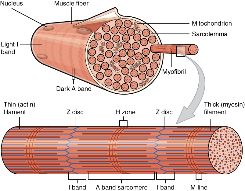
A myofibril (also known as a muscle fibril or sarcostyle) is a basic rod-like organelle of a muscle cell. Skeletal muscles are composed of long, tubular cells known as Skeletal muscle#Skeletal muscle cells, muscle fibers, and these cells contain many chains of myofibrils. Each myofibril has a diameter of 1–2 micrometres. They are created during embryogenesis, embryonic development in a process known as myogenesis. Myofibrils are composed of long proteins including actin, myosin, and titin, and other proteins that hold them together. These proteins are organized into Thick filaments, thick, thin filaments, thin, and elastic filament, elastic myofilaments, which repeat along the length of the myofibril in sections or units of contraction called sarcomeres. Muscle contraction, Muscles contract by sliding filament theory, sliding the thick myosin, and thin actin myofilaments along each other. Structure Each myofibril has a diameter of between 1 and 2 micrometres (μm). The filamen ... [...More Info...] [...Related Items...] OR: [Wikipedia] [Google] [Baidu] |
Sarcolemma
The sarcolemma (''sarco'' (from ''sarx'') from Greek; flesh, and ''lemma'' from Greek; sheath), also called the myolemma, is the cell membrane surrounding a skeletal muscle fibre or a cardiomyocyte. It consists of a lipid bilayer and a thin outer coat of polysaccharide material ( glycocalyx) that contacts the basement membrane. The basement membrane contains numerous thin collagen fibrils and specialized proteins such as laminin that provide a scaffold to which the muscle fibre can adhere. Through transmembrane proteins in the plasma membrane, the actin skeleton inside the cell is connected to the basement membrane and the cell's exterior. At each end of the muscle fibre, the surface layer of the sarcolemma fuses with a tendon fibre, and the tendon fibres, in turn, collect into bundles to form the muscle tendons that adhere to bones. The sarcolemma generally maintains the same function in muscle cells as the plasma membrane does in other eukaryote cells. It acts as a barrie ... [...More Info...] [...Related Items...] OR: [Wikipedia] [Google] [Baidu] |
Myosin
Myosins () are a Protein family, family of motor proteins (though most often protein complexes) best known for their roles in muscle contraction and in a wide range of other motility processes in eukaryotes. They are adenosine triphosphate, ATP-dependent and responsible for actin-based motility. The first myosin (M2) to be discovered was in 1864 by Wilhelm Kühne. Kühne had extracted a viscous protein from skeletal muscle that he held responsible for keeping the tension state in muscle. He called this protein ''myosin''. The term has been extended to include a group of similar ATPases found in the cell (biology), cells of both striated muscle tissue and smooth muscle tissue. Following the discovery in 1973 of enzymes with myosin-like function in ''Acanthamoeba, Acanthamoeba castellanii'', a global range of divergent myosin genes have been discovered throughout the realm of eukaryotes. Although myosin was originally thought to be restricted to muscle cells (hence ''wikt:myo-#Pr ... [...More Info...] [...Related Items...] OR: [Wikipedia] [Google] [Baidu] |
Succinate Dehydrogenase
Succinate dehydrogenase (SDH) or succinate-coenzyme Q reductase (SQR) or respiratory complex II is an enzyme complex, found in many bacterial cells and in the inner mitochondrial membrane of eukaryotes. It is the only enzyme that participates in both the citric acid cycle and oxidative phosphorylation. Histochemical analysis showing high succinate dehydrogenase in muscle demonstrates high mitochondrial content and high oxidative potential. In step 6 of the citric acid cycle, SQR catalyzes the oxidation of succinate to fumarate with the reduction of ubiquinone to ubiquinol. This occurs in the inner mitochondrial membrane by coupling the two reactions together. Structure Subunits Mitochondrial and many bacterial SQRs are composed of four structurally different subunits: two hydrophilic and two hydrophobic. The first two subunits, a flavoprotein (SDHA) and an iron-sulfur protein (SDHB), form a hydrophilic head where enzymatic activity of the complex takes place. SD ... [...More Info...] [...Related Items...] OR: [Wikipedia] [Google] [Baidu] |
Calsequestrin
Calsequestrin is a calcium-binding protein that acts as a calcium buffer within the sarcoplasmic reticulum. The protein helps hold calcium in the cisterna of the sarcoplasmic reticulum after a muscle contraction, even though the concentration of calcium in the sarcoplasmic reticulum is much higher than in the cytosol. It also helps the sarcoplasmic reticulum store an extraordinarily high amount of calcium ions. Each molecule of calsequestrin can bind 18 to 50 Ca2+ ions. Sequence analysis has suggested that calcium is not bound in distinct pockets via EF-hand motifs, but rather via presentation of a charged protein surface. Two forms of calsequestrin have been identified. The cardiac form Calsequestrin-2 (CASQ2) is present in cardiac and slow skeletal muscle and the fast skeletal form Calsequestrin-1(CASQ1) is found in fast skeletal muscle. The release of calsequestrin-bound calcium (through a calcium release channel) triggers muscle contraction. The active protein is not high ... [...More Info...] [...Related Items...] OR: [Wikipedia] [Google] [Baidu] |
ATP2A1
Sarcoplasmic/endoplasmic reticulum calcium ATPase 1 (SERCA1) also known as Calcium pump 1, is an enzyme that in humans is encoded by the ''ATP2A1'' gene. Function This gene encodes one of the SERCA Ca2+-ATPases, which are intracellular pumps located in the sarcoplasmic or endoplasmic reticula of muscle cells. This enzyme catalyzes the hydrolysis of ATP coupled with the translocation of calcium from the cytosol to the sarcoplasmic reticulum lumen, and is involved in muscular excitation and contraction. Clinical significance Mutations in this gene cause some autosomal recessive forms of Brody disease, characterized by increasing impairment of muscular relaxation during exercise. Alternative splicing results in two transcript variants encoding different isoforms. Alternative splicing of ATP2A1 is also implicated in myotonic dystrophy type 1. ATP2A1 SERCA pumps were very strongly down regulated in amyotrophic lateral sclerosis Amyotrophic lateral sclerosis (ALS), al ... [...More Info...] [...Related Items...] OR: [Wikipedia] [Google] [Baidu] |
Sarcoplasmic Reticulum
The sarcoplasmic reticulum (SR) is a membrane-bound structure found within muscle cells that is similar to the smooth endoplasmic reticulum in other cells. The main function of the SR is to store calcium ions (Ca2+). Calcium ion levels are kept relatively constant, with the concentration of calcium ions within a cell being 10,000 times smaller than the concentration of calcium ions outside the cell. This means that small increases in calcium ions within the cell are easily detected and can bring about important cellular changes (the calcium is said to be a second messenger). Calcium is used to make calcium carbonate (found in chalk) and calcium phosphate, two compounds that the body uses to make teeth and bones. This means that too much calcium within the cells can lead to hardening (calcification) of certain intracellular structures, including the mitochondria, leading to cell death. Therefore, it is vital that calcium ion levels are controlled tightly, and can be released int ... [...More Info...] [...Related Items...] OR: [Wikipedia] [Google] [Baidu] |
Multi/minicore Myopathy
Multi/minicore myopathy is a congenital myopathy usually caused by mutations in either the ''SELENON'' and ''RYR1'' genes. It is characterised the presence of multifocal, well-circumscribed areas with reduction of oxidative staining and low myofibrillar ATPase on muscle biopsy. It is also known as Minicore myopathy, Multicore myopathy, Multiminicore myopathy, Minicore myopathy with external ophthalmoplegia, Multicore myopathy with external ophthalmoplegia and Multiminicore disease with external ophthalmoplegia. Presentation There are four types of minicore myopathy Classical type (75% cases) The usual presentation is in infancy or childhood with hypotonia or proximal weakness. This weakness tends to affect the shoulder girdle and the inner thigh. The other main features are *Failure to thrive due to feeding difficulties * Axial muscle weakness, particularly affecting neck and trunk flexors * High pitched voice * Myopathic facial features A high arched or cleft palate may be ... [...More Info...] [...Related Items...] OR: [Wikipedia] [Google] [Baidu] |


