|
Concealed Conduction
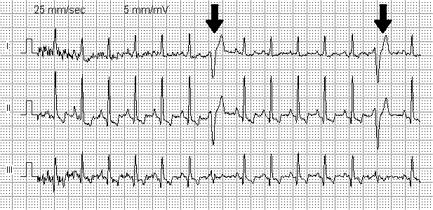
Concealed conduction is tissue stimulation without direct effect, but leading to a change in conduction characteristics. The term "concealed" is in reference to that the conduction is not observable by electrocardiogram. A common example would be an ''interpolated PVC'' (a type of premature ventricular contraction) during normal sinus rhythm; the PVC does not cause an atrial contraction, because the retrograde impulse from the PVC does not completely penetrate the AV node. However, this AV node stimulation can cause a delay in subsequent AV conduction by modifying the AV node's subsequent conduction characteristics. Hence, the P-R interval after the PVC is longer than the baseline P-R interval. Concealed conduction can be seen in cardiac aberrancy when a bundle branch temporarily blocks due to being refractory, and conduction from the other bundle branch conceals into the blocked branch retrograde thus perpetuation the bundle branch block morphology in subsequent beats. For exam ... [...More Info...] [...Related Items...] OR: [Wikipedia] [Google] [Baidu] |
Premature Ventricular Contraction
A premature ventricular contraction (PVC) is a common event where the heartbeat is initiated by Purkinje fibers in the ventricles rather than by the sinoatrial node. PVCs may cause no symptoms or may be perceived as a "skipped beat" or felt as palpitations in the chest. PVCs do not usually pose any danger. The electrical events of the heart detected by the electrocardiogram (ECG) allow a PVC to be easily distinguished from a normal heart beat. However, very frequent PVCs can be symptomatic of an underlying heart condition (such as arrhythmogenic right ventricular cardiomyopathy). Furthermore, very frequent (over 20% of all heartbeats) PVCs are considered a risk factor for arrhythmia-induced cardiomyopathy, in which the heart muscle becomes less effective and symptoms of heart failure may develop. Ultrasound of the heart is therefore recommended in people with frequent PVCs. If PVCs are frequent or troublesome, medication (beta blockers or certain calcium channel blockers) ... [...More Info...] [...Related Items...] OR: [Wikipedia] [Google] [Baidu] |
Normal Sinus Rhythm
A sinus rhythm is any cardiac rhythm in which depolarisation of the cardiac muscle begins at the sinus node. It is necessary, but not sufficient, for normal electrical activity within the heart. On the electrocardiogram (ECG), a sinus rhythm is characterised by the presence of P waves that are normal in morphology. The term normal sinus rhythm (NSR) is sometimes used to denote a specific type of sinus rhythm where all other measurements on the ECG also fall within designated normal limits, giving rise to the characteristic appearance of the ECG when the electrical conduction system of the heart is functioning normally; however, other sinus rhythms can be entirely normal in particular patient groups and clinical contexts, so the term is sometimes considered a misnomer and its use is sometimes discouraged. Other types of sinus rhythm that can be normal include sinus tachycardia, sinus bradycardia, and sinus arrhythmia. Sinus rhythms may be present together with various othe ... [...More Info...] [...Related Items...] OR: [Wikipedia] [Google] [Baidu] |
Cardiac Aberrancy
Cardiac aberrancy is a type of disruption in the shape of the electrocardiogram signal, representing abnormal activation of the ventricular heart muscle via the electrical conduction system of the heart. Normal activation utilizes the bundle of His and Purkinje fibers to produce a narrow (QRS) electrical signal. Aberration occurs when the electrical activation of the heart, which is caused by a series of action potentials, is conducting improperly which can result in temporary changes in the morphology that looks like: * Left bundle branch block ** Left anterior fascicular block ** Left posterior fascicular block * Right bundle branch block This is in contrast to a permanent dysfunction of the electrical pathways that produces wide QRS complexes in one of the above patterns or combinations of patterns (ie, bifascicular block). In the context of atrial fibrillation, the Ashman phenomenon is a form of aberrancy. Aberrancy is due to prematurity in which part of the conductio ... [...More Info...] [...Related Items...] OR: [Wikipedia] [Google] [Baidu] |
Premature Atrial Contraction
Premature atrial contraction (PAC), also known as atrial premature complexes (APC) or atrial premature beats (APB), are a common arrhythmia characterized by premature heartbeats originating in the atria. While the sinoatrial node typically regulates the heartbeat during normal sinus rhythm, PACs occur when another region of the atria depolarizes before the sinoatrial node and thus triggers a premature heartbeat, in contrast to escape beats, in which the normal sinoatrial node fails, leaving a non-nodal pacemaker to initiate a late beat. The exact cause of PACs is unclear; while several predisposing conditions exist, single isolated PACs commonly occur in healthy young and elderly people. Elderly people that get PACs usually don't need any further attention besides follow-ups due to unclear evidence. PACs are often completely asymptomatic and may be noted only with Holter monitoring, but occasionally they can be perceived as a skipped beat or a jolt in the chest. In mos ... [...More Info...] [...Related Items...] OR: [Wikipedia] [Google] [Baidu] |
Atrial Flutter
Atrial flutter (AFL) is a common abnormal heart rhythm that starts in the atrial chambers of the heart. When it first occurs, it is usually associated with a fast heart rate and is classified as a type of supraventricular tachycardia (SVT). Atrial flutter is characterized by a sudden-onset (usually) regular abnormal heart rhythm on an electrocardiogram (ECG) in which the heart rate is fast. Symptoms may include a feeling of the heart beating too fast, too hard, or skipping beats, chest discomfort, difficulty breathing, a feeling as if one's stomach has dropped, a feeling of being light-headed, or loss of consciousness. Although this abnormal heart rhythm typically occurs in individuals with cardiovascular disease (e.g., high blood pressure, coronary artery disease, and cardiomyopathy) and diabetes mellitus, it may occur spontaneously in people with otherwise normal hearts. It is typically not a stable rhythm and often degenerates into atrial fibrillation (AF). But rar ... [...More Info...] [...Related Items...] OR: [Wikipedia] [Google] [Baidu] |
Atrioventricular Node
The atrioventricular node (AV node, or Aschoff-Tawara node) electrically connects the heart's atria and ventricles to coordinate beating in the top of the heart; it is part of the electrical conduction system of the heart. The AV node lies at the lower back section of the interatrial septum near the opening of the coronary sinus, and conducts the normal electrical impulse from the atria to the ventricles. The AV node is quite compact (~1 x 3 x 5 mm).Full Size Picture triangle of-Koch.jpg Retrieved on 2008-12-22 Structure Location The AV node lies at the lower back section of the i ...[...More Info...] [...Related Items...] OR: [Wikipedia] [Google] [Baidu] |
Cardiac Electrophysiology
Cardiac electrophysiology is a branch of cardiology and Basic Science, basic science focusing on the electrical activities of the heart. The term is usually used in clinical context, to describe studies of such phenomena by invasive (intracardiac) catheter recording of spontaneous activity as well as of cardiac responses to programmed electrical stimulation - clinical cardiac electrophysiology. However, cardiac electrophysiology also encompasses basic research and translational research components. Specialists studying cardiac electrophysiology, either clinically or solely through research, are known as cardiac electrophysiologists. Description Electrophysiological (EP) studies are performed to assess complex arrhythmias, elucidate symptoms, evaluate abnormal electrocardiograms, assess risk of developing arrhythmias in the future, and design treatment. These procedures include therapeutic methods (typically radiofrequency ablation, or cryoablation) in addition to diagnostic and p ... [...More Info...] [...Related Items...] OR: [Wikipedia] [Google] [Baidu] |


