|
Collateral Ligament
{{set index ...
Collateral ligament can refer to: * Lateral collateral ligament (other): ** Fibular collateral ligament ** Lateral collateral ligament of ankle joint ** Radial collateral ligament of elbow joint * Medial collateral ligament * Collateral ligaments of interphalangeal articulations of foot * Collateral ligaments of metatarsophalangeal articulations * Ulnar collateral ligament of elbow joint * Collateral ligaments of metacarpophalangeal articulations In human anatomy, the radial (RCL) and ulnar (UCL) collateral ligaments of the metacarpophalangeal joints (MCP) of the hand A hand is a prehensile, multi-fingered appendage located at the end of the forearm or forelimb of primates such as ... [...More Info...] [...Related Items...] OR: [Wikipedia] [Google] [Baidu] |
Lateral Collateral Ligament (other)
{{Disambig ...
Lateral collateral ligament can refer to: * Fibular collateral ligament, a ligament in the knee * Lateral collateral ligament of ankle joint * Radial collateral ligament of elbow joint The radial collateral ligament (RCL), lateral collateral ligament (LCL), or external lateral ligamentAs opposed to the "internal lateral ligament", better known as the medial or ulnar collateral ligament is a ligament in the elbow on the side o ... [...More Info...] [...Related Items...] OR: [Wikipedia] [Google] [Baidu] |
Fibular Collateral Ligament
The lateral collateral ligament (LCL, long external lateral ligament or fibular collateral ligament) is a ligament located on the lateral (outer) side of the knee, and thus belongs to the extrinsic knee ligaments and posterolateral corner of the knee. Structure Rounded, more narrow and less broad than the medial collateral ligament, the lateral collateral ligament stretches obliquely downward and backward from the lateral epicondyle of the femur above, to the head of the fibula below. In contrast to the medial collateral ligament, it is fused with neither the capsular ligament nor the lateral meniscus. Because of this, the lateral collateral ligament is more flexible than its medial counterpart, and is therefore less susceptible to injury. Both collateral ligaments are taut when the knee joint is in extension. With the knee in flexion, the radius of curvatures of the condyles is decreased and the origin and insertions of the ligaments are brought closer together which make t ... [...More Info...] [...Related Items...] OR: [Wikipedia] [Google] [Baidu] |
Lateral Collateral Ligament Of Ankle Joint
The lateral collateral ligament of ankle joint (or external lateral ligament of the ankle-joint) are ligaments of the ankle which attach to the fibula. Structure Its components are: * anterior talofibular ligament The anterior talofibular ligament attaches the anterior margin of the lateral malleolus to the adjacent region of the talus bone. The most common ligament involved in ankle sprain is the anterior talofibular ligament. * posterior talofibular ligament The posterior talofibular ligament runs horizontally between the neck of the talus and the medial side of lateral malleolus * calcaneofibular ligament The calcaneofibular ligament is attached on the posteromedial side of lateral malleolus and descends posteroinferiorly below to a lateral side of the calcaneus. References See also * Sprained ankle A sprained ankle, also known as a twisted ankle or rolled ankle, is an injury where sprain occurs on one or more ligaments of the ankle. It is the most common injury to occu ... [...More Info...] [...Related Items...] OR: [Wikipedia] [Google] [Baidu] |
Radial Collateral Ligament Of Elbow Joint
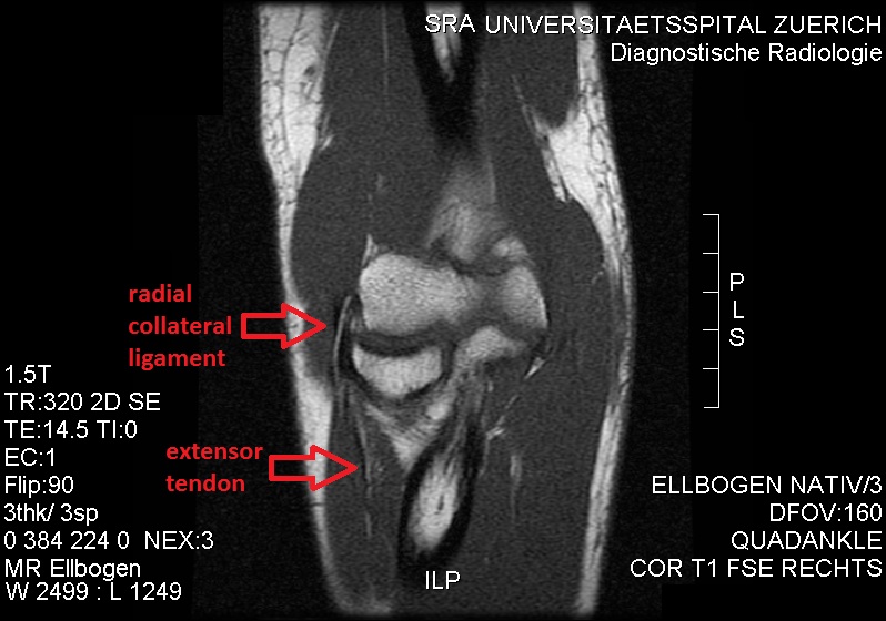
The radial collateral ligament (RCL), lateral collateral ligament (LCL), or external lateral ligamentAs opposed to the "internal lateral ligament", better known as the medial or ulnar collateral ligament is a ligament in the elbow on the side of the radius. Structure The composition of the triangular ligamentous structure on the lateral side of the elbow varies widely between individuals, see alsFigure 4/ref> and can be considered either a single ligament, in which case multiple distal attachments are generally mentioned and the annular ligament is described separately, or as several separate ligaments, in which case parts of those ligaments are often described as indistinguishable from each other. In the latter case, the ligaments are collectively referred to as the lateral collateral ligament complex (LCLC), consisting of four ligaments: * the radial collateral ligament roper(RCL), from the lateral epicondyle to the annular ligament deep to the common extensor tendon * the ... [...More Info...] [...Related Items...] OR: [Wikipedia] [Google] [Baidu] |
Medial Collateral Ligament
The medial collateral ligament (MCL), or tibial collateral ligament (TCL), is one of the four major ligaments of the knee. It is on the medial (inner) side of the knee joint in humans and other primates. Its primary function is to resist outward turning forces on the knee. Structure It is a broad, flat, membranous band, situated slightly posterior on the medial side of the knee joint. It is attached proximally to the medial epicondyle of the femur immediately below the adductor tubercle; below to the medial condyle of the tibia and medial surface of its body. It resists forces that would push the knee medially, which would otherwise produce valgus deformity. The fibers of the posterior part of the ligament are short and incline backward as they descend; they are inserted into the tibia above the groove for the semimembranosus muscle. The anterior part of the ligament is a flattened band, about 10 centimeters long, which inclines forward as it descends. It is inserted into ... [...More Info...] [...Related Items...] OR: [Wikipedia] [Google] [Baidu] |
Collateral Ligaments Of Interphalangeal Articulations Of Foot
The collateral ligaments of the interphalangeal joints of the foot are fibrous bands that are situated on both sides of the interphalangeal joints of the toes Toes are the digits (fingers) of the foot of a tetrapod. Animal species such as cats that walk on their toes are described as being ''digitigrade''. Humans, and other animals that walk on the soles of their feet, are described as being ''planti .... Ligaments of the lower limb {{ligament-stub ... [...More Info...] [...Related Items...] OR: [Wikipedia] [Google] [Baidu] |
Collateral Ligaments Of Metatarsophalangeal Articulations
The collateral ligaments of metatarsophalangeal joints are strong, rounded cords, placed one on either side of each joint, and attached, by one end, to the posterior tubercle on the side of the head of the metatarsal bone, and, by the other, to the contiguous extremity of the phalanx The phalanx ( grc, φάλαγξ; plural phalanxes or phalanges, , ) was a rectangular mass military formation, usually composed entirely of heavy infantry armed with spears, pikes, sarissas, or similar pole weapons. The term is particular .... The place of dorsal ligaments is supplied by the extensor tendons on the dorsal surfaces of the joints. References Ligaments of the lower limb {{ligament-stub ... [...More Info...] [...Related Items...] OR: [Wikipedia] [Google] [Baidu] |
Ulnar Collateral Ligament Of Elbow Joint
The ulnar collateral ligament (UCL) or internal lateral ligament is a thick triangular ligament at the medial aspect of the elbow uniting the distal aspect of the humerus to the proximal aspect of the ulna. Structure It consists of two portions, an anterior and posterior united by a thinner intermediate portion. Note that this ligament is also referred to as the medial collateral ligament and should not be confused with the lateral ulnar collateral ligament (LUCL). The ''anterior portion'', directed obliquely forward, is attached, above, by its apex, to the front part of the medial epicondyle of the humerus; and, below, by its broad base to the medial margin of the coronoid process of the ulna. The ''posterior portion'', also of triangular form, is attached, above, by its apex, to the lower and back part of the medial epicondyle; below, to the medial margin of the olecranon. Between these two bands a few intermediate fibers descend from the medial epicondyle to blend ... [...More Info...] [...Related Items...] OR: [Wikipedia] [Google] [Baidu] |
