|
Cochlosoma
''Cochlosoma'' is a genus of flagellated protozoa in the order Trichomonadida created by A. Kotlán (1923). Some of their typical features include a prominent adhesive disc, axostyle, costa, and six flagella – one of which is attached to an undulating membrane that runs laterally along the body. ''Cochlosoma'' species are parasites found in the intestines of birds and mammals. They are known to cause runting and enteritis in young turkey and ducks. The genus currently contains five species, the most notable member being ''C. anatis'', a parasite of ducks and turkeys. History of knowledge ''Cochlosoma'' was first described by Kotlán (1923) to include ''C. anatis'', a flagellate he found in the intestines of young European domestic ducks ('' Anas platyrhynchos'') suffering from coccidiosis. ''Cochlosoma rostratum'' was identified in North American domestic ducks by Kimura in 1934, although this species is now recognized as a synonym of ''C.anatis1''. Kimura was the first ... [...More Info...] [...Related Items...] OR: [Wikipedia] [Google] [Baidu] |
Trichomonadida
Trichomonadida is an order of anaerobic protists, included with the parabasalids. Members of this order are referred to as trichomonads. Some organisms in this order include: *''Trichomonas vaginalis'', an organism living inside the vagina of humans *''Dientamoeba fragilis'', parasitic ameboid in humans *''Histomonas meleagridis'', parasite that causes blackhead disease in poultry *''Mixotricha paradoxa'', a symbiotic organism inside termites, host of endosymbionts Anatomy Species in this order typically have four to six flagella at the cell's apical pole, one of which is recurrent - that is, it runs along a surface wave, giving the aspect of an undulating membrane. Like other parabasalids, they typically have an axostyle, a pelta, a costa, and parabasal bodies. In ''Histomonas'' only one flagellum and a reduced axostyle are found, and in ''Dientamoeba'', both are absent. Behavior Most species are either parasites or other endosymbionts of animals. Trichomonads repro ... [...More Info...] [...Related Items...] OR: [Wikipedia] [Google] [Baidu] |
Trichomonadidae
Trichomonadidae is a family of anaerobic protozoa. Many of its members are parasitic Parasitism is a close relationship between species, where one organism, the parasite, lives on or inside another organism, the host, causing it some harm, and is adapted structurally to this way of life. The entomologist E. O. Wilson ha ..., causing disease in humans or domestic animals. References {{Taxonbar, from=Q10384711 Excavata families ... [...More Info...] [...Related Items...] OR: [Wikipedia] [Google] [Baidu] |
Protozoa
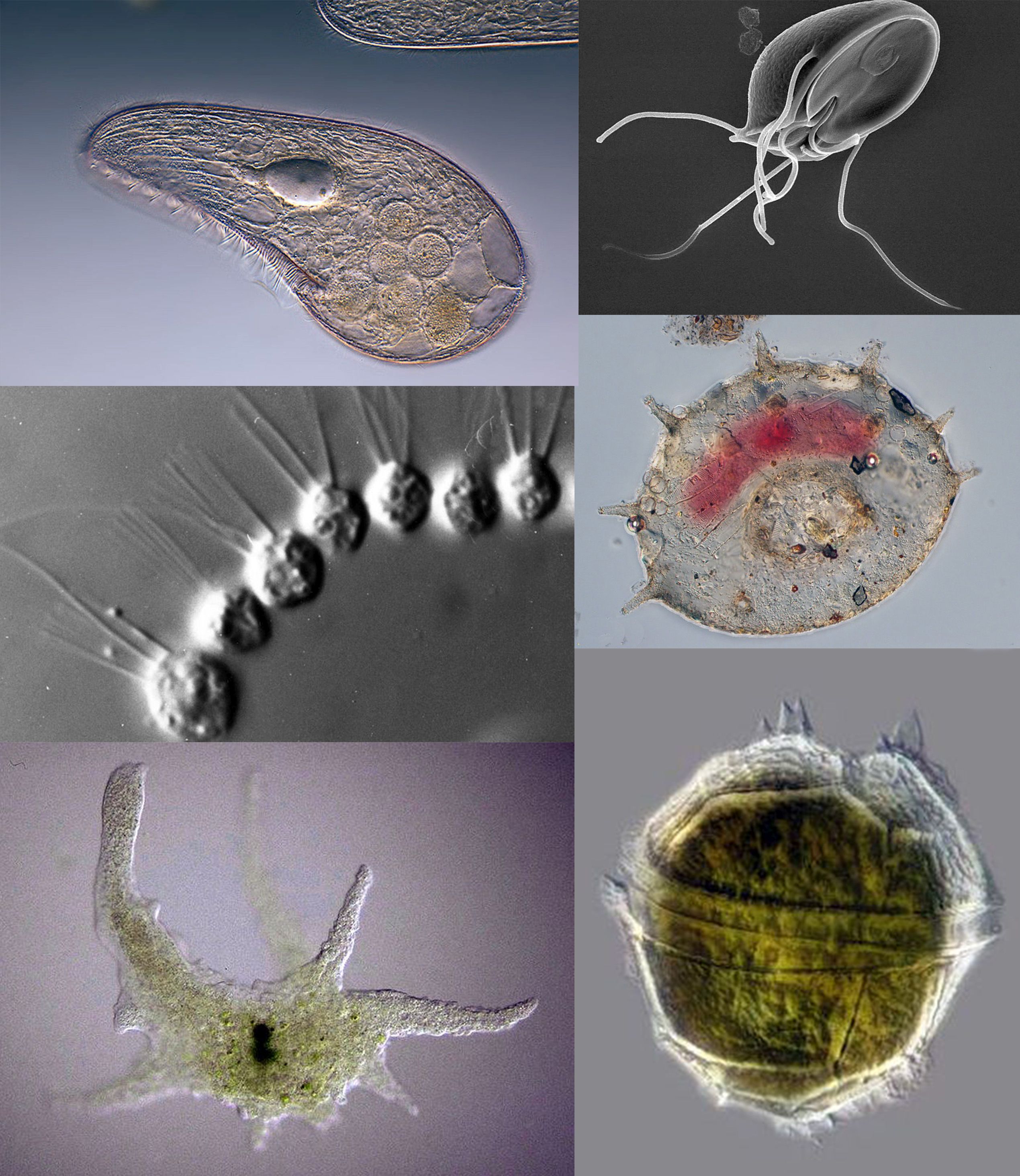
Protozoa (singular: protozoan or protozoon; alternative plural: protozoans) are a group of single-celled eukaryotes, either free-living or parasitic, that feed on organic matter such as other microorganisms or organic tissues and debris. Historically, protozoans were regarded as "one-celled animals", because they often possess animal-like behaviours, such as motility and predation, and lack a cell wall, as found in plants and many algae. When first introduced by Georg Goldfuss (originally spelled Goldfuß) in 1818, the taxon Protozoa was erected as a class within the Animalia, with the word 'protozoa' meaning "first animals". In later classification schemes it was elevated to a variety of higher ranks, including phylum, subkingdom and kingdom, and sometimes included within Protoctista or Protista. The approach of classifying Protozoa within the context of Animalia was widespread in the 19th and early 20th century, but not universal. By the 1970s, it became usual to require ... [...More Info...] [...Related Items...] OR: [Wikipedia] [Google] [Baidu] |
Trophozoite
A trophozoite (G. ''trope'', nourishment + ''zoon'', animal) is the activated, feeding stage in the life cycle of certain protozoa such as malaria-causing ''Plasmodium falciparum'' and those of the ''Giardia'' group. (The complement of the trophozoite state is the thick-walled cyst form). Life cycle stages Trophozoite and cyst stages are shown in the life cycle of '' Balantidium coli'' the causative agent of balantidiasis. In the apicomplexan life cycle the trophozoite undergoes schizogony (asexual reproduction) and develops into a schizont which contains merozoites. The trophozoite life stage of ''Giardia ''Giardia'' ( or ) is a genus of anaerobic flagellated protozoan parasites of the phylum Metamonada that colonise and reproduce in the small intestines of several vertebrates, causing the disease giardiasis. Their life cycle alternates betwee ...'' colonizes and proliferates in the small intestine. Trophozoites develop during the course of the infection into cysts which ... [...More Info...] [...Related Items...] OR: [Wikipedia] [Google] [Baidu] |
Cytoplasm
In cell biology, the cytoplasm is all of the material within a eukaryotic cell, enclosed by the cell membrane, except for the cell nucleus. The material inside the nucleus and contained within the nuclear membrane is termed the nucleoplasm. The main components of the cytoplasm are cytosol (a gel-like substance), the organelles (the cell's internal sub-structures), and various cytoplasmic inclusions. The cytoplasm is about 80% water and is usually colorless. The submicroscopic ground cell substance or cytoplasmic matrix which remains after exclusion of the cell organelles and particles is groundplasm. It is the hyaloplasm of light microscopy, a highly complex, polyphasic system in which all resolvable cytoplasmic elements are suspended, including the larger organelles such as the ribosomes, mitochondria, the plant plastids, lipid droplets, and vacuoles. Most cellular activities take place within the cytoplasm, such as many metabolic pathways including glycolysis, and proc ... [...More Info...] [...Related Items...] OR: [Wikipedia] [Google] [Baidu] |
Hydrogenosome
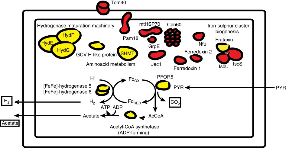
A hydrogenosome is a membrane-enclosed organelle found in some anaerobic ciliates, flagellates, and fungi. Hydrogenosomes are highly variable organelles that have presumably evolved from protomitochondria to produce molecular hydrogen and ATP in anaerobic conditions. Hydrogenosomes were discovered in 1973 by D. G. Lindmark and M. Müller. Because hydrogenosomes hold evolutionary lineage significance for organisms living in anaerobic or oxygen-stressed environments, many research institutions have since documented their findings on how the organelle differs in various sources. History Hydrogenosomes were isolated, purified, biochemically characterized and named in the early 1970s by Lindmark and Müller at Rockefeller University. In addition to this seminal study on hydrogenosomes, they also demonstrated for the first time the presence of pyruvate:ferredoxin oxido-reductase and hydrogenase in eukaryotes. Further studies were subsequently conducted on the biochemical cytology a ... [...More Info...] [...Related Items...] OR: [Wikipedia] [Google] [Baidu] |
Golgi Complex
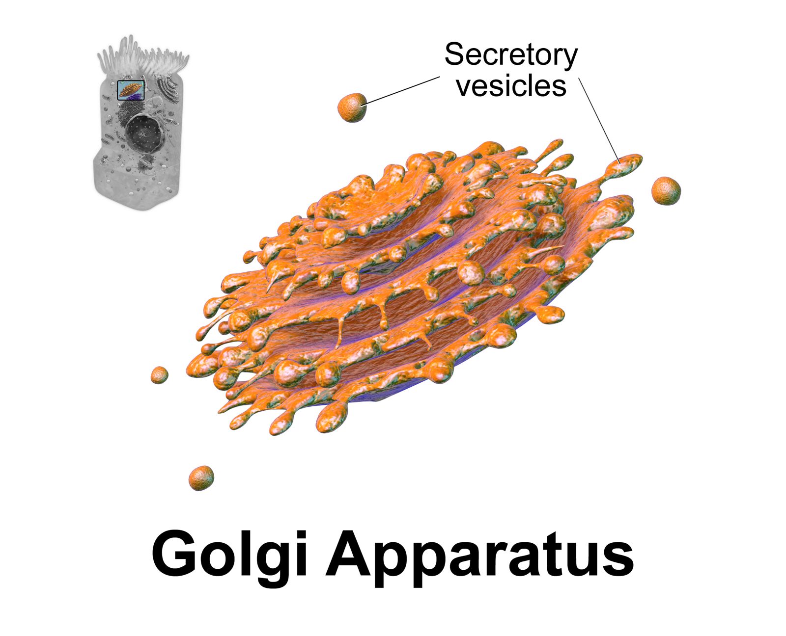
The Golgi apparatus (), also known as the Golgi complex, Golgi body, or simply the Golgi, is an organelle found in most eukaryotic cells. Part of the endomembrane system in the cytoplasm, it packages proteins into membrane-bound vesicles inside the cell before the vesicles are sent to their destination. It resides at the intersection of the secretory, lysosomal, and endocytic pathways. It is of particular importance in processing proteins for secretion, containing a set of glycosylation enzymes that attach various sugar monomers to proteins as the proteins move through the apparatus. It was identified in 1897 by the Italian scientist Camillo Golgi and was named after him in 1898. Discovery Owing to its large size and distinctive structure, the Golgi apparatus was one of the first organelles to be discovered and observed in detail. It was discovered in 1898 by Italian physician Camillo Golgi during an investigation of the nervous system. After first observing it under his m ... [...More Info...] [...Related Items...] OR: [Wikipedia] [Google] [Baidu] |
Uninucleate
{{Short pages monitor ... [...More Info...] [...Related Items...] OR: [Wikipedia] [Google] [Baidu] |
Intestinal Mucosa
The gastrointestinal wall of the gastrointestinal tract is made up of four layers of specialised tissue. From the inner cavity of the gut (the lumen) outwards, these are: # Mucosa # Submucosa # Muscular layer # Serosa or adventitia The mucosa is the innermost layer of the gastrointestinal tract. It surrounds the lumen of the tract and comes into direct contact with digested food ( chyme). The mucosa itself is made up of three layers: the epithelium, where most digestive, absorptive and secretory processes occur; the lamina propria, a layer of connective tissue, and the muscularis mucosae, a thin layer of smooth muscle. The submucosa contains nerves including the submucous plexus (also called Meissner's plexus), blood vessels and elastic fibres with collagen, that stretches with increased capacity but maintains the shape of the intestine. The muscular layer surrounds the submucosa. It comprises layers of smooth muscle in longitudinal and circular orientation that also hel ... [...More Info...] [...Related Items...] OR: [Wikipedia] [Google] [Baidu] |
Double-barred Finch
The double-barred finch (''Stizoptera bichenovii'') is an estrildid finch found in dry savannah, tropical (lowland) dry grassland and shrubland habitats in northern and eastern Australia. It is sometimes referred to as Bicheno's finch or as the owl finch, the latter of which owing to the dark ring of feathers around the face. Taxonomy Nicholas Aylward Vigors and Thomas Horsfield described the double-barred finch in 1827. The specific epithet commemorates James Ebenezer Bicheno, a colonial secretary of Van Diemen's Land appointed in September 1842. There are two subspecies: * ''Stizoptera bichenovii bichenovii'' of eastern Australia * ''Stizoptera bichenovii annulosa'' of northern and northwestern Australia, described by John Gould in 1840.Payne, R. B. (2021). Double-barred Finch (''Stizoptera bichenovii''), version 1.1. In Birds of the World (J. del Hoyo, A. Elliott, J. Sargatal, D. A. Christie, and E. de Juana, Editors). Cornell Lab of Ornithology, Ithaca, NY, USA. https: ... [...More Info...] [...Related Items...] OR: [Wikipedia] [Google] [Baidu] |
Painted Finch
The painted finch (''Emblema pictum'') is a common species of estrildid finch found in Australia. The painted finch acquired its name due to the red and white spotted and mottled underparts of both males and females. The binomial comes from emblema meaning 'mosaic or inlaid work'; and ''pictum'' derives from the Latin word ''pictus'', meaning 'painted' (from pingere, 'to paint'). Other names include Emblema finch, mountain finch, painted firetail and Emblema. The painted finch is a popular bird to be kept in captivity and in backyard aviaries. Taxonomy The painted finch is within the genus ''Emblema'' which early studies placed four species inside of. More recent research has since determined that this genus did not form a natural assemblage and three of the four species were segregated into the genus ''Stagonopleura''. The species include ''S. bella'' (Beautiful firetail), ''S. oculata'' (Red-eared firetail) and ''S. guttata'' (Diamond firetail). This species, ''Emblema pictum, ... [...More Info...] [...Related Items...] OR: [Wikipedia] [Google] [Baidu] |
Blue-faced Parrotfinch
The blue-faced parrotfinch (''Erythrura trichroa'') is a locally common species of estrildid finch found in north-eastern Australia, Japan, Indonesia, Federated States of Micronesia, France (introduced), New Caledonia, Palau, Papua New Guinea, the Solomon Islands and Vanuatu. It has an estimated global extent of occurrence of 10,000,000 km2. It is found in subtropical and tropical zones in both montane and lowland moist forest areas, where it is most often associated with forest edges and disturbed habitat. It feeds largely on seeds of grasses, including in Australia several exotic genera especially Brachiaria. The IUCN has classified the species as being of least concern. Origin and history Origin and phylogeny has been obtained by Antonio Arnaiz-Villena et al. Estrildinae may have originated in India and dispersed thereafter (towards Africa and Pacific Ocean habitats). In the past, due to less developed observation techniques, very few blue-faced parrotfinches were spotted. As ... [...More Info...] [...Related Items...] OR: [Wikipedia] [Google] [Baidu] |


