|
Cilio-spinal Centre
The ciliospinal center (also known as Budge's center) is a cluster of pre-ganglionic sympathetic neuron cell bodies located in the intermediolateral cell column (of the cornu laterale) at spinal cord segment (C8: ''Anatomic variation'') T1-T2 It receives afferents from (the posterior part of) the hypothalamus via the (ipsilateral) hypothalamospinal tract which synapse with the center's pre-ganglionic sympathetic neurons. The efferent, pre-ganglionic axons then leave the spinal cord to enter and ascend in the sympathetic trunk to reach the superior cervical ganglion (SCG) where they synapse with post-ganglionic sympathetic neurons. The post-ganglionic neurons of the SCG then join the internal carotid nerve plexus of the internal carotid artery, accompanying first this artery and subsequently its branches to reach the orbit. In the orbit, they join the long ciliary nerves and short ciliary nerves to reach and innervate the dilator pupillae muscle to mediate pupillary dilatation as ... [...More Info...] [...Related Items...] OR: [Wikipedia] [Google] [Baidu] |
Intermediolateral Nucleus
The intermediolateral nucleus (IML) is located in Rexed lamina VII of the lateral grey column, one of three grey matter columns found in the spinal cord. The intermediolateral cell column exists at vertebral levels T1 – L3. It mediates the entire sympathetic innervation of the body, but the nucleus resides in the grey matter of the spinal cord. Rexed Lamina VII contains several well defined nuclei including the posterior thoracic nucleus (Clarke's column), the intermediolateral nucleus, and the sacral autonomic nucleus. It extends from T1 to L3, and contains the autonomic motor neurons that give rise to the preganglionic fibers of the sympathetic nervous system The sympathetic nervous system (SNS or SANS, sympathetic autonomic nervous system, to differentiate it from the somatic nervous system) is one of the three divisions of the autonomic nervous system, the others being the parasympathetic nervous sy ..., (preganglionic sympathetic general visceral efferents). ... [...More Info...] [...Related Items...] OR: [Wikipedia] [Google] [Baidu] |
Lateral Grey Column
The lateral grey column (lateral column, lateral cornu, lateral horn of spinal cord, intermediolateral column) is one of the three grey columns of the spinal cord (which give the shape of a butterfly); the others being the anterior grey column, anterior and posterior grey column, posterior grey columns. The lateral grey column is primarily involved with activity in the Sympathetic nervous system, sympathetic division of the Autonomic nervous system, autonomic Motor nerve, motor system. It projects to the side as a triangular field in the thoracic and upper lumbar regions (specifically thoracic vertebrae, T1-lumbar vertebrae, L2) of the postero-lateral part of the anterior grey column. Background information Nervous system The nervous system is the system of neurons, or nerve cells that relay electrical signals through the brain and body. A nerve cell receives signals from other nerve cells through tree-branch-like extensions called dendrites and passes signals through a long ex ... [...More Info...] [...Related Items...] OR: [Wikipedia] [Google] [Baidu] |
Spinal Cord
The spinal cord is a long, thin, tubular structure made up of nervous tissue that extends from the medulla oblongata in the lower brainstem to the lumbar region of the vertebral column (backbone) of vertebrate animals. The center of the spinal cord is hollow and contains a structure called the central canal, which contains cerebrospinal fluid. The spinal cord is also covered by meninges and enclosed by the neural arches. Together, the brain and spinal cord make up the central nervous system. In humans, the spinal cord is a continuation of the brainstem and anatomically begins at the occipital bone, passing out of the foramen magnum and then enters the spinal canal at the beginning of the cervical vertebrae. The spinal cord extends down to between the first and second lumbar vertebrae, where it tapers to become the cauda equina. The enclosing bony vertebral column protects the relatively shorter spinal cord. It is around long in adult men and around long in adult women. The diam ... [...More Info...] [...Related Items...] OR: [Wikipedia] [Google] [Baidu] |
Hypothalamospinal Tract
The hypothalamospinal tract is an unmyelinated non-decussated descending nerve tract that arises in the hypothalamus and projects to the brainstem and spinal cord to synapse with pre-ganglionic autonomic (both sympathetic and parasympathetic) neurons. The direct autonomic projections of the hypothalamospinal tract represent a minority of the autonomic output of the hypothalamus; most is thought to project to various relay structures. Anatomy Origin The tract originates mainly from the paraventricular nucleus of hypothalamus, with minor contributions from the dorsomedial, ventromedial, and posterior nuclei of hypothalamus, and lateral hypothalamus. The neurons of the hypothalamospinal tract receive direct afferents from the ascending nociceptive sensory spinohypothalamic tract to mediate the autonomic response to painful stimuli. The tract terminates upon pre-ganglionic autonomic neurons in the brainstem, and spinal segments T1-L3 ( sympathetic outflow), and S2-S4 (par ... [...More Info...] [...Related Items...] OR: [Wikipedia] [Google] [Baidu] |
Sympathetic Trunk
The sympathetic trunk (sympathetic chain, gangliated cord) is a paired bundle of nerve fibers that run from the base of the skull to the coccyx. It is a major component of the sympathetic nervous system. Structure The sympathetic trunk lies just lateral to the vertebral bodies for the entire length of the vertebral column. It interacts with the anterior rami of spinal nerves by way of rami communicantes. The sympathetic trunk permits preganglionic fibers of the sympathetic nervous system to ascend to spinal levels superior to T1 and descend to spinal levels inferior to L2/3.Greenstein B., Greenstein A. (2002): Color atlas of neuroscience – Neuroanatomy and neurophysiology. Thieme, Stuttgart – New York, . The superior end of it is continued upward through the carotid canal into the skull, and forms a plexus on the internal carotid artery; the inferior part travels in front of the coccyx, where it converges with the other trunk at a structure known as the ganglion impar ... [...More Info...] [...Related Items...] OR: [Wikipedia] [Google] [Baidu] |
Superior Cervical Ganglion
The superior cervical ganglion (SCG) is the upper-most and largest of the cervical sympathetic ganglia of the sympathetic trunk. It probably formed by the union of four sympathetic ganglia of the cervical spinal nerves C1–C4. It is the only ganglion of the sympathetic nervous system that innervates the head and neck. The SCG innervates numerous structures of the head and neck. Structure The superior cervical ganglion is reddish-gray color, and usually shaped like a spindle with tapering ends. It measures about 3 cm in length. Sometimes the SCG is broad and flattened, and occasionally constricted at intervals. It formed by the coalescence of four ganglia, corresponding to the four upper-most cervical nerves C1–C4. The bodies of its preganglionic sympathetic afferent neurons are located in the lateral horn of the spinal cord. Their axons enter the SCG to synapse with postganglionic neurons whose axons then exit the rostral end of the SCG and proceed to innervate their targ ... [...More Info...] [...Related Items...] OR: [Wikipedia] [Google] [Baidu] |
Internal Carotid Nerve Plexus
The internal carotid plexus is a nerve plexus situated upon the lateral side of the internal carotid artery. It is composed of post-ganglionic sympathetic fibres which have synapsed at (i.e. have their nerve cell bodies at) the superior cervical ganglion. The plexus gives rise to the deep petrosal nerve. Anatomy Postganglionic sympathetic fibres ascend from the superior cervical ganglion, along the walls of the internal carotid artery, to enter the internal carotid plexus. These fibres are then distributed to deep structures, including the superior tarsal muscle and pupillary dilator muscle. It includes fibres destined for the pupillary dilator muscle as part of a neural circuit regulating pupillary dilatation component of the pupillary reflex. Some fibres of the plexus converge to form the deep petrosal nerve.Richard L. Drake, Wayne Vogel & Adam W M Mitchell, "Gray's Anatomy for Students", Elsevier inc., 2005 The internal carotid plexus communicates with the trigeminal gangli ... [...More Info...] [...Related Items...] OR: [Wikipedia] [Google] [Baidu] |
Internal Carotid Artery
The internal carotid artery is an artery in the neck which supplies the anterior cerebral artery, anterior and middle cerebral artery, middle cerebral circulation. In human anatomy, the internal and external carotid artery, external carotid arise from the common carotid artery, where it bifurcates at cervical vertebrae C3 or C4. The internal carotid artery supplies the brain, including the eyes, while the external carotid nourishes other portions of the head, such as the face, scalp, skull, and meninges. Classification Terminologia Anatomica in 1998 subdivided the artery into four parts: "cervical", "petrous", "cavernous", and "cerebral". In clinical settings, however, usually the classification system of the internal carotid artery follows the 1996 recommendations by Bouthillier, describing seven anatomical segments of the internal carotid artery, each with a corresponding alphanumeric identifier: C1 cervical; C2 petrous; C3 lacerum; C4 cavernous; C5 clinoid; C6 ophthalmic; ... [...More Info...] [...Related Items...] OR: [Wikipedia] [Google] [Baidu] |
Long Ciliary Nerves
The long ciliary nerves are 2-3 nerves that arise from the nasociliary nerve (itself a branch of the ophthalmic branch (CN V1) of the trigeminal nerve (CN V)). They enter the eyeball to provide sensory innervation to parts of the eye, and sympathetic visceral motor innervation to the dilator pupillae muscle. Anatomy Origin The long ciliary nerves branch from the nasociliary nerve as it crosses the optic nerve (CN II). Course Accompanied by the short ciliary nerves, the long ciliary nerves pierce and enter the posterior part of the sclera near where it is entered by the optic nerve, then run anterior-ward between the sclera and the choroid. Function The long ciliary nerves are distributed to the ciliary body, iris, and cornea. Sensory The long ciliary nerves provide sensory innervation to the eyeball, including the cornea. Sympathetic The long ciliary nerves contain post-ganglionic sympathetic fibers from the superior cervical ganglion for the dilator pupill ... [...More Info...] [...Related Items...] OR: [Wikipedia] [Google] [Baidu] |
Short Ciliary Nerves
The short ciliary nerves are nerves of the orbit around the eye. They are branches of the ciliary ganglion. They supply parasympathetic and sympathetic nerve fibers to the ciliary muscle, iris, and cornea. Damage to the short ciliary nerve may result in loss of the pupillary light reflex, or mydriasis. Structure The short ciliary nerves are branches of the ciliary ganglion. They arise from the forepart of the ganglion in two bundles connected with its superior and inferior angles. The lower bundle is the larger than the upper bundle. These split into between 6 and 10 filaments. They run forward with the ciliary arteries in a wavy course. One bundle is set above the optic nerve, while the other bundle is set below it. They are accompanied by the long ciliary nerves from the nasociliary. They pierce the sclera at the back part of the bulb of the eye, pass forward in delicate grooves on the inner surface of the sclera, and are distributed to the ciliary muscle, iris, and corne ... [...More Info...] [...Related Items...] OR: [Wikipedia] [Google] [Baidu] |
Dilator Pupillae
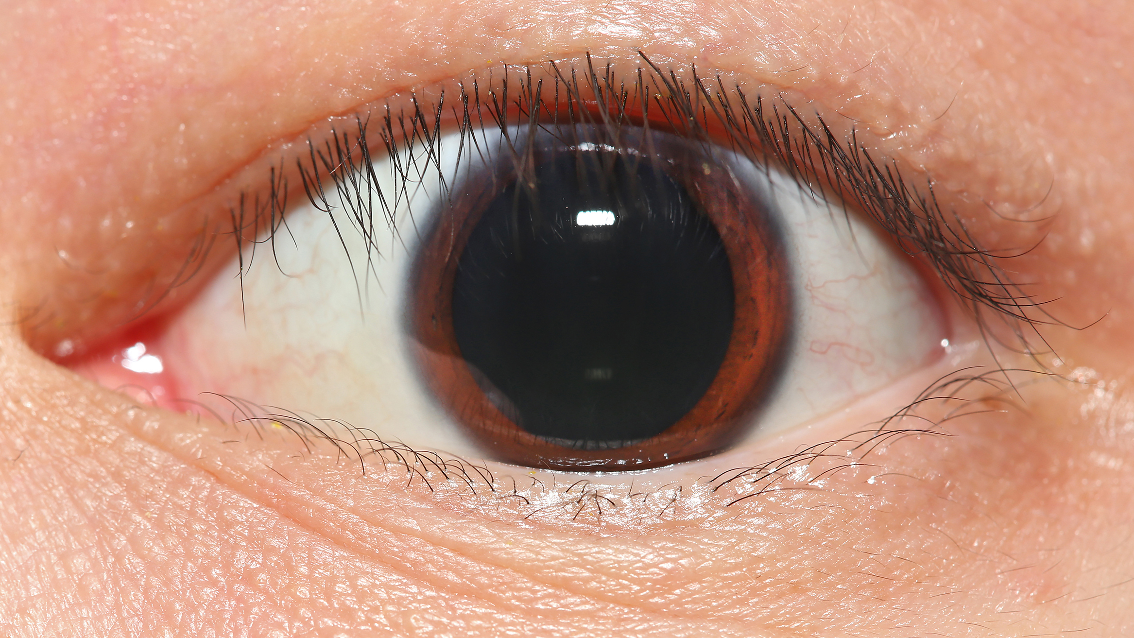
The iris dilator muscle (pupil dilator muscle, pupillary dilator, radial muscle of iris, radiating fibers), is a smooth muscle of the eye, running radially in the iris and therefore fit as a dilator. The pupillary dilator consists of a spokelike arrangement of modified contractile cells called myoepithelial cells. These cells are stimulated by the sympathetic nervous system. When stimulated, the cells contract, widening the pupil and allowing more light to enter the eye. The ciliary muscle, pupillary sphincter muscle and pupillary dilator muscle sometimes are called intrinsic ocular muscles or intraocular muscles. Structure Innervation It is innervated by the sympathetic system, which acts by releasing noradrenaline, which acts on α1-receptors. Thus, when presented with a threatening stimulus that activates the fight-or-flight response, this innervation contracts the muscle and dilates the pupil, thus temporarily letting more light reach the retina. The dilator muscle is ... [...More Info...] [...Related Items...] OR: [Wikipedia] [Google] [Baidu] |
Pupillary Dilatation
Mydriasis is the dilation of the pupil, usually having a non-physiological cause, or sometimes a physiological pupillary response. Non-physiological causes of mydriasis include disease, trauma, or the use of certain types of drugs. It may also be of unknown cause. Normally, as part of the pupillary light reflex, the pupil dilates in the dark and constricts in the light to respectively improve vividity at night and to protect the retina from sunlight damage during the day. A ''mydriatic'' pupil will remain excessively large even in a bright environment. The excitation of the radial fibres of the iris which increases the pupillary aperture is referred to as a mydriasis. More generally, mydriasis also refers to the natural dilation of pupils, for instance in low light conditions or under sympathetic stimulation. Mydriasis is frequently induced by drugs for certain ophthalmic examinations and procedures, particularly those requiring visual access to the retina. Fixed, unilateral ... [...More Info...] [...Related Items...] OR: [Wikipedia] [Google] [Baidu] |


