|
Chondrodysplasia
An osteochondrodysplasia,Etymology: . or skeletal dysplasia, is a disorder of the development of bone and cartilage. Osteochondrodysplasias are rare diseases. About 1 in 5,000 babies are born with some type of skeletal dysplasia. Nonetheless, if taken collectively, genetic skeletal dysplasias or osteochondrodysplasias comprise a recognizable group of genetically determined disorders with generalized skeletal affection. These disorders lead to disproportionate short stature and bone abnormalities, particularly in the arms, legs, and spine. Skeletal dysplasia can result in marked functional limitation and even mortality. Osteochondrodysplasias or skeletal dysplasia subtypes can overlap in clinical aspects, therefore plain radiography is absolutely necessary to establish an accurate diagnosis. Magnetic resonance imaging can provide further diagnostic insights and guide treatment strategies especially in cases of spinal involvement. As some disorders that cause skeletal dysplasia have ... [...More Info...] [...Related Items...] OR: [Wikipedia] [Google] [Baidu] |
Etymology
Etymology ( ) is the study of the origin and evolution of words—including their constituent units of sound and meaning—across time. In the 21st century a subfield within linguistics, etymology has become a more rigorously scientific study. Most directly tied to historical linguistics, philology, and semiotics, it additionally draws upon comparative semantics, morphology, pragmatics, and phonetics in order to attempt a comprehensive and chronological catalogue of all meanings and changes that a word (and its related parts) carries throughout its history. The origin of any particular word is also known as its ''etymology''. For languages with a long written history, etymologists make use of texts, particularly texts about the language itself, to gather knowledge about how words were used during earlier periods, how they developed in meaning and form, or when and how they entered the language. Etymologists also apply the methods of comparative linguistics to reconstruct in ... [...More Info...] [...Related Items...] OR: [Wikipedia] [Google] [Baidu] |
Hyperdontia
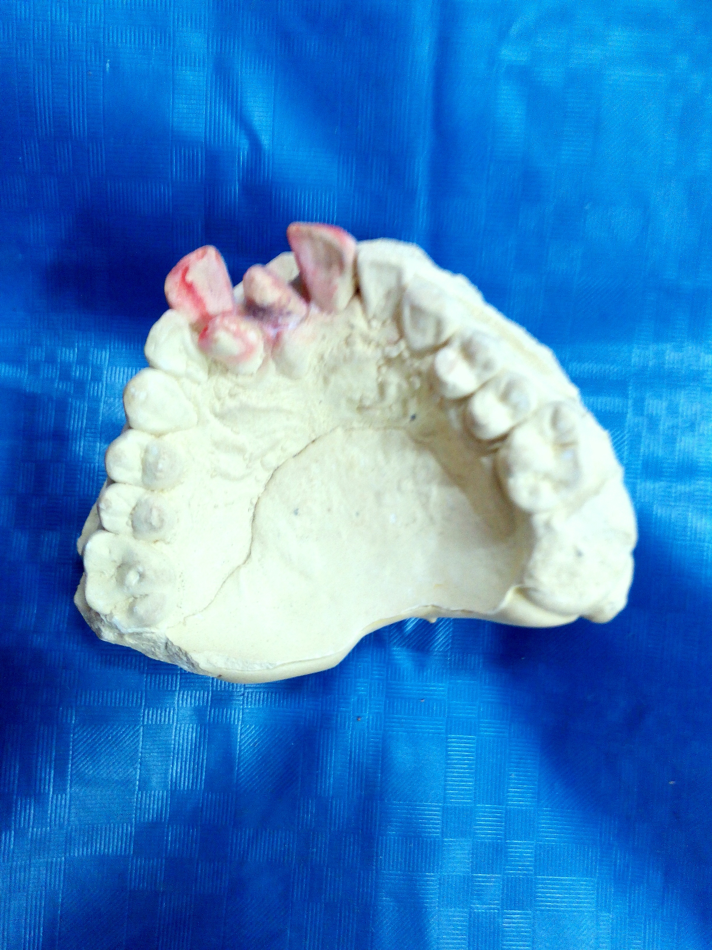
Hyperdontia is the condition of having supernumerary teeth, or teeth that appear in addition to the regular number of teeth (32 in the average adult). They can appear in any area of the dental arch and can affect any dental organ. The opposite of hyperdontia is hypodontia, where there is a congenital lack of teeth, which is a condition seen more commonly than hyperdontia.Pathology of the Hard Dental Tissues The scientific definition of hyperdontia is "any tooth or odontogenic structure that is formed from tooth germ in excess of usual number for any given region of the dental arch."R. S. Omer, R. P. Anthonappa, and N. M. King, "Determination of the optimum time for surgical removal of unerupted anterior supernumerary teeth," Pediatric Dentistry, vol. 32, no. 1, pp. 14–20, 2010. The additional teeth, which may be few or many, can occur on any place in the dental arch. Their arrangement may be symmetrical or non-symmetrical. Signs and symptoms The presence of a supernumerary ... [...More Info...] [...Related Items...] OR: [Wikipedia] [Google] [Baidu] |
X-ray
An X-ray (also known in many languages as Röntgen radiation) is a form of high-energy electromagnetic radiation with a wavelength shorter than those of ultraviolet rays and longer than those of gamma rays. Roughly, X-rays have a wavelength ranging from 10 Nanometre, nanometers to 10 Picometre, picometers, corresponding to frequency, frequencies in the range of 30 Hertz, petahertz to 30 Hertz, exahertz ( to ) and photon energies in the range of 100 electronvolt, eV to 100 keV, respectively. X-rays were discovered in 1895 in science, 1895 by the German scientist Wilhelm Röntgen, Wilhelm Conrad Röntgen, who named it ''X-radiation'' to signify an unknown type of radiation.Novelline, Robert (1997). ''Squire's Fundamentals of Radiology''. Harvard University Press. 5th edition. . X-rays can penetrate many solid substances such as construction materials and living tissue, so X-ray radiography is widely used in medical diagnostics (e.g., checking for Bo ... [...More Info...] [...Related Items...] OR: [Wikipedia] [Google] [Baidu] |
Lymphangioma
Lymphatic malformations are benign slow-flow type of vascular malformation of the lymphatic system characterized by lymphatic vessels which do not connect to the normal lymphatic circulation. The term ''lymphangioma'' is outdated and newer research reference the term ''lymphatic malformation''. Lymphatic malformations can be macrocystic, microcystic, or a combination of the two. Macrocystic have cysts greater than , and microcystic lymphatic malformation have cysts that are smaller than . These malformations can occur at any age and may involve any part of the body, but 90% occur in children less than 2 years of age and involve the head and neck. These malformations are either congenital or acquired. Congenital lymphangiomas are often associated with chromosomal abnormalities such as Turner syndrome, although they can also exist in isolation. Lymphangiomas are commonly diagnosed before birth using fetal ultrasonography. Acquired lymphangiomas may result from trauma, inflammat ... [...More Info...] [...Related Items...] OR: [Wikipedia] [Google] [Baidu] |
Hemangioma
A hemangioma or haemangioma is a usually benign vascular tumor derived from blood vessel cell types. The most common form, seen in infants, is an infantile hemangioma, known colloquially as a "strawberry mark", most commonly presenting on the skin at birth or in the first weeks of life. A hemangioma can occur anywhere on the body, but most commonly appears on the face, scalp, chest or back. They tend to grow for up to a year before gradually shrinking as the child gets older. A hemangioma may need to be treated if it interferes with vision or breathing or is likely to cause long-term disfigurement. In rare cases internal hemangiomas can cause or contribute to other medical problems. They usually disappear by 10 years of age. The first line treatment option is beta blockers, which are highly effective in the majority of cases. Hemangiomas present at birth are called ''congenital hemangiomas'', while those that form later in life are called ''infantile hemangiomas''. Types Heman ... [...More Info...] [...Related Items...] OR: [Wikipedia] [Google] [Baidu] |
Enchondroma
Enchondroma is a type of benign bone tumor belonging to the group of cartilage tumors. There may be no symptoms, or it may present typically in the short tubular bones of the hands with a swelling, pain or pathological fracture. Diagnosis is by X-ray, CT scan and sometimes MRI. Most occur as a less than three centimetre size single tumor. When several occur in one long bone or several bones, the syndrome is called enchondromatosis. Where there are no symptoms, treatment is often not needed. If treatment is required, curettage may be performed. Less than 1% become malignant, unless part of a syndrome. They comprise around 30% of cartilage tumors. 90% of tumors in the hand are enchondromas. Symptoms and signs Individuals with an enchondroma often have no symptoms at all. The following are the most common symptoms of an enchondroma. However, each individual may experience symptoms differently. Symptoms may include: * Pain that may occur at the site of the tumor if the t ... [...More Info...] [...Related Items...] OR: [Wikipedia] [Google] [Baidu] |
Human Skeleton
The human skeleton is the internal framework of the human body. It is composed of around 270 bones at birth – this total decreases to around 206 bones by adulthood after some bones get fused together. The bone mass in the skeleton makes up about 14% of the total body weight (ca. 10–11 kg for an average person) and reaches maximum mass between the ages of 25 and 30. The human skeleton can be divided into the axial skeleton and the appendicular skeleton. The axial skeleton is formed by the human vertebral column, vertebral column, the human rib cage, rib cage, the human skull, skull and other associated bones. The appendicular skeleton, which is attached to the axial skeleton, is formed by the shoulder girdle, the pelvic girdle and the bones of the upper and lower limbs. The human skeleton performs six major functions: support, movement, protection, production of blood cells, storage of minerals, and endocrine regulation. The human skeleton is not as sexually dimorphic ... [...More Info...] [...Related Items...] OR: [Wikipedia] [Google] [Baidu] |
Chromosome
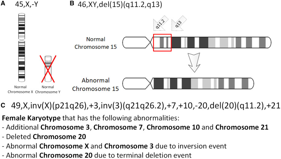
A chromosome is a package of DNA containing part or all of the genetic material of an organism. In most chromosomes, the very long thin DNA fibers are coated with nucleosome-forming packaging proteins; in eukaryotic cells, the most important of these proteins are the histones. Aided by chaperone proteins, the histones bind to and condense the DNA molecule to maintain its integrity. These eukaryotic chromosomes display a complex three-dimensional structure that has a significant role in transcriptional regulation. Normally, chromosomes are visible under a light microscope only during the metaphase of cell division, where all chromosomes are aligned in the center of the cell in their condensed form. Before this stage occurs, each chromosome is duplicated ( S phase), and the two copies are joined by a centromere—resulting in either an X-shaped structure if the centromere is located equatorially, or a two-armed structure if the centromere is located distally; the jo ... [...More Info...] [...Related Items...] OR: [Wikipedia] [Google] [Baidu] |
Genetic Deletion
In genetics, a deletion (also called gene deletion, deficiency, or deletion mutation) (sign: Δ) is a mutation (a genetic aberration) in which a part of a chromosome or a sequence of DNA is left out during DNA replication. Any number of nucleotides can be deleted, from a single base to an entire piece of chromosome. Some chromosomes have fragile spots where breaks occur, which result in the deletion of a part of the chromosome. The breaks can be induced by heat, viruses, radiation, or chemical reactions. When a chromosome breaks, if a part of it is deleted or lost, the missing piece of chromosome is referred to as a deletion or a deficiency. For synapsis to occur between a chromosome with a large intercalary deficiency and a normal complete homolog, the unpaired region of the normal homolog must loop out of the linear structure into a deletion or compensation loop. The smallest single base deletion mutations occur by a single base flipping in the template DNA, followed by temp ... [...More Info...] [...Related Items...] OR: [Wikipedia] [Google] [Baidu] |
Medullary Bone
The medullary cavity (''medulla'', innermost part) is the central cavity of bone shafts where red bone marrow and/or yellow bone marrow (adipose tissue) is stored; hence, the medullary cavity is also known as the marrow cavity. Located in the main shaft of a long bone (diaphysis) (consisting mostly of Bone#Cancellous_bone, spongy bone), the medullary cavity has walls composed of Bone#Cortical_bone, compact bone (cancellous bone) and is lined with a thin, vascular membrane (endosteum). This area is involved in the formation of red blood cells and white blood cells, and the calcium supply for bird eggshells. The area has been detected in fossil bones despite the fossilization process. Intramedullary is a medical term meaning the inside of a bone. Examples include intramedullary rods used to treat bone fractures in orthopedic surgery and intramedullary tumors occurring in some forms of cancer or benign tumors such as an enchondroma. References External links * Skelet ... [...More Info...] [...Related Items...] OR: [Wikipedia] [Google] [Baidu] |
Tumor
A neoplasm () is a type of abnormal and excessive growth of tissue. The process that occurs to form or produce a neoplasm is called neoplasia. The growth of a neoplasm is uncoordinated with that of the normal surrounding tissue, and persists in growing abnormally, even if the original trigger is removed. This abnormal growth usually forms a mass, which may be called a tumour or tumor.'' ICD-10 classifies neoplasms into four main groups: benign neoplasms, in situ neoplasms, malignant neoplasms, and neoplasms of uncertain or unknown behavior. Malignant neoplasms are also simply known as cancers and are the focus of oncology. Prior to the abnormal growth of tissue, such as neoplasia, cells often undergo an abnormal pattern of growth, such as metaplasia or dysplasia. However, metaplasia or dysplasia does not always progress to neoplasia and can occur in other conditions as well. The word neoplasm is from Ancient Greek 'new' and 'formation, creation'. Types A neopla ... [...More Info...] [...Related Items...] OR: [Wikipedia] [Google] [Baidu] |
Lesion
A lesion is any damage or abnormal change in the tissue of an organism, usually caused by injury or diseases. The term ''Lesion'' is derived from the Latin meaning "injury". Lesions may occur in both plants and animals. Types There is no designated classification or naming convention for lesions. Because lesions can occur anywhere in the body and their definition is so broad, the varieties of lesions are virtually endless. Generally, lesions may be classified by their patterns, sizes, locations, or causes. They can also be named after the person who discovered them. For example, Ghon lesions, which are found in the lungs of those with tuberculosis, are named after the lesion's discoverer, Anton Ghon. The characteristic skin lesions of a varicella zoster virus infection are called '' chickenpox''. Lesions of the teeth are usually called dental caries, or "cavities". Location Lesions are often classified by their tissue types or locations. For example, "skin lesions" or ... [...More Info...] [...Related Items...] OR: [Wikipedia] [Google] [Baidu] |


