|
Cervical Vertebral Maturation Method
Cervical vertebral maturation method is an assessment of skeletal age based on the cervical vertebrae, as seen in a cephalometric radiograph. also called as CVM. It was developed by Lamparski in 1972. Cephalometric radiographs are usually obtained for orthodontic patients, which offer the benefit of avoiding additional radiation exposure when gauging the adolescent growth spurt. Nevertheless, several studies have contested the reliability and accuracy of deriving skeletal age from cervical vertebrae, with one study contending that chronologic age is just as reliable as CVM method. Research into CVM has yielded notable findings in regards to intraobserver and interobserver reliability. Comparable results to that of hand–wrist radiographs have been recorded, which was further affirmed by the outcome of one specific prospective review of the literature. History Estimating the bone age of a living child is typically performed by comparing images of their bones to images of models of t ... [...More Info...] [...Related Items...] OR: [Wikipedia] [Google] [Baidu] |
CVM Method
CVM may refer to: Veterinary medicine * California Variegated Mutant, a sheep breed * Center for Veterinary Medicine of U.S. FDA * Cervical Vertebral Malformation or wobbler disease of dogs and horses * Complex vertebral malformation of Holstein cattle Other uses * Christian Vision for Men, a UK charity * Climate Vulnerability Monitor * Securities Commission (Brazil) The Securities and Exchange Commission of Brazil (CVM) () is the securities market authority in Brazil. It regulates the capital markets in Brazil and all of its participants. This includes stock exchanges, public companies, financial intermedi ... * Cooperation and Verification Mechanism of EU applicant state * General Pedro J. Méndez International Airport in Ciudad Victoria, Mexico, IATA code * "cvm" on Wiktionary * C. F. Vicinaux du Mayumbe (CVM), a former railway company (1898–1936) in the Congo Free State (now Democratic Republic of the Congo) {{Disambiguation ... [...More Info...] [...Related Items...] OR: [Wikipedia] [Google] [Baidu] |
Ossification Center
An ossification center is a point where ossification of the hyaline cartilage begins. The first step in ossification is that the chondrocytes at this point become hypertrophic and arrange themselves in rows. The matrix in which they are imbedded increases in quantity, so that the cells become further separated from each other. A deposit of calcareous material now takes place in this matrix, between the rows of cells, so that they become separated from each other by longitudinal columns of calcified matrix, presenting a granular and opaque appearance. Here and there the matrix between two cells of the same row also becomes calcified, and transverse bars of calcified substance stretch across from one calcareous column to another. Thus, there are longitudinal groups of the cartilage cells enclosed in oblong cavities, the walls of which are formed of calcified matrix which cuts off all nutrition from the cells; the cells, in consequence, atrophy, leaving spaces called the primary ... [...More Info...] [...Related Items...] OR: [Wikipedia] [Google] [Baidu] |
Ossification
Ossification (also called osteogenesis or bone mineralization) in bone remodeling is the process of laying down new bone material by cells named osteoblasts. It is synonymous with bone tissue formation. There are two processes resulting in the formation of normal, healthy bone tissue: Intramembranous ossification is the direct laying down of bone into the primitive connective tissue ( mesenchyme), while endochondral ossification involves cartilage as a precursor. In fracture healing, endochondral osteogenesis is the most commonly occurring process, for example in fractures of long bones treated by plaster of Paris, whereas fractures treated by open reduction and internal fixation with metal plates, screws, pins, rods and nails may heal by intramembranous osteogenesis. Heterotopic ossification is a process resulting in the formation of bone tissue that is often atypical, at an extraskeletal location. Calcification is often confused with ossification. Calcificatio ... [...More Info...] [...Related Items...] OR: [Wikipedia] [Google] [Baidu] |
Epiphysis
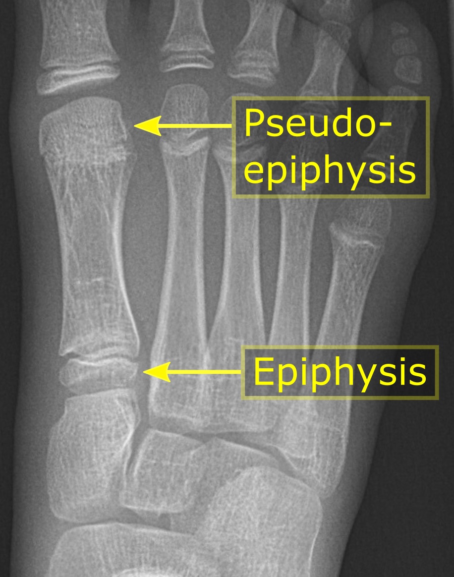
An epiphysis (; : epiphyses) is one of the rounded ends or tips of a long bone that ossify from one or more secondary centers of ossification. Between the epiphysis and diaphysis (the long midsection of the long bone) lies the metaphysis, including the epiphyseal plate (growth plate). During formation of the secondary ossification center, vascular canals (epiphysial canals) stemming from the perichondrium invade the epiphysis, supplying nutrients to the developing secondary centers of ossification. At the joint, the epiphysis is covered with articular cartilage; below that covering is a zone similar to the epiphyseal plate, known as Wikt:subchondral, subchondral bone. The epiphysis is mostly found in mammals but it is also present in some lizards. However, the secondary center of ossification may have evolved multiple times, having been found in the Jurassic sphenodont ''Sapheosaurus'' as well as in the therapsid ''Niassodon, Niassodon mfumukasi.'' The epiphysis is filled wi ... [...More Info...] [...Related Items...] OR: [Wikipedia] [Google] [Baidu] |
Metaphysis
The metaphysis (: metaphyses) is the neck portion of a long bone between the epiphysis and the diaphysis. It contains the growth plate, the part of the bone that grows during childhood, and as it grows it ossifies near the diaphysis and the epiphyses. The metaphysis contains a diverse population of cells including mesenchymal stem cells, which give rise to bone and fat cells, as well as hematopoietic stem cells which give rise to a variety of blood cells as well as bone-destroying cells called osteoclasts. Thus the metaphysis contains a highly metabolic set of tissues including trabecular (spongy) bone, blood vessels, as well as marrow adipose tissue (MAT). The metaphysis may be divided anatomically into three components based on tissue content: a cartilaginous component (epiphyseal plate), a bony component (metaphysis) and a fibrous component surrounding the periphery of the plate. The growth plate synchronizes chondrogenesis with osteogenesis or interstitial carti ... [...More Info...] [...Related Items...] OR: [Wikipedia] [Google] [Baidu] |


