|
Carol Mason
Carol Ann Mason is a Professor of Pathology and Cell Biology at Columbia University in the Mortimer B. Zuckerman Mind Brain Behavior Institute. She studies axon guidance in visual pathways in an effort to restore vision to the blind. Her research focuses on the retinal ganglion cell. She was elected a member of the National Academy of Sciences in 2018. Early life and education Mason earned her bachelor's degree at Chatham University and graduated in 1967. Mason earned her doctorate in invertebrate zoology and endocrinology at University of California, Berkeley. She was a member of Phi Beta Kappa. In 1967 she was awarded a Woodrow Wilson Foundation Fellowship. Research and career Mason was a postdoctoral fellow at the University of Wisconsin–Madison and the University of Chicago. She worked with Ray Guillery on the cellular anatomy of the visual systems of cats. She joined the New York University School of Medicine in 1980. Mason was appointed to the faculty at Columbia Uni ... [...More Info...] [...Related Items...] OR: [Wikipedia] [Google] [Baidu] |
Chatham University
Chatham University is a private university in Pittsburgh, Pennsylvania. Originally founded as a women's college, it began enrolling men in undergraduate programs in 2015. It enrolls about 2,110 students, including 1,002 undergraduate students and 1,108 graduate students. The university grants certificates and degrees including bachelor, master, first-professional, and doctorate degrees in the School of Arts, Science & Business, the School of Health Sciences, and the Falk School of Sustainability & Environment. History Founded as the Pennsylvania Female College on December 11, 1869, by Reverend William Trimble Beatty (the father of renowned operatic contralto Louise Homer), Chatham was initially situated in the Berry mansion on Woodland Road off Fifth Avenue in the neighborhood of Shadyside. Shadyside Campus today is composed of buildings and grounds from a number of former private mansions, including those of Andrew Mellon, Edward Stanton Fickes, George M. Laughlin Jr. and James ... [...More Info...] [...Related Items...] OR: [Wikipedia] [Google] [Baidu] |
Ray Guillery
Rainer Walter "Ray" Guillery FRS (28 August 1929 – 7 April 2017) was a British physiologist and neuroanatomist. He is best known for his discovery that in Siamese cats with certain genotypes of the albino gene, the wiring of the optic chiasm is disrupted, with less of the nerve-crossing than is normal. Early life and education Guillery was born in Greifswald, Germany on 28 August 1929. He began his education as a medical student at University College London (UCL) in 1948. He obtained his BSc in 1951 and his PhD in 1954. Career Guillery taught at UCL for 11 years. In 1964 he went to University of Wisconsin–Madison, where he helped to start the new graduate programme in neuroscience. In 1977, he moved to the University of Chicago to lead another new graduate neuroscience programme. In 1984, Guillery returned to the UK as head of the department of Human Anatomy and Dr. Lee's Professor of Anatomy at the University of Oxford, until 1996. He was then subsequently profes ... [...More Info...] [...Related Items...] OR: [Wikipedia] [Google] [Baidu] |
Stem Cell
In multicellular organisms, stem cells are undifferentiated or partially differentiated cells that can differentiate into various types of cells and proliferate indefinitely to produce more of the same stem cell. They are the earliest type of cell in a cell lineage. They are found in both embryonic and adult organisms, but they have slightly different properties in each. They are usually distinguished from progenitor cells, which cannot divide indefinitely, and precursor or blast cells, which are usually committed to differentiating into one cell type. In mammals, roughly 50–150 cells make up the inner cell mass during the blastocyst stage of embryonic development, around days 5–14. These have stem-cell capability. '' In vivo'', they eventually differentiate into all of the body's cell types (making them pluripotent). This process starts with the differentiation into the three germ layers – the ectoderm, mesoderm and endoderm – at the gastrulation stage. Howev ... [...More Info...] [...Related Items...] OR: [Wikipedia] [Google] [Baidu] |
Retinal Pigment Epithelium
The pigmented layer of retina or retinal pigment epithelium (RPE) is the pigmented cell layer just outside the neurosensory retina that nourishes retinal visual cells, and is firmly attached to the underlying choroid and overlying retinal visual cells. History The RPE was known in the 18th and 19th centuries as the pigmentum nigrum, referring to the observation that the RPE is dark (black in many animals, brown in humans); and as the tapetum nigrum, referring to the observation that in animals with a tapetum lucidum, in the region of the tapetum lucidum the RPE is not pigmented. Anatomy The RPE is composed of a single layer of hexagonal cells that are densely packed with pigment granules. When viewed from the outer surface, these cells are smooth and hexagonal in shape. When seen in section, each cell consists of an outer non-pigmented part containing a large oval nucleus and an inner pigmented portion which extends as a series of straight thread-like processes between the r ... [...More Info...] [...Related Items...] OR: [Wikipedia] [Google] [Baidu] |
Melanogenesis
Melanocytes are melanin-producing neural crest-derived cells located in the bottom layer (the stratum basale) of the skin's epidermis, the middle layer of the eye (the uvea), the inner ear, vaginal epithelium, meninges, bones, and heart. Melanin is a dark pigment primarily responsible for skin color. Once synthesized, melanin is contained in special organelles called melanosomes which can be transported to nearby keratinocytes to induce pigmentation. Thus darker skin tones have more melanosomes present than lighter skin tones. Functionally, melanin serves as protection against UV radiation. Melanocytes also have a role in the immune system. Function Through a process called melanogenesis, melanocytes produce melanin, which is a pigment found in the skin, eyes, hair, nasal cavity, and inner ear. This melanogenesis leads to a long-lasting pigmentation, which is in contrast to the pigmentation that originates from oxidation of already-existing melanin. There are both ba ... [...More Info...] [...Related Items...] OR: [Wikipedia] [Google] [Baidu] |
Ipsilateral
Standard anatomical terms of location are used to unambiguously describe the anatomy of animals, including humans. The terms, typically derived from Latin or Greek roots, describe something in its standard anatomical position. This position provides a definition of what is at the front ("anterior"), behind ("posterior") and so on. As part of defining and describing terms, the body is described through the use of anatomical planes and anatomical axes. The meaning of terms that are used can change depending on whether an organism is bipedal or quadrupedal. Additionally, for some animals such as invertebrates, some terms may not have any meaning at all; for example, an animal that is radially symmetrical will have no anterior surface, but can still have a description that a part is close to the middle ("proximal") or further from the middle ("distal"). International organisations have determined vocabularies that are often used as standard vocabularies for subdisciplines of anatom ... [...More Info...] [...Related Items...] OR: [Wikipedia] [Google] [Baidu] |
Contralateral Brain
The contralateral organization of the forebrain ( Latin: contra‚ against; latus‚ side; lateral‚ sided) is the property that the hemispheres of the cerebrum and the thalamus represent mainly the contralateral side of the body. Consequently, the left side of the forebrain mostly represents the right side of the body, and the right side of the brain primarily represents the left side of the body. The contralateral organization involves both executive and sensory functions (e.g., a left-sided brain lesion may cause a right-sided hemiplegia). The contralateral organization is present in all vertebrates but in no invertebrate. According to the current theory, the forebrain is twisted about the long axis of the body, so that not only the left and right sides, but also dorsal and ventral sides, are interchanged. (See below.) Anatomy Anatomically, the contralateral organization is manifested by major decussations (based upon the Latin notation for ten, 'deca,' as an up ... [...More Info...] [...Related Items...] OR: [Wikipedia] [Google] [Baidu] |
Visual Impairment
Visual impairment, also known as vision impairment, is a medical definition primarily measured based on an individual's better eye visual acuity; in the absence of treatment such as correctable eyewear, assistive devices, and medical treatment– visual impairment may cause the individual difficulties with normal daily tasks including reading and walking. Low vision is a functional definition of visual impairment that is chronic, uncorrectable with treatment or correctable lenses, and impacts daily living. As such low vision can be used as a disability metric and varies based on an individual's experience, environmental demands, accommodations, and access to services. The American Academy of Ophthalmology defines visual impairment as the best-corrected visual acuity of less than 20/40 in the better eye, and the World Health Organization defines it as a presenting acuity of less than 6/12 in the better eye. The term blindness is used for complete or nearly complete vision loss. In ... [...More Info...] [...Related Items...] OR: [Wikipedia] [Google] [Baidu] |
Three-dimensional Space
Three-dimensional space (also: 3D space, 3-space or, rarely, tri-dimensional space) is a geometric setting in which three values (called ''parameters'') are required to determine the position of an element (i.e., point). This is the informal meaning of the term dimension. In mathematics, a tuple of numbers can be understood as the Cartesian coordinates of a location in a -dimensional Euclidean space. The set of these -tuples is commonly denoted \R^n, and can be identified to the -dimensional Euclidean space. When , this space is called three-dimensional Euclidean space (or simply Euclidean space when the context is clear). It serves as a model of the physical universe (when relativity theory is not considered), in which all known matter exists. While this space remains the most compelling and useful way to model the world as it is experienced, it is only one example of a large variety of spaces in three dimensions called 3-manifolds. In this classical example, when the t ... [...More Info...] [...Related Items...] OR: [Wikipedia] [Google] [Baidu] |
Camera Lucida
A ''camera lucida'' is an optical device used as a drawing aid by artists and microscopists. The ''camera lucida'' performs an optical superimposition of the subject being viewed upon the surface upon which the artist is drawing. The artist sees both scene and drawing surface simultaneously, as in a photographic double exposure. This allows the artist to duplicate key points of the scene on the drawing surface, thus aiding in the accurate rendering of perspective. History The ''camera lucida'' was patented in 1806 by the English chemist William Hyde Wollaston. The basic optics were described 200 years earlier by the German astronomer Johannes Kepler in his ''Dioptrice'' (1611), but there is no evidence he or his contemporaries constructed a working ''camera lucida''. By the 19th century, Kepler's description had fallen into oblivion, so Wollaston's claim was never challenged. The term "''camera lucida''" (Latin "well-lit room" as opposed to ''camera obscura'' "dark room") ... [...More Info...] [...Related Items...] OR: [Wikipedia] [Google] [Baidu] |
Thalamus
The thalamus (from Greek θάλαμος, "chamber") is a large mass of gray matter located in the dorsal part of the diencephalon (a division of the forebrain). Nerve fibers project out of the thalamus to the cerebral cortex in all directions, allowing hub-like exchanges of information. It has several functions, such as the relaying of sensory signals, including motor signals to the cerebral cortex and the regulation of consciousness, sleep, and alertness. Anatomically, it is a paramedian symmetrical structure of two halves (left and right), within the vertebrate brain, situated between the cerebral cortex and the midbrain. It forms during embryonic development as the main product of the diencephalon, as first recognized by the Swiss embryologist and anatomist Wilhelm His Sr. in 1893. Anatomy The thalamus is a paired structure of gray matter located in the forebrain which is superior to the midbrain, near the center of the brain, with nerve fibers projecting out to th ... [...More Info...] [...Related Items...] OR: [Wikipedia] [Google] [Baidu] |
Mammalian Eye
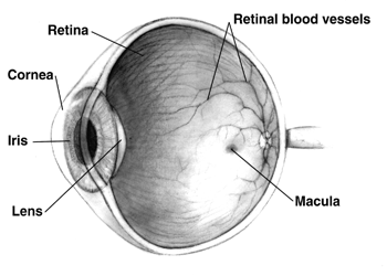
Mammals normally have a pair of eyes. Although mammalian vision is not so excellent as bird vision, it is at least dichromatic for most of mammalian species, with certain families (such as Hominidae) possessing a trichromatic color perception. The dimensions of the eyeball vary only 1–2 mm among humans. The vertical axis is 24 mm; the transverse being larger. At birth it is generally 16–17 mm, enlarging to 22.5–23 mm by three years of age. Between then and age 13 the eye attains its mature size. It weighs 7.5 grams and its volume is roughly 6.5 ml. Along a line through the nodal (central) point of the eye is the optic axis, which is slightly five degrees toward the nose from the visual axis (i.e., that going towards the focused point to the fovea). Three layers The structure of the mammalian eye has a laminar organization that can be divided into three main layers or ''tunics'' whose names reflect their basic functions: the fibrous tunic, the ... [...More Info...] [...Related Items...] OR: [Wikipedia] [Google] [Baidu] |


