|
Canalis Sinuosus
The canalis sinuosus is a passage within the maxilla through which the anterior superior alveolar nerve, artery and vein pass. The proximal opening of the canal occurs near the mid-point of the infraorbital canal. From its proximal opening, the canalis sinuosus first passes inferolaterally within the maxillary orbital floor where it is situated lateral to the infraorbital canal. It then turns to pass inferomedially and anteriorly while passing through the anterior wall of the maxillary sinus (the canal is marked by a groove upon the internal surface of the anterior wall of the sinus). It then courses to the inferior to the infraorbital foramen (of the infraorbital canal) and anterior to the anterior extremity of the inferior nasal concha to reach the margin of the anterior nasal aperture. Thereupon, it passes along the inferior rim of the anterior nasal aperture between the nasal cavity (superiorly), and the tooth sockets (dental alveoli) of the canine and incisor teeth (inferiorl ... [...More Info...] [...Related Items...] OR: [Wikipedia] [Google] [Baidu] |
Maxilla
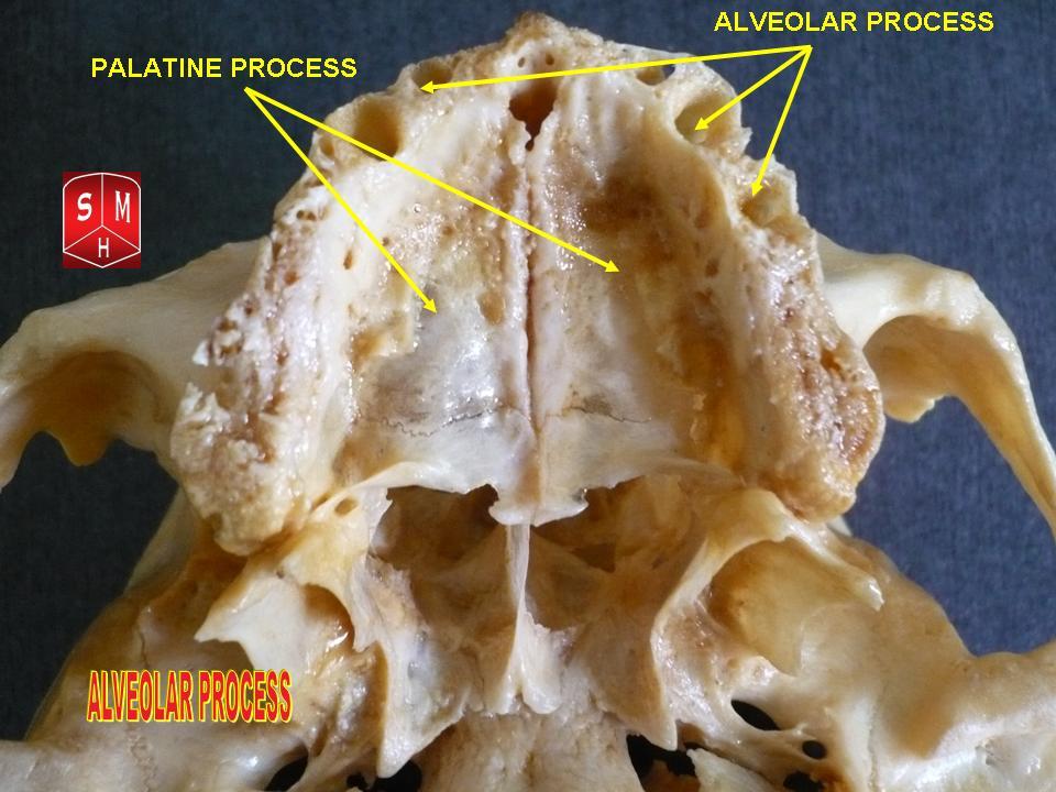
The maxilla (plural: ''maxillae'' ) in vertebrates is the upper fixed (not fixed in Neopterygii) bone of the jaw formed from the fusion of two maxillary bones. In humans, the upper jaw includes the hard palate in the front of the mouth. The two maxillary bones are fused at the intermaxillary suture, forming the anterior nasal spine. This is similar to the mandible (lower jaw), which is also a fusion of two mandibular bones at the mandibular symphysis. The mandible is the movable part of the jaw. Structure In humans, the maxilla consists of: * The body of the maxilla * Four processes ** the zygomatic process ** the frontal process of maxilla ** the alveolar process ** the palatine process * three surfaces – anterior, posterior, medial * the Infraorbital foramen * the maxillary sinus * the incisive foramen Articulations Each maxilla articulates with nine bones: * two of the cranium: the frontal and ethmoid * seven of the face: the nasal, zygomatic, lacrimal, ... [...More Info...] [...Related Items...] OR: [Wikipedia] [Google] [Baidu] |
Anterior Superior Alveolar Nerve
The anterior superior alveolar nerve (or anterior superior dental nerve), is a branch of the infraorbital nerve, itself a branch of the maxillary nerve (V2). It branches from the infraorbital nerve within the infraorbital canal before the infraorbital nerve exits through the infraorbital foramen. It descends in a canal in the anterior wall of the maxillary sinus, and divides into branches which supply the incisor and canine teeth. It communicates with the middle superior alveolar nerve, and gives off a nasal branch, which passes through a minute canal in the lateral wall of the inferior meatus, and supplies the mucous membrane of the anterior part of the inferior meatus and the floor of the nasal cavity, communicating with the nasal branches from the sphenopalatine ganglion. Dental considerations for this nerve are important. The anterior superior alveolar usually innervates all anterior teeth, loops backwards to join the middle superior alveolar nerve to form the superior denta ... [...More Info...] [...Related Items...] OR: [Wikipedia] [Google] [Baidu] |
Anterior Superior Alveolar Arteries
The anterior superior alveolar arteries originate from the infraorbital artery; they supply the upper incisors and canines; they also supply the mucous membrane of the maxillary sinus. See also * Anterior superior alveolar nerve * Posterior superior alveolar artery The posterior superior alveolar artery (posterior dental artery) is given off from the maxillary, frequently in conjunction with the infraorbital artery just as the trunk of the vessel is passing into the pterygopalatine fossa. Branches Descendi ... Arteries of the head and neck {{Circulatory-stub ... [...More Info...] [...Related Items...] OR: [Wikipedia] [Google] [Baidu] |
Infraorbital Canal
The infraorbital canal is a canal found at the base of the orbit that opens on to the maxilla. It is continuous with the infraorbital groove and opens onto the maxilla at the infraorbital foramen. The infraorbital nerve and infraorbital artery travel through the canal. Structure One of the canals of the orbital surface of the maxilla, the infraorbital canal, opens just below the margin of the orbit, the area of the skull containing the eye and related structures. It should not be confused with the infraorbital foramen, with which it is continuous. Function It transmits the infraorbital nerve as well as infraorbital artery, both of which enter this canal at the infraorbital groove and after coursing through the maxillary sinus exit via the infraorbital foramen. Before exiting, the anterior superior alveolar nerve, middle superior alveolar nerve The middle superior alveolar nerve is a nerve that drops from the infraorbital portion of the maxillary nerve to supply the sinus mu ... [...More Info...] [...Related Items...] OR: [Wikipedia] [Google] [Baidu] |
Maxillary Sinus
The pyramid-shaped maxillary sinus (or antrum of Nathaniel Highmore (surgeon), Highmore) is the largest of the paranasal sinuses, and drains into the middle meatus of the nose through the osteomeatal complex.Human Anatomy, Jacobs, Elsevier, 2008, page 209-210 Structure It is the largest air sinus in the body. Found in the body of the maxilla, this sinus has three recesses: an alveolar recess pointed inferiorly, bounded by the alveolar process of the maxilla; a zygomatic recess pointed laterally, bounded by the zygomatic bone; and an infraorbital recess pointed superiorly, bounded by the inferior Orbital surface of the body of the maxilla, orbital surface of the maxilla. The medial wall is composed primarily of cartilage. The ostia for drainage are located high on the medial wall and open into the semilunar hiatus of the lateral nasal cavity; because of the position of the ostia, gravity cannot drain the maxillary sinus contents when the head is erect (see pathology). The ostium of ... [...More Info...] [...Related Items...] OR: [Wikipedia] [Google] [Baidu] |
Infraorbital Foramen
In human anatomy, the infraorbital foramen is one of two small holes in the skull's upper jawbone ( maxillary bone), located below the eye socket and to the left and right of the nose. Both holes are used for blood vessels and nerves. In anatomical terms, it is located below the infraorbital margin of the orbit. It transmits the infraorbital artery and vein, and the infraorbital nerve, a branch of the maxillary nerve. It is typically from the infraorbital margin. Structure Forming the exterior end of the infraorbital canal, the infraorbital foramen communicates with the infraorbital groove, the canal's opening on the interior side. The ramifications of the three principal branches of the trigeminal nerve—at the supraorbital, infraorbital, and mental foramen—are distributed on a vertical line (in anterior view) passing through the middle of the pupil The pupil is a black hole located in the center of the iris of the eye that allows light to strike the retina.Cassin ... [...More Info...] [...Related Items...] OR: [Wikipedia] [Google] [Baidu] |
Inferior Nasal Concha
The inferior nasal concha (inferior turbinated bone or inferior turbinal/turbinate) is one of the three paired nasal conchae in the nose. It extends horizontally along the lateral wall of the nasal cavity and consists of a lamina of spongy bone, curled upon itself like a scroll, (''turbinate'' meaning inverted cone). The inferior nasal conchae are considered a pair of facial bones. As the air passes through the turbinates, the air is churned against these mucosa-lined bones in order to receive warmth, moisture and cleansing. Superior to inferior nasal concha are the middle nasal concha and superior nasal concha which both arise from the ethmoid bone, of the cranial portion of the skull. Hence, these two are considered as a part of the cranial bones. It has two surfaces, two borders, and two extremities. Structure Surfaces The medial surface is convex, perforated by numerous apertures, and traversed by longitudinal grooves for the lodgement of vessels. The lateral surface is con ... [...More Info...] [...Related Items...] OR: [Wikipedia] [Google] [Baidu] |
Anterior Nasal Aperture
The piriform aperture, pyriform aperture, or anterior nasal aperture, is a pear-shaped opening in the human skull. Its long axis is vertical, and narrow end upward; in the recent state it is much contracted by the lateral nasal cartilage and the greater and lesser alar cartilages of the nose. It is bounded above by the inferior borders of the nasal bones; laterally by the thin, sharp margins which separate the anterior from the nasal surfaces of the maxilla; and below by the same borders, where they curve medialward to join each other at the anterior nasal spine The anterior nasal spine, or anterior nasal spine of maxilla, is a bony projection in the skull that serves as a cephalometric landmark. The anterior nasal spine is the projection formed by the fusion of the two maxillary bones at the intermaxill .... References Nose {{musculoskeletal-stub ... [...More Info...] [...Related Items...] OR: [Wikipedia] [Google] [Baidu] |
Nasal Cavity
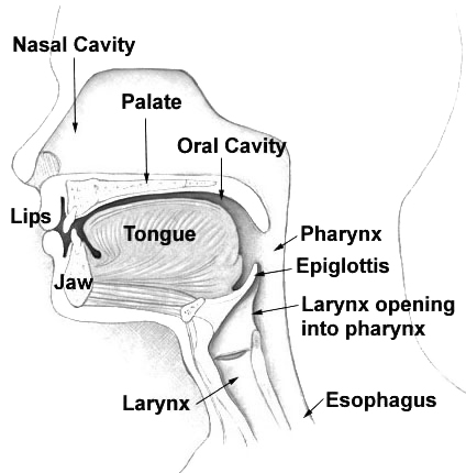
The nasal cavity is a large, air-filled space above and behind the nose in the middle of the face. The nasal septum divides the cavity into two cavities, also known as fossae. Each cavity is the continuation of one of the two nostrils. The nasal cavity is the uppermost part of the respiratory system and provides the nasal passage for inhaled air from the nostrils to the nasopharynx and rest of the respiratory tract. The paranasal sinuses surround and drain into the nasal cavity. Structure The term "nasal cavity" can refer to each of the two cavities of the nose, or to the two sides combined. The lateral wall of each nasal cavity mainly consists of the maxilla. However, there is a deficiency that is compensated for by the perpendicular plate of the palatine bone, the medial pterygoid plate, the labyrinth of ethmoid and the inferior concha. The paranasal sinuses are connected to the nasal cavity through small orifices called ostia. Most of these ostia communicate with ... [...More Info...] [...Related Items...] OR: [Wikipedia] [Google] [Baidu] |
Tooth Socket
Dental alveoli (singular ''alveolus'') are sockets in the jaws in which the roots of teeth are held in the alveolar process with the periodontal ligament. The lay term for dental alveoli is tooth sockets. A joint that connects the roots of the teeth and the alveolus is called ''gomphosis'' (plural ''gomphoses''). Alveolar bone is the bone that surrounds the roots of the teeth forming bone sockets. In mammals, tooth sockets are found in the maxilla, the premaxilla, and the mandible. Etymology 1706, "a hollow," especially "the socket of a tooth," from Latin alveolus "a tray, trough, basin; bed of a small river; small hollow or cavity," diminutive of alvus "belly, stomach, paunch, bowels; hold of a ship," from PIE root *aulo- "hole, cavity" (source also of Greek aulos "flute, tube, pipe;" Serbo-Croatian, Polish, Russian ulica "street," originally "narrow opening;" Old Church Slavonic uliji, Lithuanian aulys "beehive" (hollow trunk), Armenian yli "pregnant"). The word was extended i ... [...More Info...] [...Related Items...] OR: [Wikipedia] [Google] [Baidu] |
Nasal Septum
The nasal septum () separates the left and right airways of the nasal cavity, dividing the two nostrils. It is depressed by the depressor septi nasi muscle. Structure The fleshy external end of the nasal septum is called the columella or columella nasi, and is made up of cartilage and soft tissue. The nasal septum contains bone and hyaline cartilage. It is normally about 2 mm thick. The nasal septum is composed of four structures: * Perpendicular plate of ethmoid bone * Vomer bone * Septal nasal cartilage * Maxillary bone (the crest) The lowest part of the septum is a narrow strip of bone that projects from the maxilla and the palatine bones, and is the length of the septum. This strip of bone is called the maxillary crest; it articulates in front with the septal nasal cartilage, and at the back with the vomer. The maxillary crest is described in the anatomy of the nasal septum as having a maxillary component and a palatine component. Development At an early peri ... [...More Info...] [...Related Items...] OR: [Wikipedia] [Google] [Baidu] |
Incisive Canals
The incisive canals (also: "''nasopalatine canals''") are two bony canals of the anterior hard palate connecting the nasal cavity and the oral cavity. An incisive canal courses through each maxilla. Below, the two incisive canals typically converge medially. Each incisive canal transmits a nasopalatine nerve, and an anastomosis of the greater palatine artery and a posterior septal branch of the sphenopalatine artery. Anatomy An incisive canal has an average length of 10 mm, and an average width of up to 6 mm at the incisive fossa (the dimensions of the canal change with age, trauma, and loss of teeth). Course and openings The two incisive canals usually (in 60% of individuals) have a characteristic "Y"-shaped or "V"-shaped morphology: above, each incisive canal opens into the nasal cavity on either side of the nasal septum as the nasal foramina; below, the two incisive canals converge medially to open into the oral cavity at midline at the incisive fossa as several incisive ... [...More Info...] [...Related Items...] OR: [Wikipedia] [Google] [Baidu] |


