|
Calyx Of Held
The calyx of Held is a particularly large excitatory synapse in the mammalian auditory nervous system, so named after Hans Held who first described it in his 1893 article ''Die centrale Gehörleitung''Held, H. "Die centrale Gehörleitung" Arch. Anat. Physiol. Anat. Abt, 1893 because of its resemblance to the calyx of a flower. Globular bushy cells in the anteroventral cochlear nucleus (AVCN) send axons to the contralateral medial nucleus of the trapezoid body (MNTB), where they synapse via these calyces on MNTB principal cells. These principal cells then project to the ipsilateral lateral superior olive (LSO), where they inhibit postsynaptic neurons and provide a basis for interaural level detection (ILD), required for high frequency sound localization. This synapse has been described as the largest in the brain. The related endbulb of Held is also a large axon terminal synapse (15–30 μm in diameter) found in another auditory brainstem structure, namely the anterov ... [...More Info...] [...Related Items...] OR: [Wikipedia] [Google] [Baidu] |
Calyx Of Held V3
CALYX, Inc. is a non-profit publisher of art and literature by women founded in 1976 based in Corvallis, Oregon. CALYX publishes both ''Calyx (journal), CALYX, A Journal of Art and Literature by Women'' twice a year and CALYX Books, which publishes one to three books annually. CALYX is one of the nation's oldest feminist presses. CALYX uses a collective model to make its editorial decisions. Several volunteer editors review and edit an author's work and often work with authors to improve their pieces. All manuscripts are read by a minimum of two editors. Manuscripts held for further consideration are read by all of the editors. ''CALYX Journal'' usually receives 1,000 submissions annually from women across the United States and internationally during their reading period, October 1-December 31. Mission statement The CALYX mission statement is to introduce a wide audience to high quality literature and art by women, provide a forum for diversity and underrepresented writers and ... [...More Info...] [...Related Items...] OR: [Wikipedia] [Google] [Baidu] |
Hyperpolarization (biology)
Hyperpolarization is a change in a cell's membrane potential that makes it more negative. Cells typically have a negative resting potential, with neuronal action potentials depolarizing the membrane. When the resting membrane potential is made more negative, it increases the minimum stimulus needed to surpass the needed threshold. Neurons naturally become hyperpolarized at the end of an action potential, which is often referred to as the relative refractory period. Relative refractory periods typically last 2 milliseconds, during which a stronger stimulus is needed to trigger another action potential. Cells can also become hyperpolarized depending on channels and receptors present on the membrane, which can have an inhibitory effect. Hyperpolarization is often caused by efflux of K+ (a cation) through K+ channels, or influx of Cl– (an anion) through Cl– channels. On the other hand, influx of cations, e.g. Na+ through Na+ channels or Ca2+ through Ca2+ cha ... [...More Info...] [...Related Items...] OR: [Wikipedia] [Google] [Baidu] |
Fibroblast Growth Factor
Fibroblast growth factors (FGF) are a family of cell signalling proteins produced by the macrophages. They are involved in a wide variety of processes, most notably as crucial elements for normal development in animal cells. Any irregularities in their function will lead to a range of developmental defects. These growth factors typically act as a systemic or locally circulating molecules of extracellular origin that activate cell surface receptors. A defining property of FGFs is that they bind to heparin and to heparan sulfate. Thus, some are sequestered in the extracellular matrix of tissues that contains heparan sulfate proteoglycans, and released locally upon injury or tissue remodeling. Families In humans, 23 members of the FGF family have been identified, all of which are ''structurally'' related signaling molecules: * Members FGF1 through FGF10 all bind fibroblast growth factor receptors (FGFRs). FGF1 is also known as ''acidic fibroblast growth factor'', and FGF2 is al ... [...More Info...] [...Related Items...] OR: [Wikipedia] [Google] [Baidu] |
Morphology (biology)
Morphology (from Ancient Greek μορφή (morphḗ) "form", and λόγος (lógos) "word, study, research") is the study of the form and structure of organisms and their specific structural features. This includes aspects of the outward appearance (shape, structure, color, pattern, size), as well as the form and structure of internal parts like bones and organs, i.e., anatomy. This is in contrast to physiology, which deals primarily with function. Morphology is a branch of life science dealing with the study of the overall structure of an organism or taxon and its component parts. History The etymology of the word "morphology" is from the Ancient Greek (), meaning "form", and (), meaning "word, study, research". While the concept of form in biology, opposed to function, dates back to Aristotle (see Aristotle's biology), the field of morphology was developed by Johann Wolfgang von Goethe (1790) and independently by the German anatomist and physiologist Karl Fried ... [...More Info...] [...Related Items...] OR: [Wikipedia] [Google] [Baidu] |
NG2 Glia
Oligodendrocyte progenitor cells (OPCs), also known as oligodendrocyte precursor cells, NG2-glia, O2A cells, or polydendrocytes, are a subtype of glia in the central nervous system named for their essential role as precursors to oligodendrocytes and myelin. They are typically identified in the human by co-expression of PDGFRA and CSPG4. OPCs play a critical role in developmental and adult myelinogenesis. They give rise to oligodendrocytes, which then wrap around axons and provide electrical insulation by forming a myelin sheath. This enables faster action potential propagation and high fidelity transmission without a need for an increase in axonal diameter. The loss or lack of OPCs, and consequent lack of differentiated oligodendrocytes, is associated with a loss of myelination and subsequent impairment of neurological functions. In addition, OPCs express receptors for various neurotransmitters and undergo membrane depolarization when they receive synaptic inputs from neurons. S ... [...More Info...] [...Related Items...] OR: [Wikipedia] [Google] [Baidu] |
Astrocyte
Astrocytes (from Ancient Greek , , "star" and , , "cavity", "cell"), also known collectively as astroglia, are characteristic star-shaped glial cells in the brain and spinal cord. They perform many functions, including biochemical control of endothelial cells that form the blood–brain barrier, provision of nutrients to the nervous tissue, maintenance of extracellular ion balance, regulation of cerebral blood flow, and a role in the repair and scarring process of the brain and spinal cord following infection and traumatic injuries. The proportion of astrocytes in the brain is not well defined; depending on the counting technique used, studies have found that the astrocyte proportion varies by region and ranges from 20% to around 40% of all glia. Another study reports that astrocytes are the most numerous cell type in the brain. Astrocytes are the major source of cholesterol in the central nervous system. Apolipoprotein E transports cholesterol from astrocytes to neurons and ot ... [...More Info...] [...Related Items...] OR: [Wikipedia] [Google] [Baidu] |
Neuroglia
Glia, also called glial cells (gliocytes) or neuroglia, are non- neuronal cells in the central nervous system (the brain and the spinal cord) and in the peripheral nervous system that do not produce electrical impulses. The neuroglia make up more than one half the volume of neural tissue in the human body. They maintain homeostasis, form myelin, and provide support and protection for neurons. In the central nervous system, glial cells include oligodendrocytes (that produce myelin), astrocytes, ependymal cells and microglia, and in the peripheral nervous system they include Schwann cells (that produce myelin), and satellite cells. Function They have four main functions: * to surround neurons and hold them in place * to supply nutrients and oxygen to neurons * to insulate one neuron from another * to destroy pathogens and remove dead neurons. They also play a role in neurotransmission and synaptic connections, and in physiological processes such as breathing. While ... [...More Info...] [...Related Items...] OR: [Wikipedia] [Google] [Baidu] |
Calcium Buffering
Calcium buffering describes the processes which help stabilise the concentration of free calcium ions within cells, in a similar manner to how pH buffers maintain a stable concentration of hydrogen ions. The majority of calcium ions within the cell are bound to intracellular proteins, leaving a minority freely dissociated. When calcium is added to or removed from the cytoplasm by transport across the cell membrane or sarcoplasmic reticulum, calcium buffers minimise the effect on changes in cytoplasmic free calcium concentration by binding calcium to or releasing calcium from intracellular proteins. As a result, 99% of the calcium added to the cytosol of a cardiomyocyte during each cardiac cycle becomes bound to calcium buffers, creating a relatively small change in free calcium. The regulation of free calcium is of particular importance in excitable cells like cardiomyocytes and neurons. Within these cells, many intracellular proteins can act as calcium buffers. In cardiac mu ... [...More Info...] [...Related Items...] OR: [Wikipedia] [Google] [Baidu] |
Depolarization
In biology, depolarization or hypopolarization is a change within a cell (biology), cell, during which the cell undergoes a shift in electric charge distribution, resulting in less negative charge inside the cell compared to the outside. Depolarization is essential to the function of many cells, communication between cells, and the overall physiology of an organism. Most cells in higher organisms maintain an internal environment that is negatively charged relative to the cell's exterior. This difference in charge is called the cell's membrane potential. In the process of depolarization, the negative internal charge of the cell temporarily becomes more positive (less negative). This shift from a negative to a more positive membrane potential occurs during several processes, including an action potential. During an action potential, the depolarization is so large that the potential difference across the cell membrane briefly reverses polarity, with the inside of the cell becoming p ... [...More Info...] [...Related Items...] OR: [Wikipedia] [Google] [Baidu] |
Mitochondrion
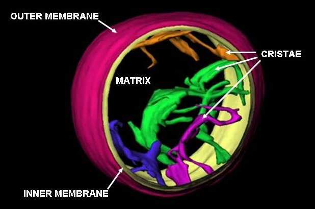
A mitochondrion () is an organelle found in the cell (biology), cells of most eukaryotes, such as animals, plants and fungi. Mitochondria have a double lipid bilayer, membrane structure and use aerobic respiration to generate adenosine triphosphate (ATP), which is used throughout the cell as a source of chemical energy. They were discovered by Albert von Kölliker in 1857 in the voluntary muscles of insects. The term ''mitochondrion'', meaning a thread-like granule, was coined by Carl Benda in 1898. The mitochondrion is popularly nicknamed the "powerhouse of the cell", a phrase popularized by Philip Siekevitz in a 1957 ''Scientific American'' article of the same name. Some cells in some multicellular organisms lack mitochondria (for example, mature mammalian red blood cells). The multicellular animal ''Henneguya zschokkei, Henneguya salminicola'' is known to have retained mitochondrion-related organelles despite a complete loss of their mitochondrial genome. A large number ... [...More Info...] [...Related Items...] OR: [Wikipedia] [Google] [Baidu] |
Microtubules
Microtubules are polymers of tubulin that form part of the cytoskeleton and provide structure and shape to eukaryotic cells. Microtubules can be as long as 50 micrometres, as wide as 23 to 27 nm and have an inner diameter between 11 and 15 nm. They are formed by the polymerization of a dimer of two globular proteins, alpha and beta tubulin into protofilaments that can then associate laterally to form a hollow tube, the microtubule. The most common form of a microtubule consists of 13 protofilaments in the tubular arrangement. Microtubules play an important role in a number of cellular processes. They are involved in maintaining the structure of the cell and, together with microfilaments and intermediate filaments, they form the cytoskeleton. They also make up the internal structure of cilia and flagella. They provide platforms for intracellular transport and are involved in a variety of cellular processes, including the movement of secretory vesicles, organel ... [...More Info...] [...Related Items...] OR: [Wikipedia] [Google] [Baidu] |
Neuropeptides
Neuropeptides are chemical messengers made up of small chains of amino acids that are synthesized and released by neurons. Neuropeptides typically bind to G protein-coupled receptors (GPCRs) to modulate neural activity and other tissues like the gut, muscles, and heart. Neuropeptides are synthesized from large precursor proteins which are cleaved and post-translationally processed then packaged into large dense core vesicles. Neuropeptides are often co-released with other neuropeptides and neurotransmitters in a single neuron, yielding a multitude of effects. Once released, neuropeptides can diffuse widely to affect a broad range of targets. Neuropeptides are extremely ancient and highly diverse chemical messengers. Placozoa, Placozoans such as ''Trichoplax'', extremely basal animals which do not possess neurons, use peptides for cell-to-cell communication in a way similar to the neuropeptides of higher animals. Examples Peptide signals play a role in information processing t ... [...More Info...] [...Related Items...] OR: [Wikipedia] [Google] [Baidu] |


