|
Cadherin Superfamily
Cadherins (named for "calcium-dependent adhesion") are a type of cell adhesion molecule (CAM) that is important in the formation of adherens junctions to allow cells to adhere to each other . Cadherins are a class of type-1 transmembrane proteins, and they are dependent on calcium (Ca2+) ions to function, hence their name. Cell-cell adhesion is mediated by extracellular cadherin domains, whereas the intracellular cytoplasmic tail associates with numerous adaptors and signaling proteins, collectively referred to as the cadherin adhesome. The cadherin family is essential in maintaining the cell-cell contact and regulating cytoskeletal complexes. The cadherin superfamily includes cadherins, protocadherins, desmogleins, desmocollins, and more. In structure, they share ''cadherin repeats'', which are the extracellular Ca2+-binding domains. There are multiple classes of cadherin molecules, each designated with a prefix (in general, noting the types of tissue with which it is associated ... [...More Info...] [...Related Items...] OR: [Wikipedia] [Google] [Baidu] |
Catenin
Catenins are a family of proteins found in complexes with cadherin cell adhesion molecules of animal cells. The first two catenins that were identified became known as α-catenin and β-catenin. α-Catenin can bind to β-catenin and can also bind filamentous actin (F-actin). β-Catenin binds directly to the cytoplasmic tail of classical cadherins. Additional catenins such as γ-catenin and δ-catenin have been identified. The name "catenin" was originally selected ('catena' means 'chain' in Latin) because it was suspected that catenins might link cadherins to the cytoskeleton. Types * α-catenin * β-catenin * γ-catenin * δ-catenin All but α-catenin contain armadillo repeats. They exhibit a high degree of protein dynamics, alone or in complex. Function Several types of catenins work with N-cadherins to play an important role in learning and memory. Cell-cell adhesion complexes are required for simple epithelia in higher organisms to maintain structure, function a ... [...More Info...] [...Related Items...] OR: [Wikipedia] [Google] [Baidu] |
CDH2
Cadherin-2 also known as Neural cadherin (N-cadherin), is a protein that in humans is encoded by the ''CDH2'' gene. CDH2 has also been designated as CD325 (cluster of differentiation 325). Cadherin-2 is a transmembrane protein expressed in multiple tissues and functions to mediate cell–cell adhesion. In cardiac muscle, Cadherin-2 is an integral component in adherens junctions residing at intercalated discs, which function to mechanically and electrically couple adjacent cardiomyocytes. Alterations in expression and integrity of Cadherin-2 has been observed in various forms of disease, including human dilated cardiomyopathy. Variants in ''CDH2'' have also been identified to cause a syndromic neurodevelopmental disorder. Structure Cadherin-2 is a protein with molecular weight of 99.7 kDa, and 906 amino acids in length. Cadherin-2, a classical cadherin from the cadherin superfamily, is composed of five extracellular cadherin repeats, a transmembrane region and a highly conserve ... [...More Info...] [...Related Items...] OR: [Wikipedia] [Google] [Baidu] |
APC/C Activator Protein CDH1
Cdh1 (cdc20 homolog 1) is one of the substrate adaptor protein of the anaphase-promoting complex (APC) in the budding yeast ''Saccharomyces cerevisiae''. Functioning as an activator of the APC/C, Cdh1 regulates the activity and substrate specificity of this ubiquitin E3-ligase. The human homolog is encoded by the FZR1 gene, which is not to be confused with the CDH1 gene. Introduction Cdh1 plays a pivotal role in controlling cell division at the end of mitosis (telophase) and in the subsequent G1 phase of cell cycle: By recognizing and binding proteins (like mitotic cyclins) which contain a destruction box (D-box) and an additional degradation signal (KEN box), Cdh1 recruits them in a C-box-dependent mechanism to the APC for ubiquination and subsequent proteolysis. Cdh1 is required for the exit of mitosis. Furthermore, it is thought to be a possible target of a BUB2-dependent spindle checkpoint pathway. Function The anaphase-promoting complex/cyclosome (APC/c) is an ubiqu ... [...More Info...] [...Related Items...] OR: [Wikipedia] [Google] [Baidu] |
Catenins
Catenins are a family of proteins found in complexes with cadherin cell adhesion molecules of animal cells. The first two catenins that were identified became known as α-catenin and β-catenin. α-Catenin can bind to β-catenin and can also bind filamentous actin (F-actin). β-Catenin binds directly to the cytoplasmic tail of classical cadherins. Additional catenins such as γ-catenin and δ-catenin have been identified. The name "catenin" was originally selected ('catena' means 'chain' in Latin) because it was suspected that catenins might link cadherins to the cytoskeleton. Types * α-catenin * β-catenin * γ-catenin * δ-catenin All but α-catenin contain armadillo repeats. They exhibit a high degree of protein dynamics, alone or in complex. Function Several types of catenins work with N-cadherins to play an important role in learning and memory. Cell-cell adhesion complexes are required for simple epithelia in higher organisms to maintain structure, function and p ... [...More Info...] [...Related Items...] OR: [Wikipedia] [Google] [Baidu] |
Cell Adhesion Molecule
Cell adhesion molecules (CAMs) are a subset of cell surface proteins that are involved in the binding of cells with other cells or with the extracellular matrix (ECM), in a process called cell adhesion. In essence, CAMs help cells stick to each other and to their surroundings. CAMs are crucial components in maintaining tissue structure and function. In fully developed animals, these molecules play an integral role in generating force and movement and consequently ensuring that organs are able to execute their functions normally. In addition to serving as "molecular glue", CAMs play important roles in the cellular mechanisms of growth, contact inhibition, and apoptosis. Aberrant expression of CAMs may result in a wide range of pathologies, ranging from frostbite to cancer. Structure CAMs are typically single-pass transmembrane receptors and are composed of three conserved domains: an intracellular domain that interacts with the cytoskeleton, a transmembrane domain, and an ext ... [...More Info...] [...Related Items...] OR: [Wikipedia] [Google] [Baidu] |
Catenin
Catenins are a family of proteins found in complexes with cadherin cell adhesion molecules of animal cells. The first two catenins that were identified became known as α-catenin and β-catenin. α-Catenin can bind to β-catenin and can also bind filamentous actin (F-actin). β-Catenin binds directly to the cytoplasmic tail of classical cadherins. Additional catenins such as γ-catenin and δ-catenin have been identified. The name "catenin" was originally selected ('catena' means 'chain' in Latin) because it was suspected that catenins might link cadherins to the cytoskeleton. Types * α-catenin * β-catenin * γ-catenin * δ-catenin All but α-catenin contain armadillo repeats. They exhibit a high degree of protein dynamics, alone or in complex. Function Several types of catenins work with N-cadherins to play an important role in learning and memory. Cell-cell adhesion complexes are required for simple epithelia in higher organisms to maintain structure, function a ... [...More Info...] [...Related Items...] OR: [Wikipedia] [Google] [Baidu] |
Induced Pluripotent Stem Cell
Induced pluripotent stem cells (also known as iPS cells or iPSCs) are a type of pluripotent stem cell that can be generated directly from a somatic cell. The iPSC technology was pioneered by Shinya Yamanaka's lab in Kyoto, Japan, who showed in 2006 that the introduction of four specific genes (named Myc, Oct3/4, Sox2 and Klf4), collectively known as Yamanaka factors, encoding transcription factors could convert somatic cells into pluripotent stem cells. He was awarded the 2012 Nobel Prize along with Sir John Gurdon "for the discovery that mature cells can be reprogrammed to become pluripotent." Pluripotent stem cells hold promise in the field of regenerative medicine. Because they can propagate indefinitely, as well as give rise to every other cell type in the body (such as neurons, heart, pancreatic, and liver cells), they represent a single source of cells that could be used to replace those lost to damage or disease. The most well-known type of pluripotent stem cel ... [...More Info...] [...Related Items...] OR: [Wikipedia] [Google] [Baidu] |
Epithelial–mesenchymal Transition
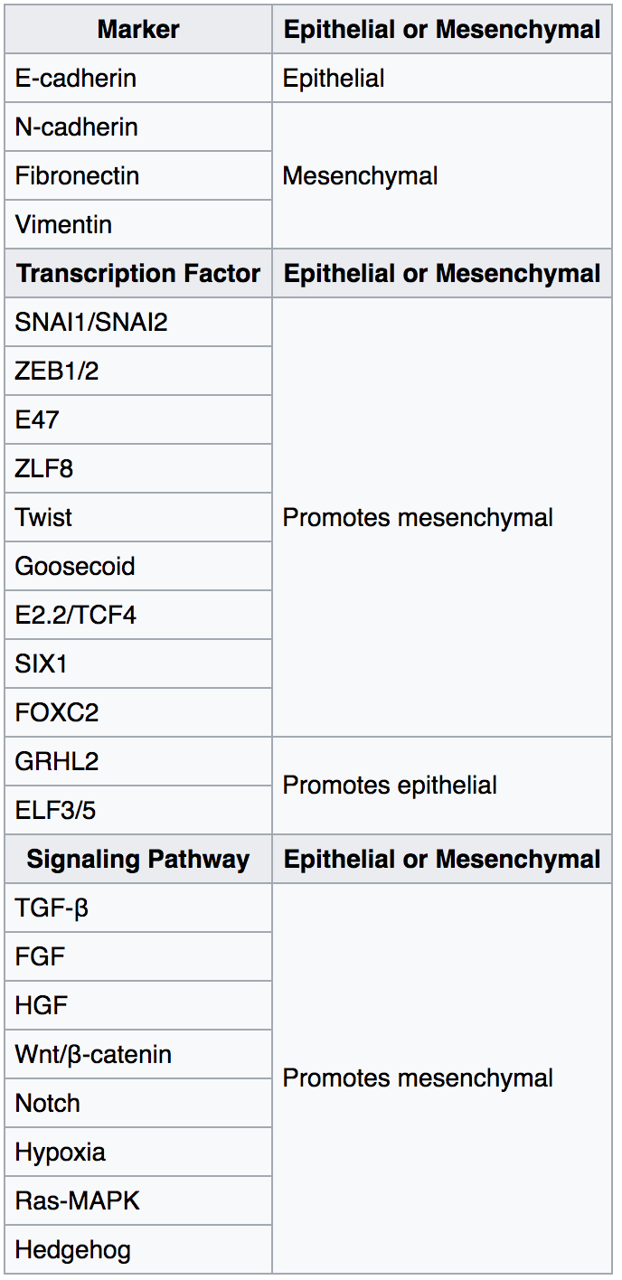
The epithelial–mesenchymal transition (EMT) is a process by which epithelial cells lose their cell polarity and cell–cell adhesion, and gain migratory and invasive properties to become mesenchymal stem cells; these are multipotent stromal cells that can differentiate into a variety of cell types. EMT is essential for numerous developmental processes including mesoderm formation and neural tube formation. EMT has also been shown to occur in wound healing, in organ fibrosis and in the initiation of metastasis in cancer progression. Introduction Epithelial–mesenchymal transition was first recognized as a feature of embryogenesis by Betty Hay in the 1980s. EMT, and its reverse process, MET ( mesenchymal-epithelial transition) are critical for development of many tissues and organs in the developing embryo, and numerous embryonic events such as gastrulation, neural crest formation, heart valve formation, secondary palate development, and myogenesis. Epithelial and mesenchym ... [...More Info...] [...Related Items...] OR: [Wikipedia] [Google] [Baidu] |
Animal Embryonic Development
In developmental biology, animal embryonic development, also known as animal embryogenesis, is the developmental stage of an animal embryo. Embryonic development starts with the fertilization of an egg cell (ovum) by a sperm cell, (spermatozoon). Once fertilized, the ovum becomes a single diploid cell known as a zygote. The zygote undergoes mitotic divisions with no significant growth (a process known as cleavage) and cellular differentiation, leading to development of a multicellular embryo after passing through an organizational checkpoint during mid-embryogenesis. In mammals, the term refers chiefly to the early stages of prenatal development, whereas the terms fetus and fetal development describe later stages. The main stages of animal embryonic development are as follows: * The zygote undergoes a series of cell divisions (called cleavage) to form a structure called a morula. * The morula develops into a structure called a blastula through a process called blastulation. * ... [...More Info...] [...Related Items...] OR: [Wikipedia] [Google] [Baidu] |
Cardiac Muscle Cell
Cardiac muscle (also called heart muscle, myocardium, cardiomyocytes and cardiac myocytes) is one of three types of vertebrate muscle tissues, with the other two being skeletal muscle and smooth muscle. It is an involuntary, striated muscle that constitutes the main tissue of the wall of the heart. The cardiac muscle (myocardium) forms a thick middle layer between the outer layer of the heart wall (the pericardium) and the inner layer (the endocardium), with blood supplied via the coronary circulation. It is composed of individual cardiac muscle cells joined by intercalated discs, and encased by collagen fibers and other substances that form the extracellular matrix. Cardiac muscle contracts in a similar manner to skeletal muscle, although with some important differences. Electrical stimulation in the form of a cardiac action potential triggers the release of calcium from the cell's internal calcium store, the sarcoplasmic reticulum. The rise in calcium causes the ... [...More Info...] [...Related Items...] OR: [Wikipedia] [Google] [Baidu] |
Neural Plate
The neural plate is a key developmental structure that serves as the basis for the nervous system. Cranial to the primitive node of the embryonic primitive streak, ectodermal tissue thickens and flattens to become the neural plate. The region anterior to the primitive node can be generally referred to as the neural plate. Cells take on a columnar appearance in the process as they continue to lengthen and narrow. The ends of the neural plate, known as the neural folds, push the ends of the plate up and together, folding into the neural tube, a structure critical to brain and spinal cord development. This process as a whole is termed primary neurulation. Signaling proteins are also important in neural plate development, and aid in differentiating the tissue destined to become the neural plate. Examples of such proteins include bone morphogenetic proteins and cadherins. Expression of these proteins is essential to neural plate folding and subsequent neural tube formation. Invol ... [...More Info...] [...Related Items...] OR: [Wikipedia] [Google] [Baidu] |

.jpg)


