|
CTL-mediated Cytotoxicity
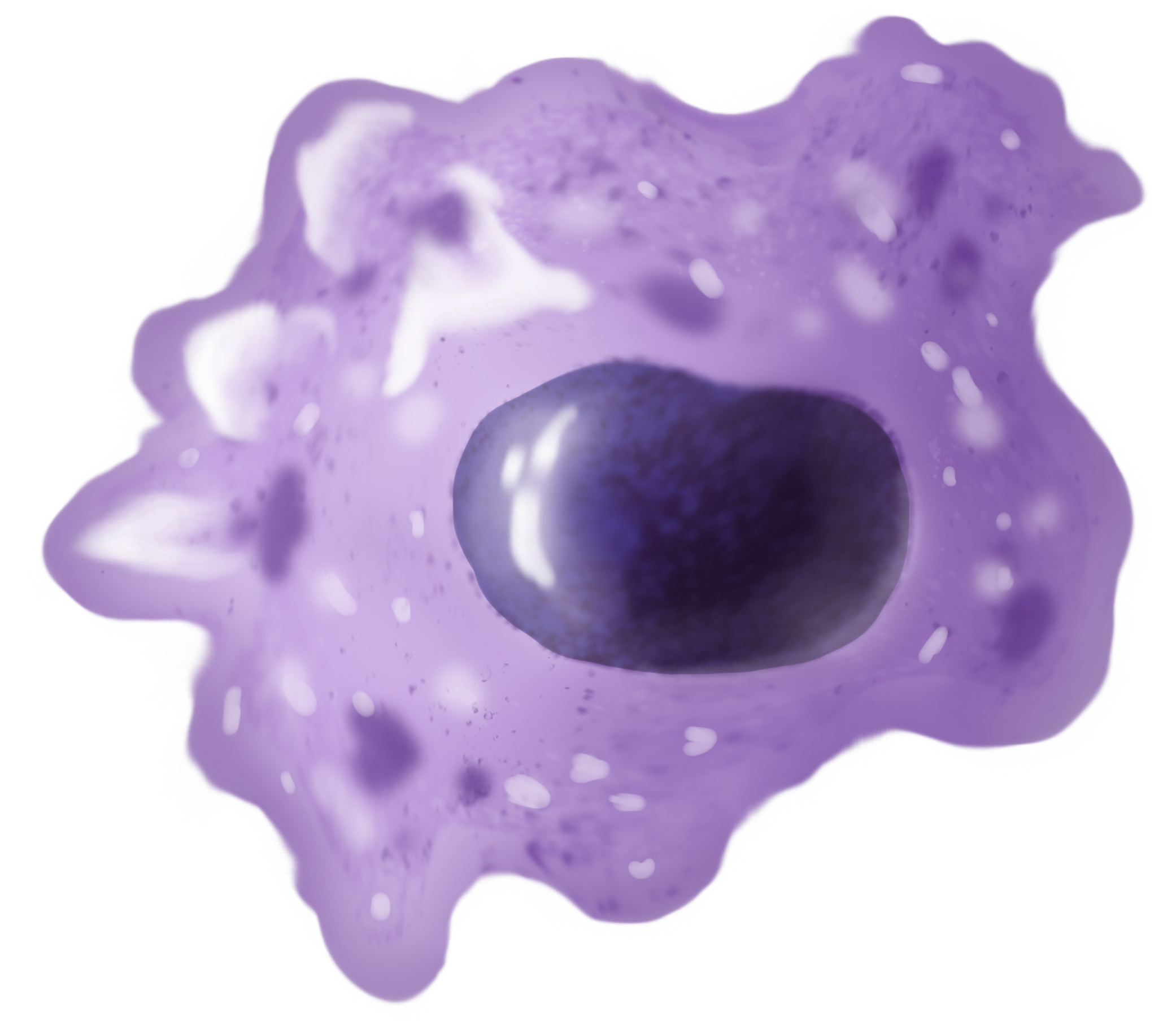
Within the scientific discipline of toxicology, Cytotoxic T lymphocytes (CTLs) are generated by immune activation of cytotoxic T cells (Tc cells). They are generally CD8+, which makes them MHC class I restricted. CTLs are able to eliminate most cells in the body since most nucleated cells express class I MHC molecules. The CTL-mediated immune system can be divided into two phases. In the first phase, functional effector CTLs are generated from naive Tc cells through activation and differentiation. In the second phase, affector CTLs destroy target cells by recognizing the antigen-MHC class I complex. Phase 1 In phase one, effector CTLs are generated from CTL precursors. The CTL precursors include naive Tc cells since they are incapable of killing target cells. After a precursor cell has been activated, it can then differentiate into a functional CTL with cytotoxic activity. There are three sequential signals that are required to complete this process. First, there is TCR recogniti ... [...More Info...] [...Related Items...] OR: [Wikipedia] [Google] [Baidu] |
Toxicology
Toxicology is a scientific discipline, overlapping with biology, chemistry, pharmacology, and medicine, that involves the study of the adverse effects of chemical substances on living organisms and the practice of diagnosing and treating exposures to toxins and toxicants. The relationship between dose and its effects on the exposed organism is of high significance in toxicology. Factors that influence chemical toxicity include the dosage, duration of exposure (whether it is acute or chronic), route of exposure, species, age, sex, and environment. Toxicologists are experts on poisons and poisoning. There is a movement for evidence-based toxicology as part of the larger movement towards evidence-based practices. Toxicology is currently contributing to the field of cancer research, since some toxins can be used as drugs for killing tumor cells. One prime example of this is ribosome-inactivating proteins, tested in the treatment of leukemia. The word ''toxicology'' () is a neoclas ... [...More Info...] [...Related Items...] OR: [Wikipedia] [Google] [Baidu] |
Cytotoxic T Cells
A cytotoxic T cell (also known as TC, cytotoxic T lymphocyte, CTL, T-killer cell, cytolytic T cell, CD8+ T-cell or killer T cell) is a T lymphocyte (a type of white blood cell) that kills cancer cells, cells that are infected by intracellular pathogens (such as viruses or bacteria), or cells that are damaged in other ways. Most cytotoxic T cells express T-cell receptors (TCRs) that can recognize a specific antigen. An antigen is a molecule capable of stimulating an immune response and is often produced by cancer cells, viruses, bacteria or intracellular signals. Antigens inside a cell are bound to class I MHC molecules, and brought to the surface of the cell by the class I MHC molecule, where they can be recognized by the T cell. If the TCR is specific for that antigen, it binds to the complex of the class I MHC molecule and the antigen, and the T cell destroys the cell. In order for the TCR to bind to the class I MHC molecule, the former must be accompanied by a glycoprotein c ... [...More Info...] [...Related Items...] OR: [Wikipedia] [Google] [Baidu] |
MHC Class I
MHC class I molecules are one of two primary classes of major histocompatibility complex (MHC) molecules (the other being MHC class II) and are found on the cell surface of all nucleated cells in the bodies of vertebrates. They also occur on platelets, but not on red blood cells. Their function is to display peptide fragments of proteins from within the cell to cytotoxic T cells; this will trigger an immediate response from the immune system against a particular non-self antigen displayed with the help of an MHC class I protein. Because MHC class I molecules present peptides derived from cytosolic proteins, the pathway of MHC class I presentation is often called ''cytosolic'' or ''endogenous pathway''. In humans, the HLAs corresponding to MHC class I are HLA-A, HLA-B, and HLA-C. Function Class I MHC molecules bind peptides generated mainly from degradation of cytosolic proteins by the proteasome. The MHC I:peptide complex is then inserted via endoplasmic reticulum in ... [...More Info...] [...Related Items...] OR: [Wikipedia] [Google] [Baidu] |
T-cell Receptor
The T-cell receptor (TCR) is a protein complex found on the surface of T cells, or T lymphocytes, that is responsible for recognizing fragments of antigen as peptides bound to major histocompatibility complex (MHC) molecules. The binding between TCR and antigen peptides is of relatively low affinity and is degenerate: that is, many TCRs recognize the same antigen peptide and many antigen peptides are recognized by the same TCR. The TCR is composed of two different protein chains (that is, it is a hetero dimer). In humans, in 95% of T cells the TCR consists of an alpha (α) chain and a beta (β) chain (encoded by ''TRA'' and ''TRB'', respectively), whereas in 5% of T cells the TCR consists of gamma and delta (γ/δ) chains (encoded by '' TRG'' and '' TRD'', respectively). This ratio changes during ontogeny and in diseased states (such as leukemia). It also differs between species. Orthologues of the 4 loci have been mapped in various species. Each locus can produce a ... [...More Info...] [...Related Items...] OR: [Wikipedia] [Google] [Baidu] |
Antigen-presenting Cell
An antigen-presenting cell (APC) or accessory cell is a cell that displays antigen bound by major histocompatibility complex (MHC) proteins on its surface; this process is known as antigen presentation. T cells may recognize these complexes using their T cell receptors (TCRs). APCs process antigens and present them to T-cells. Almost all cell types can present antigens in some way. They are found in a variety of tissue types. Professional antigen-presenting cells, including macrophages, B cells and dendritic cells, present foreign antigens to helper T cells, while virus-infected cells (or cancer cells) can present antigens originating inside the cell to cytotoxic T cells. In addition to the MHC family of proteins, antigen presentation relies on other specialized signaling molecules on the surfaces of both APCs and T cells. Antigen-presenting cells are vital for effective adaptive immune response, as the functioning of both cytotoxic and helper T cells is dependent on APCs. A ... [...More Info...] [...Related Items...] OR: [Wikipedia] [Google] [Baidu] |
CD28
CD28 (Cluster of Differentiation 28) is one of the proteins expressed on T cells that provide co-stimulatory signals required for T cell activation and survival. T cell stimulation through CD28 in addition to the T-cell receptor ( TCR) can provide a potent signal for the production of various interleukins ( IL-6 in particular). CD28 is the receptor for CD80 (B7.1) and CD86 (B7.2) proteins. When activated by Toll-like receptor ligands, the CD80 expression is upregulated in antigen-presenting cells (APCs). The CD86 expression on antigen-presenting cells is constitutive (expression is independent of environmental factors). CD28 is the only B7 receptor constitutively expressed on naive T cells. Association of the TCR of a naive T cell with MHC:antigen complex without CD28:B7 interaction results in a T cell that is anergic. Furthermore, CD28 was also identified on bone marrow stromal cells, plasma cells, neutrophils and eosinophils, but the functional importance of CD28 o ... [...More Info...] [...Related Items...] OR: [Wikipedia] [Google] [Baidu] |
B7 (protein)
B7 is a type of integral membrane protein found on activated antigen-presenting cells (APC) that, when paired with either a CD28 or CD152 (CTLA-4) surface protein on a T cell, can produce a costimulatory signal or a coinhibitory signal to enhance or decrease the activity of a MHC- TCR signal between the APC and the T cell, respectively. Binding of the B7 of APC to CTLA-4 of T-cells causes inhibition of the activity of T-cells. There are two major types of B7 proteins: B7-1 or CD80, and B7-2 or CD86. It is not known if they differ significantly from each other. So far CD80 is found on dendritic cells, macrophages, and activated B cells, CD86 (B7-2) on B cells. The proteins CD28 and CTLA-4 (CD152) each interact with both B7-1 and B7-2. Costimulation There are several steps to activation of the immune system against a pathogen. The T-cell receptor must first interact with the Major histocompatibility complex (MHC) surface protein. The CD4 or CD8 proteins on the T-cell surfac ... [...More Info...] [...Related Items...] OR: [Wikipedia] [Google] [Baidu] |
Interleukin 2
Interleukin-2 (IL-2) is an interleukin, a type of cytokine signaling molecule in the immune system. It is a 15.5–16 kDa protein that regulates the activities of white blood cells (leukocytes, often lymphocytes) that are responsible for immunity. IL-2 is part of the body's natural response to microbial infection, and in discriminating between foreign ("non-self") and "self". IL-2 mediates its effects by binding to IL-2 receptors, which are expressed by lymphocytes. The major sources of IL-2 are activated CD4+ T cells and activated CD8+ T cells. IL-2 receptor IL-2 is a member of a cytokine family, each member of which has a four alpha helix bundle; the family also includes IL-4, IL-7, IL-9, IL-15 and IL-21. IL-2 signals through the IL-2 receptor, a complex consisting of three chains, termed alpha ( CD25), beta ( CD122) and gamma ( CD132). The gamma chain is shared by all family members. The IL-2 receptor (IL-2R) α subunit binds IL-2 with low affinity ... [...More Info...] [...Related Items...] OR: [Wikipedia] [Google] [Baidu] |
IL-2 Receptor
The interleukin-2 receptor (IL-2R) is a heterotrimeric protein expressed on the surface of certain immune cells, such as lymphocytes, that binds and responds to a cytokine called IL-2. Composition IL-2 binds to the IL-2 receptor, which has three forms, generated by different combinations of three different proteins, often referred to as "chains": α (alpha) (also called IL-2Rα, CD25, or Tac antigen), β (beta) (also called IL-2Rβ, or CD122), and γ (gamma) (also called IL-2Rγ, γc, common gamma chain, or CD132); these subunits are also parts of receptors for other cytokines. The β and γ chains of the IL-2R are members of the type I cytokine receptor family. Structure-activity relationships of the IL-2/IL-2R interaction The three receptor chains are expressed separately and differently on various cell types and can assemble in different combinations and orders to generate low, intermediate, and high affinity IL-2 receptors. The α chain binds IL-2 with low a ... [...More Info...] [...Related Items...] OR: [Wikipedia] [Google] [Baidu] |
Apoptosis
Apoptosis (from grc, ἀπόπτωσις, apóptōsis, 'falling off') is a form of programmed cell death that occurs in multicellular organisms. Biochemical events lead to characteristic cell changes ( morphology) and death. These changes include blebbing, cell shrinkage, nuclear fragmentation, chromatin condensation, DNA fragmentation, and mRNA decay. The average adult human loses between 50 and 70 billion cells each day due to apoptosis. For an average human child between eight and fourteen years old, approximately twenty to thirty billion cells die per day. In contrast to necrosis, which is a form of traumatic cell death that results from acute cellular injury, apoptosis is a highly regulated and controlled process that confers advantages during an organism's life cycle. For example, the separation of fingers and toes in a developing human embryo occurs because cells between the digits undergo apoptosis. Unlike necrosis, apoptosis produces cell fragments called apopt ... [...More Info...] [...Related Items...] OR: [Wikipedia] [Google] [Baidu] |
Granzymes
Granzymes are serine proteases released by cytoplasmic granules within cytotoxic T cells and natural killer (NK) cells. They induce programmed cell death (apoptosis) in the target cell, thus eliminating cells that have become cancerous or are infected with viruses or bacteria. Granzymes also kill bacteria and inhibit viral replication. In NK cells and T cells, granzymes are packaged in cytotoxic granules along with perforin. Granzymes can also be detected in the rough endoplasmic reticulum, golgi complex, and the trans-golgi reticulum. The contents of the cytotoxic granules function to permit entry of the granzymes into the target cell cytosol. The granules are released into an immune synapse formed with a target cell, where perforin mediates the delivery of the granzymes into endosomes in the target cell, and finally into the target cell cytosol. Granzymes are part of the serine esterase family. They are closely related to other immune serine proteases expressed by innate immune ce ... [...More Info...] [...Related Items...] OR: [Wikipedia] [Google] [Baidu] |
Macrophages
Macrophages (abbreviated as M φ, MΦ or MP) ( el, large eaters, from Greek ''μακρός'' (') = large, ''φαγεῖν'' (') = to eat) are a type of white blood cell of the immune system that engulfs and digests pathogens, such as cancer cells, microbes, cellular debris, and foreign substances, which do not have proteins that are specific to healthy body cells on their surface. The process is called phagocytosis, which acts to defend the host against infection and injury. These large phagocytes are found in essentially all tissues, where they patrol for potential pathogens by amoeboid movement. They take various forms (with various names) throughout the body (e.g., histiocytes, Kupffer cells, alveolar macrophages, microglia, and others), but all are part of the mononuclear phagocyte system. Besides phagocytosis, they play a critical role in nonspecific defense ( innate immunity) and also help initiate specific defense mechanisms ( adaptive immunity) by recruiting oth ... [...More Info...] [...Related Items...] OR: [Wikipedia] [Google] [Baidu] |
_(6009043040).jpg)


