|
Bursae
A synovial bursa, usually simply bursa (: bursae or bursas), is a small fluid-filled sac lined by synovial membrane with an inner capillary layer of viscous synovial fluid (similar in consistency to that of a raw egg white). It provides a cushion between bones and tendons and/or muscles around a joint. This helps to reduce friction between the bones and allows free movement. Bursae are found around most major joints of the body. Structure Based on location, there are three types of bursa: subcutaneous, submuscular and subtendinous. A subcutaneous bursa is located between the skin and an underlying bone. It allows skin to move smoothly over the bone. Examples include the prepatellar bursa located over the kneecap and the olecranon bursa at the tip of the elbow. A submuscular bursa is found between a muscle and an underlying bone, or between adjacent muscles. These prevent rubbing of the muscle during movements. A large submuscular bursa, the trochanteric bursa, is found at the ... [...More Info...] [...Related Items...] OR: [Wikipedia] [Google] [Baidu] |
Suprapatellar Bursa
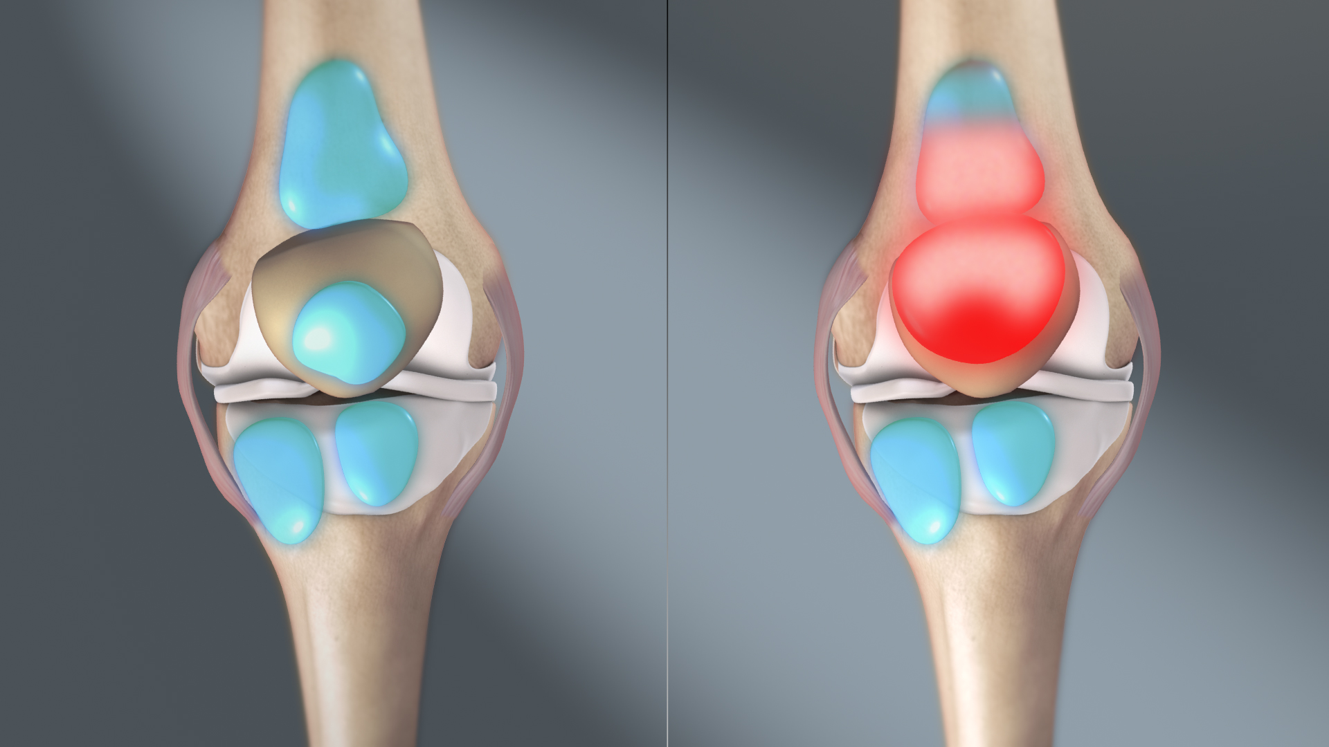
The knee bursae are the fluid-filled sacs and synovial pockets that surround and sometimes communicate with the knee joint cavity. The bursae are thin-walled, and filled with synovial fluid. They represent the weak point of the joint, but also provide enlargements to the joint space. They can be grouped into either ''communicating'' and ''non-communicating'' bursae or, after their location – frontal, lateral, or medial. Frontal In front, there are five bursae: # the suprapatellar bursa or recess between the anterior surface of the lower part of the femur and the deep surface of the quadriceps femoris.Burgener (2002), p 390 It allows for movement of the quadriceps tendon over the distal end of the femur. In about 85% of individuals, this bursa communicates with the knee joint. A distension of this bursa is therefore generally an indication of knee effusion. # the prepatellar bursa between the patella and the skin It allows movement of the skin over the underlying patella. # t ... [...More Info...] [...Related Items...] OR: [Wikipedia] [Google] [Baidu] |
Bursitis
Bursitis is the inflammation of one or more bursae (synovial sacs) of synovial fluid in the body. They are lined with a synovial membrane that secretes a lubricating synovial fluid. There are more than 150 bursae in the human body. The bursae (bur-see) rest at the points where internal functionaries, such as muscles and tendons, slide across bone. Healthy bursae create a smooth, almost frictionless functional gliding surface making normal movement painless. When bursitis occurs, however, movement relying on the inflamed bursa becomes difficult and painful. Moreover, movement of tendons and muscles over the inflamed bursa aggravates its inflammation, perpetuating the problem. Muscle can also be stiffened. Signs and symptoms Bursitis commonly affects superficial bursae. These include the subacromial, prepatellar, retrocalcaneal, and ''pes anserinus'' bursae of the shoulder, knee, heel and shin, etc. (see below). Symptoms vary from localized warmth and erythema (redness) to joi ... [...More Info...] [...Related Items...] OR: [Wikipedia] [Google] [Baidu] |
Knee Bursae
The knee bursae are the fluid-filled sacs and synovial pockets that surround and sometimes communicate with the knee joint cavity. The bursae are thin-walled, and filled with synovial fluid. They represent the weak point of the joint, but also provide enlargements to the joint space. They can be grouped into either ''communicating'' and ''non-communicating'' bursae or, after their location – frontal, lateral, or medial. Frontal In front, there are five bursae: # the suprapatellar bursa or recess between the anterior surface of the lower part of the femur and the deep surface of the quadriceps femoris.Burgener (2002), p 390 It allows for movement of the quadriceps tendon over the distal end of the femur. In about 85% of individuals, this bursa communicates with the knee joint. A distension of this bursa is therefore generally an indication of knee effusion. # the prepatellar bursa between the patella and the skin It allows movement of the skin over the underlying patella. # ... [...More Info...] [...Related Items...] OR: [Wikipedia] [Google] [Baidu] |
Synovial Bursae
A synovial bursa, usually simply bursa (: bursae or bursas), is a small fluid-filled sac lined by synovial membrane with an inner capillary layer of viscous synovial fluid (similar in consistency to that of a raw egg white). It provides a cushion between bones and tendons and/or muscles around a joint. This helps to reduce friction between the bones and allows free movement. Bursae are found around most major joints of the body. Structure Based on location, there are three types of bursa: subcutaneous, submuscular and subtendinous. A subcutaneous bursa is located between the skin and an underlying bone. It allows skin to move smoothly over the bone. Examples include the prepatellar bursa located over the kneecap and the olecranon bursa at the tip of the elbow. A submuscular bursa is found between a muscle and an underlying bone, or between adjacent muscles. These prevent rubbing of the muscle during movements. A large submuscular bursa, the trochanteric bursa, is found at the ... [...More Info...] [...Related Items...] OR: [Wikipedia] [Google] [Baidu] |
Prepatellar Bursa
The prepatellar bursa is a frontal bursa of the knee joint. It is a superficial bursa with a thin synovial lining located between the skin and the patella. Pathology Prepatellar bursitis, also known as housemaid's knee, is a common cause of swelling and pain above the patella (kneecap), and is due to inflammation of the prepatellar bursa. It is common in people who frequently kneel, such as roofers, plumbers, carpet layers, and gardeners. It is also common in wrestlers due to the repeated impact on the knee when shooting. Symptoms Symptoms include knee pain, swelling, redness and inability to flex the knee on the affected side. Rest usually relieves symptoms. Physical exam reveals erythema, tenderness to touch, fluctuant edema Edema (American English), also spelled oedema (British English), and also known as fluid retention, swelling, dropsy and hydropsy, is the build-up of fluid in the body's tissue (biology), tissue. Most commonly, the legs or arms are affected. S ... [...More Info...] [...Related Items...] OR: [Wikipedia] [Google] [Baidu] |
Olecranon Bursa
Olecranon bursitis is a condition characterized by swelling, redness, and pain at the tip of the elbow. If the underlying cause is due to an infection, fever may be present. The condition is relatively common and is one of the most frequent types of bursitis. It usually occurs as a result of trauma or pressure to the elbow, infection, or certain medical conditions such as rheumatoid arthritis or gout. Olecranon bursitis is associated with certain types of work including plumbing, mining, gardening, and mechanics. The underlying mechanism is inflammation of the fluid filled sac between the olecranon and skin. Diagnosis is usually based on symptoms. Treatment involves avoiding further trauma, a compression bandage, and NSAIDs. If there is concern of infection the fluid should be drained and tested and antibiotics are typically recommended. The use of steroid injections is controversial. Surgery may be done if other measures are not effective. Signs and symptoms Symptoms inc ... [...More Info...] [...Related Items...] OR: [Wikipedia] [Google] [Baidu] |
Trochanteric Bursa
Greater trochanteric pain syndrome (GTPS), a form of bursitis, is inflammation of the trochanteric bursa, a part of the hip. This bursa is at the top, outer side of the femur, between the insertion of the gluteus medius and gluteus minimus muscles into the greater trochanter of the femur and the femoral shaft. It has the function, in common with other bursae, of working as a shock absorber and as a lubricant for the movement of the muscles adjacent to it. Occasionally, this bursa can become inflamed and clinically painful and tender. This condition can be a manifestation of an injury (often resulting from a twisting motion or from overuse), but sometimes arises for no obviously definable cause. The symptoms are pain in the hip region on walking, and tenderness over the upper part of the femur, which may result in the inability to lie in comfort on the affected side. More often the lateral hip pain is caused by disease of the gluteal tendons that secondarily inflames the bursa ... [...More Info...] [...Related Items...] OR: [Wikipedia] [Google] [Baidu] |
Subacromial Bursa
The subacromial bursa is the synovial cavity located just below the acromion, which communicates with the subdeltoid bursa in most individuals, forming the so-called subacromial-subdeltoid bursa (SSB). The SSB bursa is located deep to the deltoid muscle and the coracoacromial arch and extends laterally beyond the humeral attachment of the rotator cuff, anteriorly to overlie the intertubercular groove, medially to the acromioclavicular joint, and posteriorly over the rotator cuff. The SSB decreases friction, and allows free motion of the rotator cuff relative to the coracoacromial arch and the deltoid muscle. French anatomist and surgeon Jean-François Jarjavay is credited as the first to describe morbid processes of the SSB in 1867. Since then, histologic studies have documented that synovial membrane may undergo inflammatory and/or degenerative changes and many now believe that they correspond to different stages in the spectrum of disease, with long-lasting inflammation leadin ... [...More Info...] [...Related Items...] OR: [Wikipedia] [Google] [Baidu] |
Olecranon Bursitis
Olecranon bursitis is a condition characterized by swelling, redness, and pain at the tip of the elbow. If the underlying cause is due to an infection, fever may be present. The condition is relatively common and is one of the most frequent types of bursitis. It usually occurs as a result of trauma or pressure to the elbow, infection, or certain medical conditions such as rheumatoid arthritis or gout. Olecranon bursitis is associated with certain types of work including plumbing, mining, gardening, and mechanics. The underlying mechanism is inflammation of the fluid filled sac between the olecranon and skin. Diagnosis is usually based on symptoms. Treatment involves avoiding further trauma, a elastic bandage, compression bandage, and NSAIDs. If there is concern of infection the fluid should be drained and tested and antibiotics are typically recommended. The use of steroid injections is controversial. Surgery may be done if other measures are not effective. Signs and symp ... [...More Info...] [...Related Items...] OR: [Wikipedia] [Google] [Baidu] |
Bursectomy
A bursectomy is the removal of a bursa, which is a small sac filled with synovial fluid that cushions adjacent bone structures and reduces friction in joint movement. This procedure is usually carried out to relieve chronic inflammation (bursitis) or infection, when conservative management has failed to improve patient outcomes. See also * List of surgeries by type Many Surgery, surgical procedure names can be broken into parts to indicate the meaning. For example, in gastrectomy, "ectomy" is a suffix (linguistics), suffix meaning the removal of a part of the body. "Gastro-" means stomach. Thus, ''gastrectom ... References Further reading * * * Orthopedic surgical procedures Synovial bursae {{Treatment-stub ... [...More Info...] [...Related Items...] OR: [Wikipedia] [Google] [Baidu] |
Bursa Of Fabricius
In birds, the bursa of Fabricius (Latin: ''bursa cloacalis'' or ''bursa Fabricii'') is the site of hematopoiesis. It is a specialized organ that, as first demonstrated by Bruce Glick and later by Max Dale Cooper and Robert Good, is necessary for B cell (part of the immune system) development in birds. Mammals generally do not have an equivalent organ; the bone marrow is often the site of both hematopoiesis and B cell development. The bursa is present in the cloaca of birds and is named after Hieronymus Fabricius, who described it in 1621. Description The bursa is an epithelial and lymphoid organ that is found only in birds. The bursa develops as a dorsal diverticulum of the proctodeal region of the cloaca. The luminal (interior) surface of the bursa is plicated with as many as 15 primary and 7 secondary plicae or folds. These plicae have hundreds of bursal follicles containing follicle-associated epithelial cells, lymphocytes, macrophages, and plasma cells. Lymphoid stem ce ... [...More Info...] [...Related Items...] OR: [Wikipedia] [Google] [Baidu] |
Shoulder Joint
The shoulder joint (or glenohumeral joint from Greek ''glene'', eyeball, + -''oid'', 'form of', + Latin ''humerus'', shoulder) is structurally classified as a synovial joint, synovial ball-and-socket joint and functionally as a diarthrosis and multiaxial joint. It involves an articulation between the glenoid fossa of the scapula (shoulder blade) and the head of the humerus (upper arm bone). Due to the very loose joint capsule, it gives a limited interface of the humerus and scapula, it is the most mobile joint of the human body. Structure The shoulder joint is a ball-and-socket joint between the scapula and the humerus. The socket of the glenoid fossa of the scapula is itself quite shallow, but it is made deeper by the addition of the glenoid labrum. The glenoid labrum is a ring of cartilage, cartilaginous fibre attached to the circumference of the cavity. This ring is continuous with the tendon of the Biceps, biceps brachii above. Spaces Significant joint spaces are: * The ... [...More Info...] [...Related Items...] OR: [Wikipedia] [Google] [Baidu] |


