|
Bulbocavernosus Reflex
The bulbocavernosus reflex (BCR), bulbospongiosus reflex (BSR) or "Osinski reflex" is a polysynaptic reflex that is useful in testing for spinal shock and gaining information about the state of spinal cord injuries (SCI). ''Bulbocavernosus'' is an older term for ''bulbospongiosus'', thus this reflex may also be referred to as the bulbospongiosus reflex. Procedure The test involves monitoring internal/ external anal sphincter contraction in response to squeezing the glans penis or clitoris, or tugging on an indwelling Foley catheter. This reflex can also be tested electrophysiologically, by stimulating the penis or vulva and recording from the anal sphincter. This testing modality is used in intraoperative neurophysiology monitoring to verify the function of sensory and motor sacral roots as well as the conus medullaris. Trauma The reflex is spinal mediated and involves S2– S4. The absence of the reflex in a person with acute paralysis from trauma indicates spinal shoc ... [...More Info...] [...Related Items...] OR: [Wikipedia] [Google] [Baidu] |
Bulbospongiosus Muscle
The bulbospongiosus muscles (in older texts bulbocavernosus and, for female muscle, constrictor cunni) are a subgroup of the superficial muscles of the perineum. They have a slightly different origin, insertion and function in males and females. In males, these muscles cover the bulb of the penis, while in females, they cover the vestibular bulbs. In both sexes, they are innervated by the deep or muscular branch of the perineal nerve, which is a branch of the pudendal nerve. Structure In males, the bulbospongiosus is located in the middle line of the perineum, in front of the anus. It consists of two symmetrical parts, united along the median line by a tendinous perineal raphe. It arises from the central tendinous point of the perineum and from the median perineal raphe in front. In females, there is no union, nor a tendinous perineal raphe; the parts are disjoint primarily and arise from the same central tendinous point of the perineum, which is the tendon that is forme ... [...More Info...] [...Related Items...] OR: [Wikipedia] [Google] [Baidu] |
Human Penis
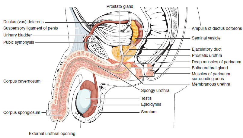
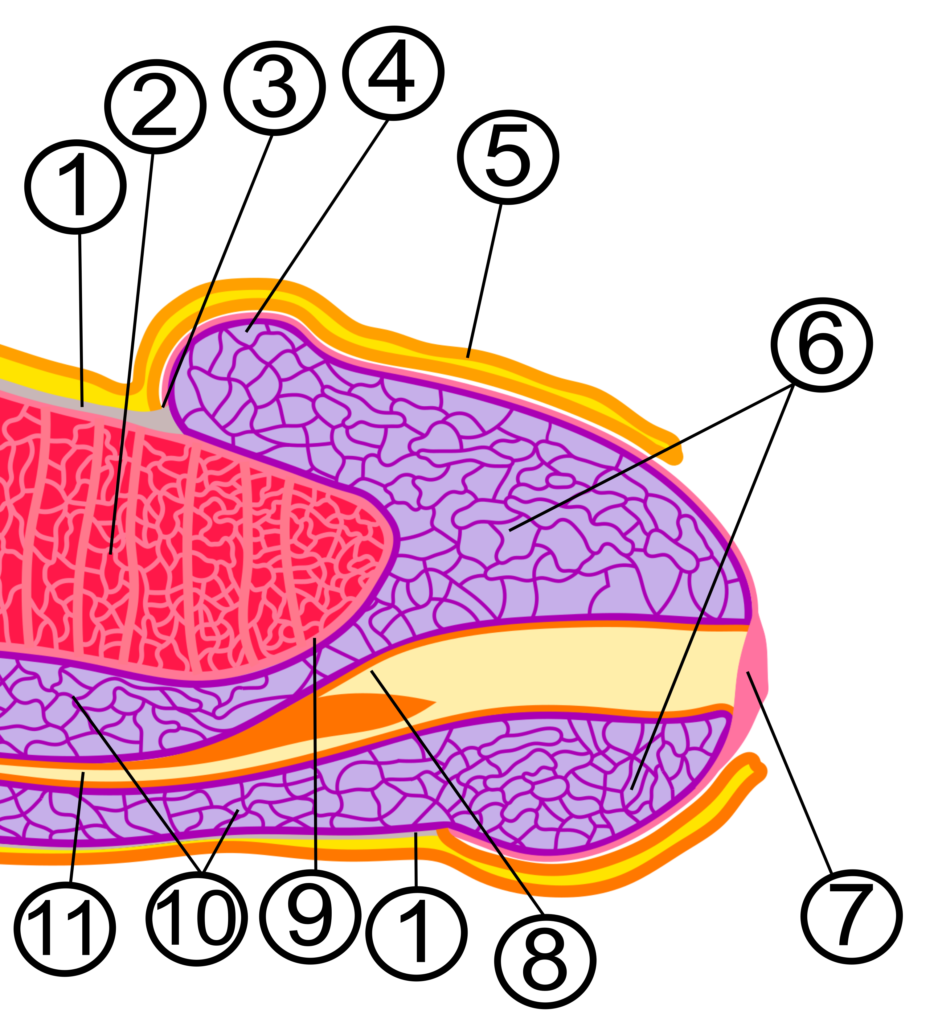
In Human body, human anatomy, the penis (; : penises or penes; from the Latin ''pēnis'', initially 'tail') is an external sex organ (intromittent organ) through which males urination, urinate and ejaculation, ejaculate, as Penis, on other animals. Together with the testes and surrounding structures, the penis functions as part of the male reproductive system. The main parts of the penis are the Root of penis, root, Body of penis, body, the epithelium of the penis, including the shaft skin, and the foreskin covering the glans penis, glans. The body of the penis is made up of three columns of tissue (biology), tissue: two Corpus cavernosum penis, corpora cavernosa on the dorsal side and corpus spongiosum penis, corpus spongiosum between them on the ventral side. The Urethra#Male, urethra passes through the prostate gland, where it is joined by the ejaculatory ducts, and then through the penis. The urethra goes across the corpus spongiosum and ends at the tip of the glans as the o ... [...More Info...] [...Related Items...] OR: [Wikipedia] [Google] [Baidu] |
Sacral Nerve
A spinal nerve is a mixed nerve, which carries motor, sensory, and autonomic signals between the spinal cord and the body. In the human body there are 31 pairs of spinal nerves, one on each side of the vertebral column. These are grouped into the corresponding cervical, thoracic, lumbar, sacral and coccygeal regions of the spine. There are eight pairs of cervical nerves, twelve pairs of thoracic nerves, five pairs of lumbar nerves, five pairs of sacral nerves, and one pair of coccygeal nerves. The spinal nerves are part of the peripheral nervous system. Structure Each spinal nerve is a mixed nerve, formed from the combination of nerve root fibers from its dorsal and ventral roots. The dorsal root is the afferent sensory root and carries sensory information to the brain. The ventral root is the efferent motor root and carries motor information from the brain. The spinal nerve emerges from the spinal column through an opening (intervertebral foramen) between adjacent ve ... [...More Info...] [...Related Items...] OR: [Wikipedia] [Google] [Baidu] |
Sacral Spinal Nerve 4
The sacral spinal nerve 4 (S4) is a spinal nerve of the sacral segment. Nervous System -- Groups of Nerves It originates from the from below the 4th body of the 
Muscles S4 supplies many muscles, either directly or through nerves originating from S4. They are not innervated with S4 as single origin, but partly by S4 and partly by other spinal nerves. The muscl ...[...More Info...] [...Related Items...] OR: [Wikipedia] [Google] [Baidu] |
Sacral Spinal Nerve 2
The sacral spinal nerve 2 (S2) is a spinal nerve of the sacral segment. Nervous System -- Groups of Nerves It originates from the from below the 2nd body of the 
Muscles S2 supplies many muscles, either directly or through nerves originating from S2. They are not innervated with S2 as single origin, but partly by S2 and partly by other spinal nerves. They are ...[...More Info...] [...Related Items...] OR: [Wikipedia] [Google] [Baidu] |
Conus Medullaris
The conus medullaris (Latin for "medullary cone") or conus terminalis is the tapered, lower end of the spinal cord. It occurs near lumbar vertebral levels 1 (L1) and 2 (L2), occasionally lower. The upper end of the conus medullaris is usually not well defined, however, its corresponding spinal cord segments are usually S1–S5. After the spinal cord tapers out, the spinal nerves continue to branch out diagonally, forming the cauda equina. The pia mater that surrounds the spinal cord, however, projects directly downward, forming a slender filament called the filum terminale, which connects the conus medullaris to the back of the coccyx. The filum terminale provides a connection between the conus medullaris and the coccyx which stabilizes the entire spinal cord. Blood supply The blood supply consists of three spinal arterial vessels—the anterior median longitudinal arterial trunk and the right and left posterior spinal arteries. Other less prominent sources of blood supply in ... [...More Info...] [...Related Items...] OR: [Wikipedia] [Google] [Baidu] |
Intraoperative
The perioperative period is the period of a patient's surgical procedure. It commonly includes ward admission, anesthesia, surgery, and recovery. Perioperative may refer to the three phases of surgery: preoperative, intraoperative, and postoperative, though it is a term most often used for the first and third of these only - a term which is often specifically utilized to imply 'around' the time of the surgery. The primary concern of perioperative care is to provide better conditions for patients before an operation (sometimes construed as during operation) and after an operation. Perioperative care Perioperative care is the care that is given before and after surgery. It takes place in hospitals, in surgical centers attached to hospitals, in freestanding surgical centers, or health care providers' offices. This period prepares the patient both physically and psychologically for the surgical procedure and after surgery. For emergency surgeries this period can be short and the pat ... [...More Info...] [...Related Items...] OR: [Wikipedia] [Google] [Baidu] |
Vulva
In mammals, the vulva (: vulvas or vulvae) comprises mostly external, visible structures of the female sex organ, genitalia leading into the interior of the female reproductive tract. For humans, it includes the mons pubis, labia majora, labia minora, clitoris, vulval vestibule, vestibule, urinary meatus, vaginal introitus, hymen, and openings of the vestibular glands (Bartholin's gland, Bartholin's and Skene's gland, Skene's). The folds of the outer and inner labia provide a double layer of protection for the vagina (which leads to the uterus). Pelvic floor muscles support the structures of the vulva. Other muscles of the urogenital triangle also give support. Blood supply to the vulva comes from the three pudendal arteries. The internal pudendal veins give drainage. Lymphatic vessel#Afferent vessels, Afferent lymph vessels carry lymph away from the vulva to the inguinal lymph nodes. The nerves that supply the vulva are the pudendal nerve, perineal nerve, ilioinguinal nerve ... [...More Info...] [...Related Items...] OR: [Wikipedia] [Google] [Baidu] |
Foley Catheter
In urology, a Foley catheter is one of many types of urinary catheters (UC). The Foley UC was named after Frederic Foley, who produced the original design in 1929. Foleys are indwelling UC, often referred to as an IDCs (sometimes IDUCs). This differs from in/out catheters (with only a single tube and no valves, designed to go into the bladder, drain it, and come straight back out). The UC is a flexible tube if it is indwelling and stays put, or rigid (glass or rigid plastic) if it is in/out, that a clinician, or the client themselves, often in the case of in/out UC, passes it through the urethra and into the Urinary bladder, bladder to drain urine. Foley and similar brand catheters usually have two separated channels, or Lumen (anatomy), ''lumina'' (or ''lumen''), running down its length. One lumen, opens at both ends, drains urine into a collection bag. The other has a valve on the outside end and connects to a balloon at the inside tip. The balloon is inflated with sterile water ... [...More Info...] [...Related Items...] OR: [Wikipedia] [Google] [Baidu] |
Polysynaptic
A reflex arc is a neural pathway that controls a reflex. In vertebrates, most sensory neurons synapse in the spinal cord and the signal then travels through it into the brain. This allows for faster reflex actions to occur by activating spinal motor neurons without the delay of routing signals through the brain. The brain will receive the input while the reflex is being carried out and the analysis of the signal takes place after the reflex action. There are two types: autonomic reflex arc (affecting inner organs) and somatic reflex arc (affecting muscles). Autonomic reflexes sometimes involve the spinal cord and some somatic reflexes are mediated more by the brain than the spinal cord. During a somatic reflex, nerve signals travel along the following pathway: # ''Somatic receptors'' in the skin, muscles and tendons # ''Afferent nerve fibers'' carry signals from the somatic receptors to the posterior horn of the spinal cord or to the brainstem # An ''integrating center'', the p ... [...More Info...] [...Related Items...] OR: [Wikipedia] [Google] [Baidu] |
Clitoris
In amniotes, the clitoris ( or ; : clitorises or clitorides) is a female sex organ. In humans, it is the vulva's most erogenous zone, erogenous area and generally the primary anatomical source of female Human sexuality, sexual pleasure. The clitoris is a complex structure, and its size and sensitivity can vary. The visible portion, the glans, of the clitoris is typically roughly the size and shape of a pea and is estimated to have at least 8,000 Nerve, nerve endings. * * Peters, B; Uloko, M; Isabey, PHow many Nerve Fibers Innervate the Human Clitoris? A Histomorphometric Evaluation of the Dorsal Nerve of the Clitoris 2 p.m. ET 27 October 2022, 23rd annual joint scientific meeting of Sexual Medicine Society of North America and International Society for Sexual Medicine Sexology, Sexological, medical, and psychological debate has focused on the clitoris, and it has been subject to social constructionist analyses and studies. Such discussions range from anatomical accuracy, g ... [...More Info...] [...Related Items...] OR: [Wikipedia] [Google] [Baidu] |
Glans Penis
In male human anatomy, the glans penis or penile glans, commonly referred to as the glans, (; from Latin ''glans'' meaning "acorn") is the bulbous structure at the Anatomical terms of location#Proximal and distal, distal end of the human penis that is the human male's most sensitive erogenous zone and primary anatomical source of Human sexuality, sexual pleasure. The glans penis is present in the male reproductive system, reproductive organs of humans and most other mammals where it may appear smooth, spiny, elongated or divided. It is externally lined with Mucosa, mucosal tissue, which creates a smooth texture and glossy appearance. In humans, the glans is located over the distal ends of the Corpus cavernosum penis, corpora cavernosa and is a continuation of the Corpus spongiosum (penis), corpus spongiosum of the penis. At the summit appears the urinary meatus and at the base forms the Corona of glans penis, corona glandis. An elastic band of tissue, known as the Penile frenulum ... [...More Info...] [...Related Items...] OR: [Wikipedia] [Google] [Baidu] |


