|
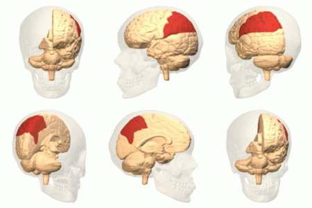
Brodmann Area 14
Brodmann Area 14 is one of Brodmann's subdivisions of the cerebral cortex in the brain. It was defined by Brodmann in the guenon monkey . While Brodmann, writing in 1909, argued that no equivalent structure existed in humans, later work demonstrated that area 14 has a clear homologue in the human ventromedial prefrontal cortex. Anatomy Brodmann areas were defined based on cytoarchitecture rather than function. Area 14 differs most clearly from Brodmann area 13-1905 in that it lacks a distinct internal granular layer (IV). Other differences are a less distinct external granular layer (II), a widening of the relatively cell-free zone of the external pyramidal layer (III); cells in the internal pyramidal layer (V) are denser and rounded; and the cells of the multiform layer (VI) assume a more distinct tangential orientation. Function According to one theory, Area 14 is believed to serve as association cortex for the visceral senses and olfaction along with Area 51. Its an ... [...More Info...] [...Related Items...] OR: [Wikipedia] [Google] [Baidu] |
Sapajus
Robust capuchin monkeys are capuchin monkeys in the genus ''Sapajus''. Formerly, all capuchin monkeys were placed in the genus ''Cebus''. ''Sapajus'' was erected in 2012 by Jessica Lynch Alfaro et al. to differentiate the robust (tufted) capuchin monkeys (formerly the ''C. apella'' group) from the gracile capuchin monkeys (formerly the ''C. capucinus'' group), which remain in ''Cebus''. Taxonomy Based on the species and subspecies proposed by Groves in 2001 and 2005, robust capuchin monkey taxa include: ''S. flavius'' was only rediscovered in 2006. The specific species and subspecies within ''Sapajus'' are not universally agreed upon. For example, Silva (2001) proposed a slightly different species and subspecies split in which, for example Azara's capuchun, ''Sapajus libidinosus paraguayanus'', is considered a separate species, ''Sapajus cay'', as are the large-headed capuchin and the crested capuchin. Taxonomic history Philip Hershkovitz and William Ch ... [...More Info...] [...Related Items...] OR: [Wikipedia] [Google] [Baidu] |
Parietal Lobe
The parietal lobe is one of the four Lobes of the brain, major lobes of the cerebral cortex in the brain of mammals. The parietal lobe is positioned above the temporal lobe and behind the frontal lobe and central sulcus. The parietal lobe integrates sensory information among various sensory modality, modalities, including spatial sense and navigation (proprioception), the main sensory receptive area for the sense of touch in the somatosensory cortex which is just posterior to the central sulcus in the postcentral gyrus, and the two-streams hypothesis#Dorsal stream, dorsal stream of the visual system. The major sensory inputs from the skin (mechanoreceptor, touch, thermoreceptor, temperature, and nociceptor, pain receptors), relay through the thalamus to the parietal lobe. Several areas of the parietal lobe are important in language processing in the brain, language processing. The somatosensory cortex can be illustrated as a distorted figure – the cortical homunculus (Latin: "li ... [...More Info...] [...Related Items...] OR: [Wikipedia] [Google] [Baidu] |
Autonomic Nervous System
The autonomic nervous system (ANS), sometimes called the visceral nervous system and formerly the vegetative nervous system, is a division of the nervous system that operates viscera, internal organs, smooth muscle and glands. The autonomic nervous system is a control system that acts largely unconsciously and regulates bodily functions, such as the heart rate, its Myocardial contractility, force of contraction, digestion, respiratory rate, pupillary dilation, pupillary response, Micturition, urination, and Animal sexual behaviour, sexual arousal. The fight-or-flight response, also known as the acute stress response, is set into action by the autonomic nervous system. The autonomic nervous system is regulated by integrated reflexes through the brainstem to the spinal cord and organ (anatomy), organs. Autonomic functions include control of respiration, heart rate, cardiac regulation (the cardiac control center), vasomotor activity (the vasomotor center), and certain reflex, reflex ... [...More Info...] [...Related Items...] OR: [Wikipedia] [Google] [Baidu] |
Olfaction
The sense of smell, or olfaction, is the special sense through which smells (or odors) are perceived. The sense of smell has many functions, including detecting desirable foods, hazards, and pheromones, and plays a role in taste. In humans, it occurs when an odor binds to a receptor within the nasal cavity, transmitting a signal through the olfactory system. Glomeruli aggregate signals from these receptors and transmit them to the olfactory bulb, where the sensory input will start to interact with parts of the brain responsible for smell identification, memory, and emotion. There are many different things which can interfere with a normal sense of smell, including damage to the nose or smell receptors, anosmia, upper respiratory infections, traumatic brain injury, and neurodegenerative disease. History of study Early scientific study of the sense of smell includes the extensive doctoral dissertation of Eleanor Gamble, published in 1898, which compared olfactory to ... [...More Info...] [...Related Items...] OR: [Wikipedia] [Google] [Baidu] |
Visceral
In a multicellular organism, an organ is a collection of Tissue (biology), tissues joined in a structural unit to serve a common function. In the biological organization, hierarchy of life, an organ lies between Tissue (biology), tissue and an organ system. Tissues are formed from same type Cell (biology), cells to act together in a function. Tissues of different types combine to form an organ which has a specific function. The Gastrointestinal tract, intestinal wall for example is formed by epithelial tissue and smooth muscle tissue. Two or more organs working together in the execution of a specific body function form an organ system, also called a biological system or body system. An organ's tissues can be broadly categorized as parenchyma, the functional tissue, and stroma (tissue), stroma, the structural tissue with supportive, connective, or ancillary functions. For example, the gland's tissue that makes the hormones is the parenchyma, whereas the stroma includes the nerve t ... [...More Info...] [...Related Items...] OR: [Wikipedia] [Google] [Baidu] |
Multiform Layer
The cerebral cortex, also known as the cerebral mantle, is the outer layer of neural tissue of the cerebrum of the brain in humans and other mammal A mammal () is a vertebrate animal of the Class (biology), class Mammalia (). Mammals are characterised by the presence of milk-producing mammary glands for feeding their young, a broad neocortex region of the brain, fur or hair, and three ...s. It is the largest site of Neuron, neural integration in the central nervous system, and plays a key role in attention, perception, awareness, thought, memory, language, and consciousness. The six-layered neocortex makes up approximately 90% of the Cortex (anatomy), cortex, with the allocortex making up the remainder. The cortex is divided into left and right parts by the longitudinal fissure, which separates the two cerebral hemispheres that are joined beneath the cortex by the corpus callosum and other commissural fibers. In most mammals, apart from small mammals that have small ... [...More Info...] [...Related Items...] OR: [Wikipedia] [Google] [Baidu] |
Pyramidal Layer
The cerebral cortex, also known as the cerebral mantle, is the outer layer of neural tissue of the cerebrum of the brain in humans and other mammals. It is the largest site of neural integration in the central nervous system, and plays a key role in attention, perception, awareness, thought, memory, language, and consciousness. The six-layered neocortex makes up approximately 90% of the cortex, with the allocortex making up the remainder. The cortex is divided into left and right parts by the longitudinal fissure, which separates the two cerebral hemispheres that are joined beneath the cortex by the corpus callosum and other commissural fibers. In most mammals, apart from small mammals that have small brains, the cerebral cortex is folded, providing a greater surface area in the confined volume of the cranium. Apart from minimising brain and cranial volume, cortical folding is crucial for the brain circuitry and its functional organisation. In mammals with small brains, ther ... [...More Info...] [...Related Items...] OR: [Wikipedia] [Google] [Baidu] |
Granular Layer (cerebral Cortex)
The internal granular layer of the cerebral cortex, also commonly referred to as the granular layer of the cortex, is the layer IV in the subdivision of the mammalian cerebral cortex into 6 layers. The adjective internal is used in opposition to the external granular layer of the cortex, the term granular refers to the granule cells found here. This layer receives the afferent connections from the thalamus The thalamus (: thalami; from Greek language, Greek Wikt:θάλαμος, θάλαμος, "chamber") is a large mass of gray matter on the lateral wall of the third ventricle forming the wikt:dorsal, dorsal part of the diencephalon (a division of ... and from other cortical regions and sends connections to the other layers. The line of Gennari (occipital stripe) is also present in this layer. See also * Granular layer * External granular layer (cerebral cortex) Cerebral cortex {{neuroanatomy-stub ... [...More Info...] [...Related Items...] OR: [Wikipedia] [Google] [Baidu] |
Brodmann Area 13
Area 13 is part of the Orbitofrontal cortex, a subdivision of the cerebral cortex as defined by cytoarchitecture. Location Area 13 is located in the posterior part of the Orbitofrontal cortex, and can be subdivided into areas 13a, 13b, 13m, 13l. Area 13a is anterior to the junction of olfactory tract and area 13b occupies a region just anterior to 13a along the olfactory sulcus. Area 13m is on the medial part of the middle orbital gyrus, whereas 13l is in the lateral part of the gyrus. Subregions Areas 13m and 13l are dysgranular regions of cortex. These areas are differentiated from the more anterior area 11 by a lack of continuous granular layer, and from the more posterior agranular Insular cortex. Area 13b is a thin and dysgranular cortical area, often characterized by crossing patterns of striations in layers III and V. Area 13a has an agranular structure. See also * Brodmann areas * List of regions in the human brain The human brain anatomical regions are ordered f ... [...More Info...] [...Related Items...] OR: [Wikipedia] [Google] [Baidu] |
Cytoarchitecture
Cytoarchitecture (from Greek κύτος 'cell' and ἀρχιτεκτονική 'architecture'), also known as cytoarchitectonics, is the study of the cellular composition of the central nervous system's tissues under the microscope. Cytoarchitectonics is one of the ways to parse the brain, by obtaining sections of the brain using a microtome and staining them with chemical agents which reveal where different neurons are located. The study of the parcellation of ''nerve fibers'' (primarily axons) into layers forms the subject of myeloarchitectonics (from Greek μυελός 'marrow' and ἀρχιτεκτονική 'architecture'), an approach complementary to cytoarchitectonics. History of the cerebral cytoarchitecture Defining cerebral cytoarchitecture began with the advent of histology—the science of slicing and staining brain slices for examination. It is credited to the Viennese psychiatrist Theodor Meynert (1833–1892), who in 1867 noticed regional variations in the ... [...More Info...] [...Related Items...] OR: [Wikipedia] [Google] [Baidu] |
Frontal Lobe
The frontal lobe is the largest of the four major lobes of the brain in mammals, and is located at the front of each cerebral hemisphere (in front of the parietal lobe and the temporal lobe). It is parted from the parietal lobe by a Sulcus (neuroanatomy), groove between tissues called the central sulcus and from the temporal lobe by a deeper groove called the lateral sulcus (Sylvian fissure). The most anterior rounded part of the frontal lobe (though not well-defined) is known as the frontal pole, one of the three Cerebral hemisphere#Poles, poles of the cerebrum. The frontal lobe is covered by the frontal cortex. The frontal cortex includes the premotor cortex and the primary motor cortex – parts of the motor cortex. The front part of the frontal cortex is covered by the prefrontal cortex. The nonprimary motor cortex is a functionally defined portion of the frontal lobe. There are four principal Gyrus, gyri in the frontal lobe. The precentral gyrus is directly anterior to the ... [...More Info...] [...Related Items...] OR: [Wikipedia] [Google] [Baidu] |
Coronal Section
The dorsal plane (also known as the coronal plane or frontal plane, especially in human anatomy) is an anatomical plane that divides the body into dorsal and ventral sections. It is perpendicular to the sagittal and transverse planes. Human anatomy The coronal plane is an example of a longitudinal plane. For a human, the mid-coronal plane would transect a standing body into two halves (front and back, or anterior and posterior) in an imaginary line that cuts through both shoulders. The sternal plane (''planum sternale'') is a coronal plane which transects the front of the sternum. Etymology The term is derived from Latin ''corona'' ('garland, crown'), from Ancient Greek κορώνη (''korōnē'', 'garland, wreath'). The coronal plane is so called because it lies in the same direction as the coronal suture. Additional images File:Coronal plane CT scan of the paranasal sinuses illustrative image.jpg, CT scan of the paranasal sinuses with coronal reconstruction (right) and ax ... [...More Info...] [...Related Items...] OR: [Wikipedia] [Google] [Baidu] |


