|
Breast Ultrasound
Breast ultrasound is a medical imaging technique that uses medical ultrasonography to perform imaging of the breast. It can be performed for either diagnostic or screening purposes and can be used with or without a mammogram. In particular, breast ultrasound may be useful for younger women who have denser fibrous breast tissue that may make mammograms more challenging to interpret. Automated whole-breast ultrasound (AWBU) is a technique that produces volumetric images of the breast and is largely independent of operator skill. It utilizes high-frequency ultrasound to help perform a diagnostic evaluation of the lactiferous ducts (duct sonography) and make dilated ducts and intraductal masses visible. Galactography is another technique that can be used to visualize the system of lactiferous ducts and allows a wider area to be visualized. Elastography is a type of ultrasound examination that measures tissue stiffness and can be used to detect tumours. Breast ultrasound is als ... [...More Info...] [...Related Items...] OR: [Wikipedia] [Google] [Baidu] |
Medical Ultrasound
Medical ultrasound includes Medical diagnosis, diagnostic techniques (mainly medical imaging, imaging) using ultrasound, as well as therapeutic ultrasound, therapeutic applications of ultrasound. In diagnosis, it is used to create an image of internal body structures such as tendons, muscles, joints, blood vessels, and internal organs, to measure some characteristics (e.g., distances and velocities) or to generate an informative audible sound. The usage of ultrasound to produce visual images for medicine is called medical ultrasonography or simply sonography, or echography. The practice of examining pregnant women using ultrasound is called obstetric ultrasonography, and was an early development of clinical ultrasonography. The machine used is called an ultrasound machine, a sonograph or an echograph. The visual image formed using this technique is called an ultrasonogram, a sonogram or an echogram. Ultrasound is composed of sound waves with frequency, frequencies greater than ... [...More Info...] [...Related Items...] OR: [Wikipedia] [Google] [Baidu] |
Galactography
Galactography or ductography (or ''galactogram'', ''ductogram'') is a medical diagnostic procedure for viewing the milk ducts. The procedure involves the radiography of the ducts after injection of a radiopaque substance into the duct system through the nipple. The procedure is used for investigating the pathology of nipple discharge. Galactography is capable of detecting smaller abnormalities than mammograms, MRI or ultrasound tests. With galactography, a larger part of the ductal system can be visualized than with the endoscopic investigation of a duct (called galactoscopy or ductoscopy). Causes for nipple discharge include duct ectasia, intraductal papilloma, and occasionally ductal carcinoma in situ or invasive ductal carcinoma. The standard treatment of galactographically suspicious breast lesions is to perform a surgical intervention on the concerned duct or ducts: if the discharge clearly stems from a single duct, then the excision of the duct (microdochectomy) is indicat ... [...More Info...] [...Related Items...] OR: [Wikipedia] [Google] [Baidu] |
Acorn Cyst Sign
Acorn cyst sign is a radiologic sign indicating the presence of a benign uncomplicated cyst in ultrasound examinations of the breast. It consists of a deep anechoic fluid portion resembling an acorn The acorn is the nut (fruit), nut of the oaks and their close relatives (genera ''Quercus'', ''Notholithocarpus'' and ''Lithocarpus'', in the family Fagaceae). It usually contains a seedling surrounded by two cotyledons (seedling leaves), en ..., and a superficial echogenic layer resembling an acorn cap. This sign is helpful for radiologists to differentiate a benign uncomplicated cyst from a complex mass. References Radiologic signs Cysts Medical ultrasonography Breast imaging {{Neoplasm-stub ... [...More Info...] [...Related Items...] OR: [Wikipedia] [Google] [Baidu] |
Axillary Lymph Nodes
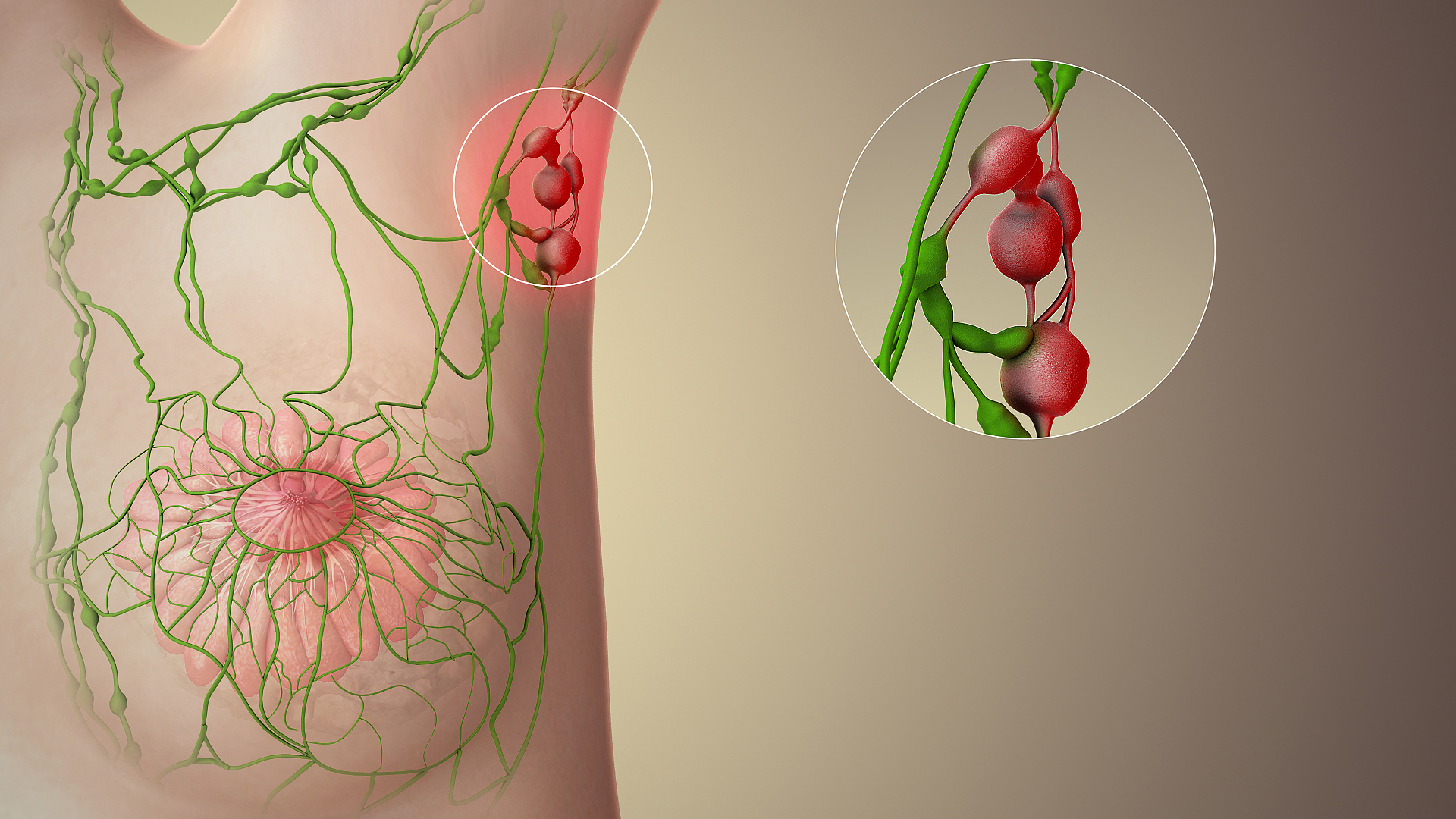
The axillary lymph nodes or armpit lymph nodes are lymph nodes in the human armpit. Between 20 and 49 in number, they drain lymph vessels from the lateral quadrants of the breast, the superficial lymph vessels from thin walls of the chest and the abdomen above the level of the navel, and the vessels from the upper limb. They are divided in several groups according to their location in the armpit. These lymph nodes are clinically significant in breast cancer, and metastases from the breast to the axillary lymph nodes are considered in the Cancer staging, staging of the disease. Structure The axillary lymph nodes are arranged in six groups: #Pectoral axillary lymph nodes, Anterior (pectoral) group: Lying along the lower border of the pectoralis minor behind the pectoralis major, these nodes receive lymph vessels from the lateral quadrants of the breast and superficial vessels from the anterolateral abdominal wall above the level of the umbilicus. #Subscapular axillary lymph nodes, ... [...More Info...] [...Related Items...] OR: [Wikipedia] [Google] [Baidu] |
Tail Of Spence
The tail of Spence (Spence's tail, axillary process, axillary tail) has historically been described as an extension of the tissue of the upper outer quadrant of the breast traveling into the axilla. The "axillary tail" has been reported to pass into the axilla through an opening in the deep fascia called foramen of Langer. The "tail of Spence" was named after the Scottish surgeon James Spence, who served as a President of the Royal College of Surgeons in Edinburgh in the latter half of the 19th Century. A recent publication has presented an updated description of the anatomy of the breast and upper outer chest, calling into question the concept of an axillary tail. The report does not challenge that lymphatic drainage consistently extends from the primary breast into the axilla through the foramen of Langer, but does demonstrate that a superolaterally oriented "tail" of breast fat (with or without ductal tissue) is rarely if ever present. Instead, upper lateral chest anatomy is ... [...More Info...] [...Related Items...] OR: [Wikipedia] [Google] [Baidu] |
Breast Abscess
Mastitis is inflammation of the breast or udder, usually associated with breastfeeding. Symptoms typically include local pain and redness. There is often an associated fever and general soreness. Onset is typically fairly rapid and usually occurs within the first few months of delivery. Complications can include abscess formation. Risk factors include poor latch, cracked nipples, and weaning. Use of a breast pump has historically been associated with Mastitis, but has been determined as an indirect association. The bacteria most commonly involved are ''Staphylococcus'' and ''Streptococci''. Diagnosis is typically based on symptoms. Ultrasound may be useful for detecting a potential abscess. Prevention of this breastfeeding difficulty is by proper breastfeeding techniques. When infection is present, antibiotics such as cephalexin may be recommended. Breastfeeding should typically be continued, as emptying the breast is important for healing. Tentative evidence supports ben ... [...More Info...] [...Related Items...] OR: [Wikipedia] [Google] [Baidu] |
Fine-needle Aspiration
Fine-needle aspiration (FNA) is a diagnostic procedure used to investigate lumps or masses. In this technique, a thin (23–25 gauge (0.52 to 0.64 mm outer diameter)), hollow needle is inserted into the mass for sampling of cells that, after being stained, are examined under a microscope (biopsy). The sampling and biopsy considered together are called fine-needle aspiration biopsy (FNAB) or fine-needle aspiration cytology (FNAC) (the latter to emphasize that any aspiration biopsy involves cytopathology, not histopathology). Fine-needle aspiration biopsies are very safe for minor surgical procedures. Often, a major surgical (excisional or open) biopsy can be avoided by performing a needle aspiration biopsy instead, eliminating the need for hospitalization. In 1981, the first fine-needle aspiration biopsy in the United States was done at Maimonides Medical Center. The modern procedure is widely used to diagnose cancer and inflammatory conditions. Fine needle aspiration is g ... [...More Info...] [...Related Items...] OR: [Wikipedia] [Google] [Baidu] |
Elastography
Elastography is any of a class of medical imaging diagnostic methods that map the elastic properties and stiffness of soft tissue. The main idea is that whether the tissue is hard or soft will give diagnostic information about the presence or status of disease. For example, cancerous tumours will often be harder than the surrounding tissue, and diseased livers are stiffer than healthy ones. The most prominent techniques use ultrasound or magnetic resonance imaging (MRI) to make both the stiffness map and an anatomical image for comparison. Historical background Palpation is the practice of feeling the stiffness of a person's or animal's tissues with the health practitioner's hands. Manual palpation dates back at least to 1500 BC, with the Egyptian Ebers Papyrus and Edwin Smith Papyrus both giving instructions on diagnosis with palpation. In ancient Greece, Hippocrates gave instructions on many forms of diagnosis using palpation, including palpation of the breasts, wounds, bowe ... [...More Info...] [...Related Items...] OR: [Wikipedia] [Google] [Baidu] |
Lactiferous Duct
Lactiferous ducts are ducts that converge and form a Morphogenesis#Branching morphogenesis, branched system connecting the nipple to the lobules of the mammary gland. When lactogenesis occurs, under the influence of hormones, the breast milk, milk is moved to the nipple by the action of smooth muscle contractions along the ductal system to the tip of the nipple. They are also referred to as ''galactophores'', ''galactophorous ducts'', ''mammary ducts'', ''mamillary ducts'' or ''milk ducts''. Structure Lactiferous ducts are lined by a columnar epithelium supported by myoepithelial cells. Prior to 2005, it was thought within the areola the lactiferous duct would dilate to form the lactiferous sinus in which milk accumulates between breastfeeding sessions. However past studies have shown that the lactiferous sinus does not exist. Function The columnar epithelium plays a key role in balancing milk production, milk stasis and reabsorption. The cells of the columnar epithelium form tig ... [...More Info...] [...Related Items...] OR: [Wikipedia] [Google] [Baidu] |
Breast Imaging
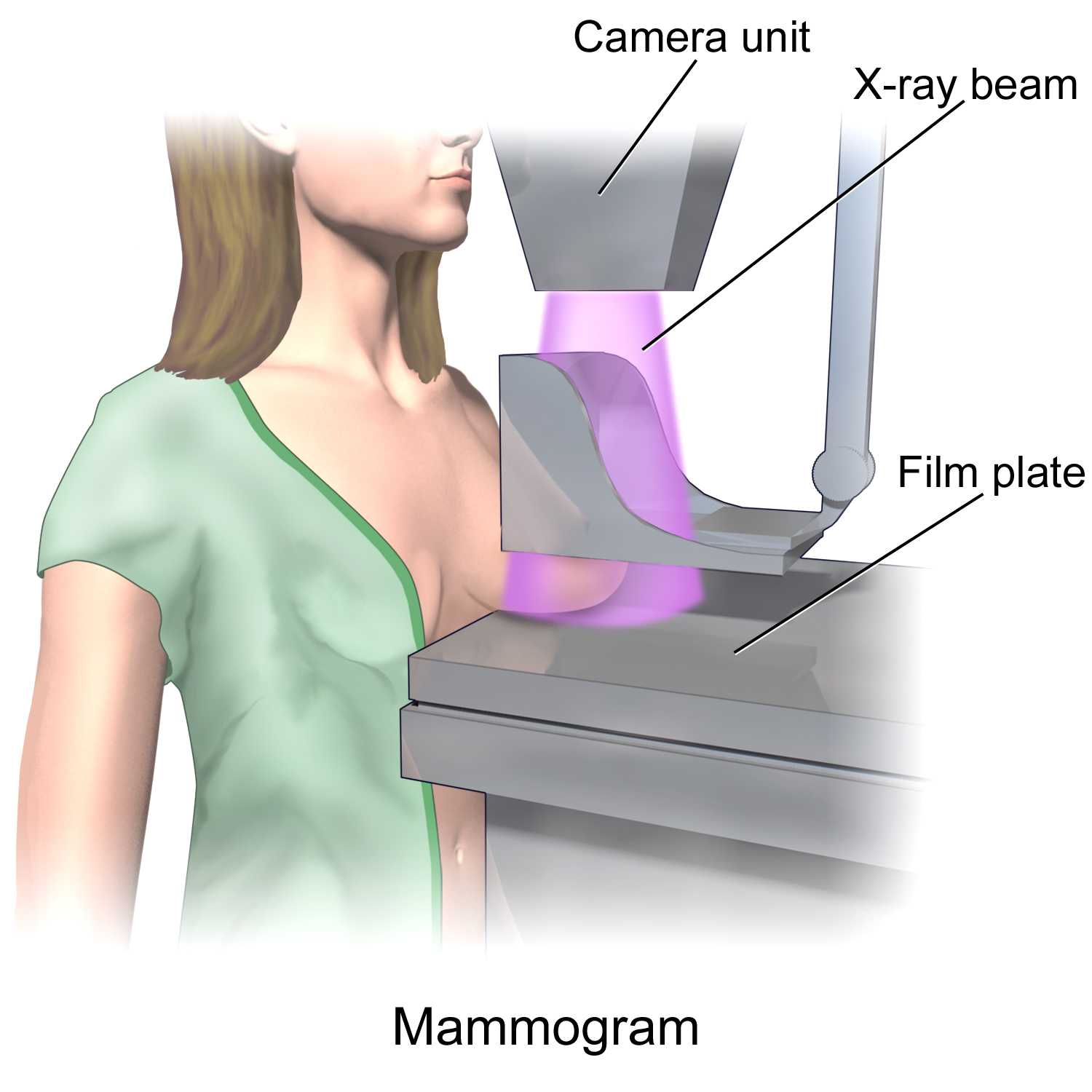
In medicine, breast imaging is a sub-speciality of diagnostic radiology that involves imaging of the breasts for screening or diagnostic purposes. There are various methods of breast imaging using a variety of technologies as described in detail below. Traditional screening and diagnostic mammography ("2D mammography") uses x-ray technology and has been the mainstay of breast imaging for many decades. Breast tomosynthesis ("3D mammography") is a relatively new digital x-ray mammography technique that produces multiple image slices of the breast similar to, but distinct from, CT scan, computed tomography (CT). Xeromammography and galactography are somewhat outdated technologies that also use x-ray technology and are now used infrequently in the detection of breast cancer. Breast ultrasound is another technology employed in diagnosis and screening that can help differentiate between fluid filled and solid lesions, an important factor to determine if a lesion may be cancerous. Breast ... [...More Info...] [...Related Items...] OR: [Wikipedia] [Google] [Baidu] |
Automated Whole-breast Ultrasound
Automated whole-breast ultrasound (AWBU) is a medical imaging technique used in radiology to obtain volumetric ultrasound data of the entire breast. How it works Similarly to the 3D ultrasound technique used for pregnant women, AWBU allows volumetric image data to be obtained from ultrasound sonography. With automated whole-breast ultrasound, the ultrasound transducer is guided over the breast in an automatic manner. The position and speed of the transducer is regulated automatically, whereas the angle of incidence and the amount of pressure applied is set by the human operator. The entire breast is scanned in an automated manner, and the procedure yields volumetric image data of the breast. The resulting image data can be read at any convenient time by the radiologist, who is freed from performing the scan. This allows selected scan planes to be visualized, and also allows the data to be displayed as a volumetric image. Applications AWBU has been proposed as an additional c ... [...More Info...] [...Related Items...] OR: [Wikipedia] [Google] [Baidu] |
Food And Drug Administration
The United States Food and Drug Administration (FDA or US FDA) is a List of United States federal agencies, federal agency of the United States Department of Health and Human Services, Department of Health and Human Services. The FDA is responsible for protecting and promoting public health through the control and supervision of food safety, tobacco products, caffeine products, dietary supplements, Prescription drug, prescription and Over-the-counter drug, over-the-counter pharmaceutical drugs (medications), vaccines, biopharmaceuticals, blood transfusions, medical devices, electromagnetic radiation emitting devices (ERED), cosmetics, Animal feed, animal foods & feed and Veterinary medicine, veterinary products. The FDA's primary focus is enforcement of the Federal Food, Drug, and Cosmetic Act (FD&C). However, the agency also enforces other laws, notably Section 361 of the Public Health Service Act as well as associated regulations. Much of this regulatory-enforcement work is ... [...More Info...] [...Related Items...] OR: [Wikipedia] [Google] [Baidu] |


