|
Automated Whole-breast Ultrasound
Automated whole-breast ultrasound (AWBU) is a medical imaging technique used in radiology to obtain volumetric ultrasound data of the entire breast. How it works Similarly to the 3D ultrasound 3D ultrasound is a medical ultrasound technique, often used in fetal, cardiac, trans-rectal and intra-vascular applications. 3D ultrasound refers specifically to the volume rendering of ultrasound data. When involving a series of 3D volumes collec ... technique used for pregnant women, AWBU allows volumetric image data to be obtained from ultrasound sonography. With automated whole-breast ultrasound, the ultrasound transducer is guided over the breast in an automatic manner. The position and speed of the transducer is regulated automatically, whereas the angle of incidence and the amount of pressure applied is set by the human operator. The entire breast is scanned in an automated manner, and the procedure yields volumetric image data of the breast. The resulting image data can be read ... [...More Info...] [...Related Items...] OR: [Wikipedia] [Google] [Baidu] |
Medical Imaging
Medical imaging is the technique and process of imaging the interior of a body for clinical analysis and medical intervention, as well as visual representation of the function of some organs or tissues ( physiology). Medical imaging seeks to reveal internal structures hidden by the skin and bones, as well as to diagnose and treat disease. Medical imaging also establishes a database of normal anatomy and physiology to make it possible to identify abnormalities. Although imaging of removed organs and tissues can be performed for medical reasons, such procedures are usually considered part of pathology instead of medical imaging. Measurement and recording techniques that are not primarily designed to produce images, such as electroencephalography (EEG), magnetoencephalography (MEG), electrocardiography (ECG), and others, represent other technologies that produce data susceptible to representation as a parameter graph versus time or maps that contain data about the measurement ... [...More Info...] [...Related Items...] OR: [Wikipedia] [Google] [Baidu] |
Breast Ultrasound
Breast ultrasound is the use of medical ultrasonography to perform imaging of the breast. It can be considered either a diagnostic or a screening procedure. It may be used either with or without a mammogram. It may be useful in younger women, where the denser fibrous tissue of the breast may make mammograms more difficult to interpret. Automated whole-breast ultrasound (AWBU) is an ultrasound investigation of the breast that is largely independent of the operator skill and that allows the reconstruction of volumetric images of the breast. Using high-frequency ultrasound, a diagnostic evaluation of the lactiferous ducts by means of ultrasound (duct sonography) can be performed. In this manner, dilated ducts and intraductal masses can be made visible. Another technique for visualizing the system of lactiferous ducts is galactography, which allows a wider area of the lactiferous duct system to be visualized. A type of ultrasound examination to measure tissue stiffness, which ... [...More Info...] [...Related Items...] OR: [Wikipedia] [Google] [Baidu] |
Breast
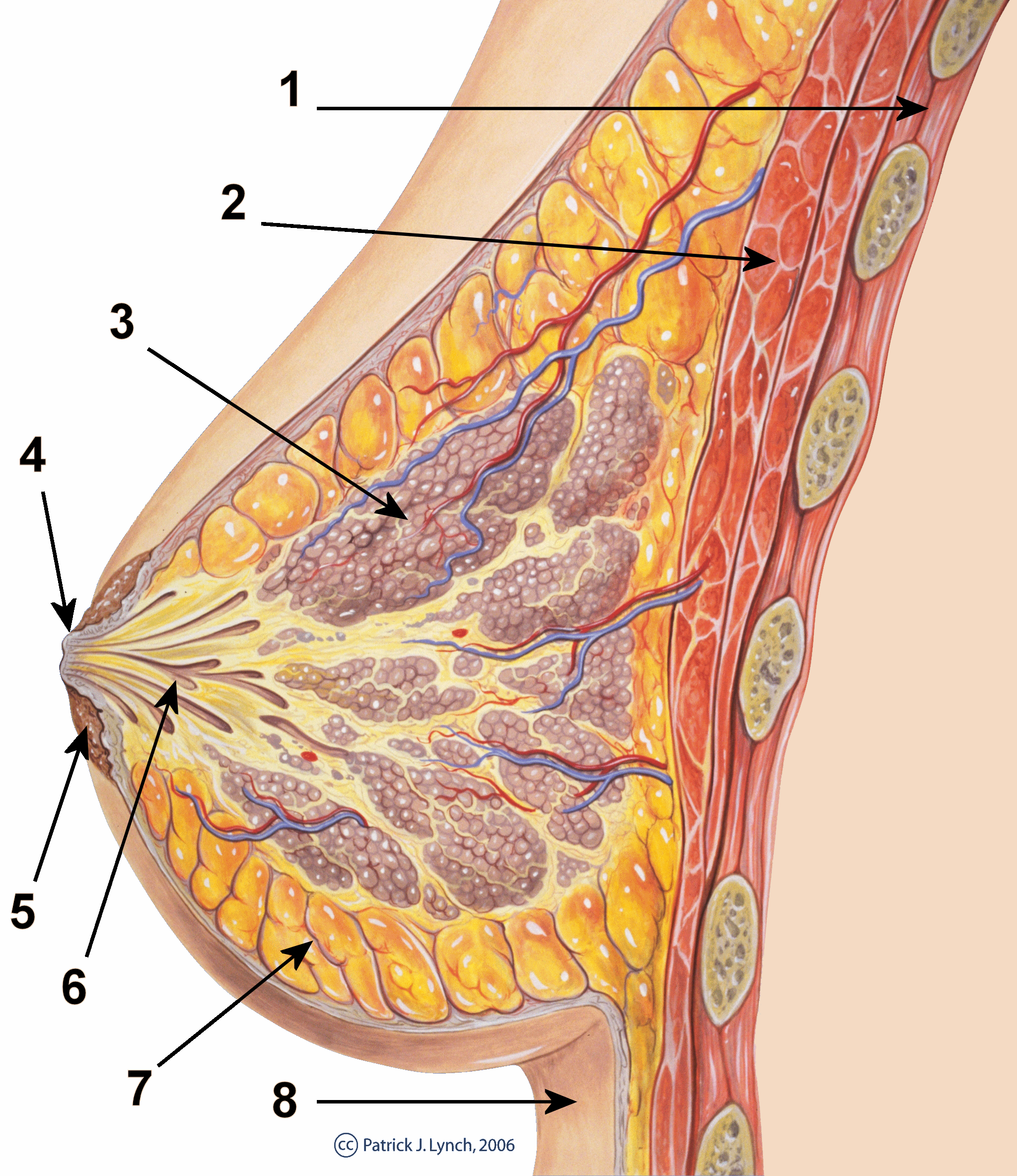
The breast is one of two prominences located on the upper ventral region of a primate's torso. Both females and males develop breasts from the same embryological tissues. In females, it serves as the mammary gland, which produces and secretes milk to feed infants. Subcutaneous fat covers and envelops a network of ducts that converge on the nipple, and these tissues give the breast its size and shape. At the ends of the ducts are lobules, or clusters of alveoli, where milk is produced and stored in response to hormonal signals. During pregnancy, the breast responds to a complex interaction of hormones, including estrogens, progesterone, and prolactin, that mediate the completion of its development, namely lobuloalveolar maturation, in preparation of lactation and breastfeeding. Humans are the only animals with permanent breasts. At puberty, estrogens, in conjunction with growth hormone, cause permanent breast growth in female humans. This happens only to ... [...More Info...] [...Related Items...] OR: [Wikipedia] [Google] [Baidu] |
3D Ultrasound
3D ultrasound is a medical ultrasound technique, often used in fetal, cardiac, trans-rectal and intra-vascular applications. 3D ultrasound refers specifically to the volume rendering of ultrasound data. When involving a series of 3D volumes collected over time, it can also be referred to as 4D ultrasound (three spatial dimensions plus one time dimension) or real-time 3D ultrasound. Methods When generating a 3D volume, the ultrasound data can be collected in four common ways by a sonographer: *Freehand, which involves tilting the probe and capturing a series of ultrasound images and recording the transducer orientation for each slice. *Mechanically, where the internal linear probe tilt is handled by a motor inside the probe. *Using an endoprobe, which generates the volume by inserting a probe and then removing the transducer in a controlled manner. *A matrix array transducer, which uses beam steering to sample points throughout a pyramid shaped volume. Risks The general risks of u ... [...More Info...] [...Related Items...] OR: [Wikipedia] [Google] [Baidu] |
Breast Cancer Screening
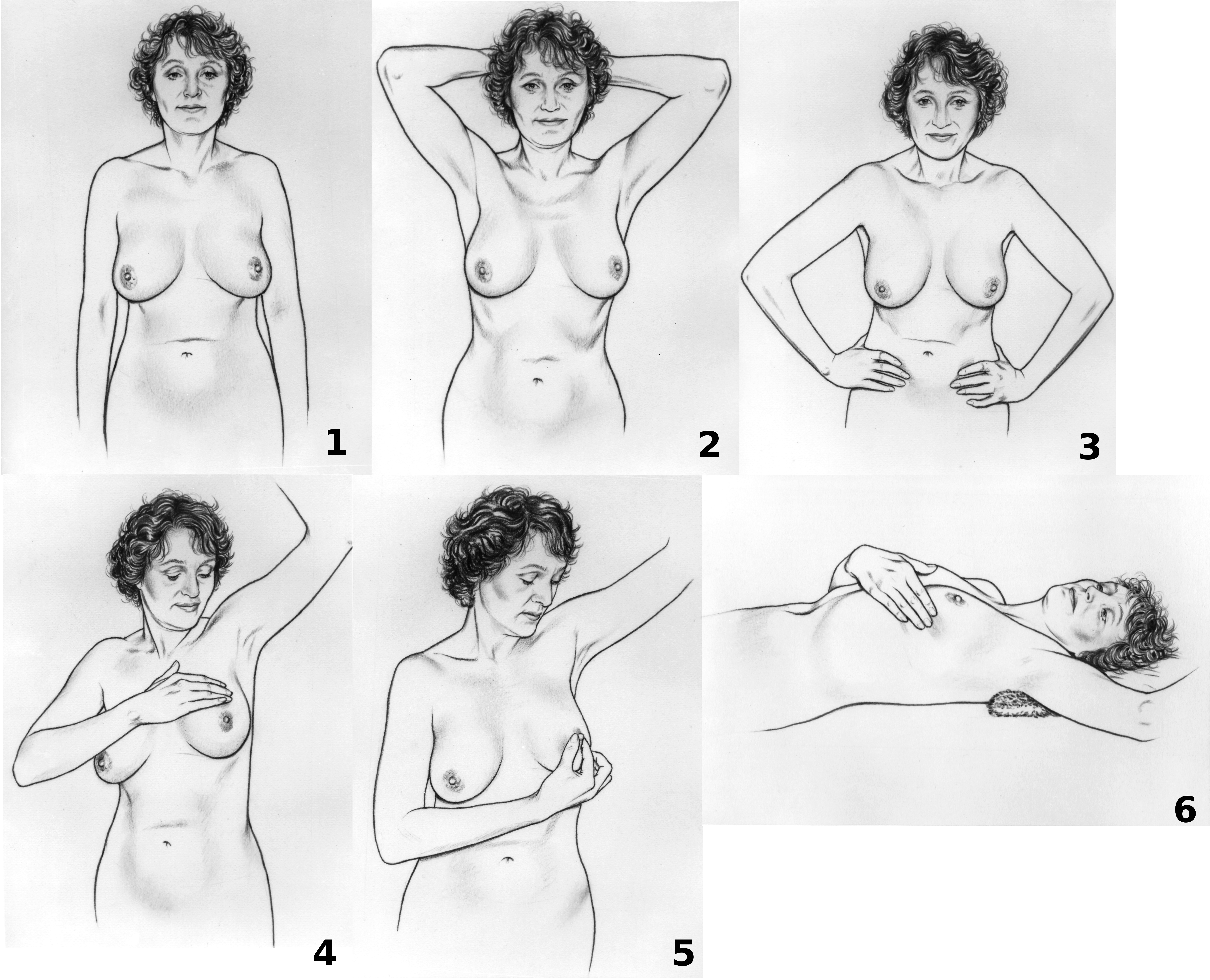
Breast cancer screening is the medical screening of asymptomatic, apparently healthy women for breast cancer in an attempt to achieve an earlier diagnosis. The assumption is that early detection will improve outcomes. A number of screening tests have been employed, including clinical and self breast exams, mammography, genetic screening, ultrasound, and magnetic resonance imaging. A clinical or self breast exam involves feeling the breast for lumps or other abnormalities. Medical evidence, however, does not support its use in women with a typical risk for breast cancer. Universal screening with mammography is controversial as it may not reduce all-cause mortality and may cause harms through unnecessary treatments and medical procedures. Many national organizations recommend it for most older women. The United States Preventive Services Task Force recommends screening mammography in women at normal risk for breast cancer, every two years between the ages of 50 and 74. Other ... [...More Info...] [...Related Items...] OR: [Wikipedia] [Google] [Baidu] |
Dense Breasts
Dense breast tissue, also known as dense breasts, is a condition of the breasts where a higher proportion of the breasts are made up of glandular tissue and fibrous tissue than fatty tissue. Around 40–50% of women have dense breast tissue and one of the main medical components of the condition is that mammograms are unable to differentiate tumorous tissue from the surrounding dense tissue. This increases the risk of late diagnosis of breast cancer in women with dense breast tissue. Additionally, women with such tissue have a higher likelihood of developing breast cancer in general, though the reasons for this are poorly understood. Definition Dense breast tissue is defined based on the amount of glandular and fibrous tissue as compared to the percentage of fatty tissue. The current mammography classifications split up the density of breasts into four categories. Approximately 10% of women have almost entirely fatty breasts, 40% with small pockets of dense tissue, 40% with even d ... [...More Info...] [...Related Items...] OR: [Wikipedia] [Google] [Baidu] |
Breast Biopsy
A breast biopsy is usually done after a suspicious lesion is discovered on either mammography or ultrasound to get tissue for pathological diagnosis. Several methods for a breast biopsy now exist. The most appropriate method of biopsy for a patient depends upon a variety of factors, including the size, location, appearance and characteristics of the abnormality. The different types of breast biopsies include fine-needle aspiration (FNA), vacuum-assisted biopsy, core needle biopsy, and surgical excision biopsy. Breast biopsies can be done utilizing ultrasound, MRI or a stereotactic biopsy imaging guidance. Vacuum assisted biopsies are typically done using stereotactic techniques when the suspicious lesion can only be seen on mammography. On average, 5-10 biopsies of a suspicious breast lesion will lead to the diagnosis of one case of breast cancer. Needle biopsies have largely replaced open surgical biopsies in the initial assessment of imaging as well as palpable abnormalities in ... [...More Info...] [...Related Items...] OR: [Wikipedia] [Google] [Baidu] |
Medical Ultrasonography
Medical ultrasound includes diagnostic techniques (mainly imaging techniques) using ultrasound, as well as therapeutic applications of ultrasound. In diagnosis, it is used to create an image of internal body structures such as tendons, muscles, joints, blood vessels, and internal organs, to measure some characteristics (e.g. distances and velocities) or to generate an informative audible sound. Its aim is usually to find a source of disease or to exclude pathology. The usage of ultrasound to produce visual images for medicine is called medical ultrasonography or simply sonography. The practice of examining pregnant women using ultrasound is called obstetric ultrasonography, and was an early development of clinical ultrasonography. Ultrasound is composed of sound waves with frequencies which are significantly higher than the range of human hearing (>20,000 Hz). Ultrasonic images, also known as sonograms, are created by sending pulses of ultrasound into tissue usin ... [...More Info...] [...Related Items...] OR: [Wikipedia] [Google] [Baidu] |


