|
Brasilodontidae
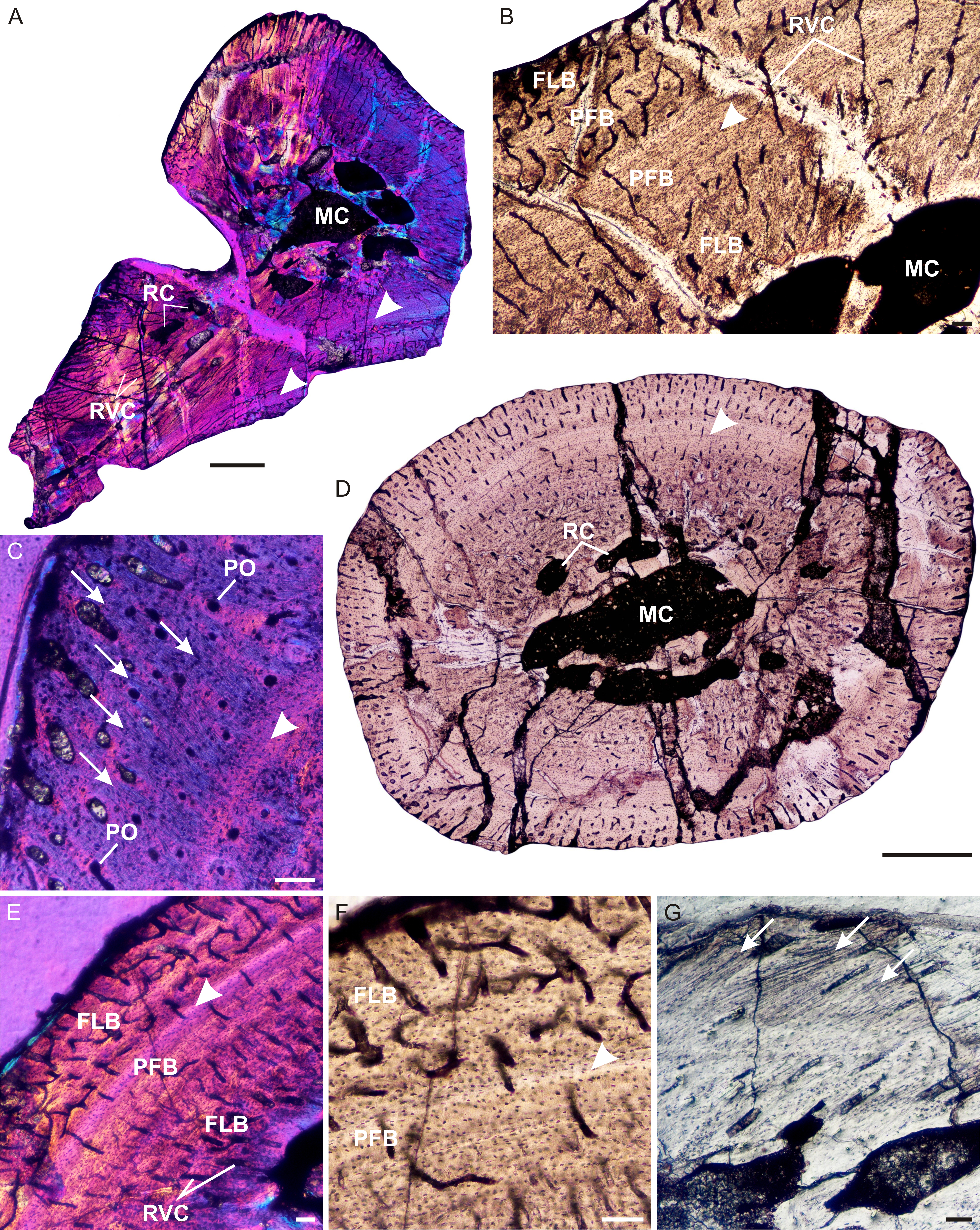
''Brasilodon'' ("tooth from Brazil") is an extinct genus of small, mammal-like cynodonts that lived in what is now Brazil during the Norian age of the Late Triassic epoch, about 225.42 million years ago. While no complete skeletons have been found, the length of ''Brasilodon'' has been estimated at . Its dentition shows that it was most likely an insectivore. The genus is monotypic, containing only the species ''B. quadrangularis''. ''Brasilodon'' belongs to the family Brasilodontidae, whose members were some of the closest relatives of mammals, the only cynodonts alive today. Two other brasilodontid genera, ''Brasilitherium'' and ''Minicynodon'', are now considered to be junior synonyms of ''Brasilodon''. Discovery and naming The first three specimens referred to ''Brasilodon quadrangularis'' were found at the Linha São Luiz site, a quarry near the town of Faxinal do Soturno in the state of Rio Grande do Sul. The rocks where ''Brasilodon'' was found belong to the upper part ... [...More Info...] [...Related Items...] OR: [Wikipedia] [Google] [Baidu] |
Cynodont
Cynodontia () is a clade of eutheriodont therapsids that first appeared in the Late Permian (approximately 260 Megaannum, mya), and extensively diversified after the Permian–Triassic extinction event. Mammals are cynodonts, as are their extinct ancestors and close relatives (Mammaliaformes), having evolved from advanced probainognathian cynodonts during the Late Triassic. Non-mammalian cynodonts occupied a variety of ecological niches, both as carnivores and as herbivores. Following the emergence of mammals, most other cynodont lines went extinct, with the last known non-mammaliaform cynodont group, the Tritylodontidae, having its youngest records in the Early Cretaceous. Description Early cynodonts have many of the skeletal characteristics of mammals. The teeth were fully differentiated and the braincase bulged at the back of the head. Outside of some Crown group, crown-group mammals (notably the therians), all cynodonts probably laid eggs. The temporal fenestrae#Fenestra ... [...More Info...] [...Related Items...] OR: [Wikipedia] [Google] [Baidu] |
Secondary Palate
The secondary palate is an anatomical structure that divides the nasal cavity from the oral cavity in many vertebrates. In human embryology, it refers to that portion of the hard palate that is formed by the growth of the two palatine shelves medially and their mutual fusion in the midline. It forms the majority of the adult palate and meets the primary palate at the incisive foramen. Clinical significance Secondary palate development begins in the sixth week of pregnancy and can lead to cleft palate when development goes awry. There are three major mechanisms known to cause this failure: #Growth retardation — Palatal shelves do not grow enough to meet each other. #Mechanical obstruction — Improper mouth size, or abnormal anatomical structures in the embryonic mouth prevent fully grown shelves from meeting each other. #Midline epithelial dysfunction (MED) - The surface mucosa of embryonic shelves is impaired, which causes a failure of palatal fusion. Evolution T ... [...More Info...] [...Related Items...] OR: [Wikipedia] [Google] [Baidu] |
Mandibular Symphysis
In human anatomy, the facial skeleton of the skull the external surface of the mandible is marked in the median line by a faint ridge, indicating the mandibular symphysis (Latin: ''symphysis menti'') or line of junction where the two lateral halves of the mandible typically fuse in the first year of life (6–9 months after birth). It is not a true symphysis as there is no cartilage between the two sides of the mandible. This ridge divides below and encloses a triangular eminence, the mental protuberance, the base of which is depressed in the center but raised on either side to form the mental tubercle. The lowest (most inferior) end of the mandibular symphysis — the point of the chin — is called the "menton". It serves as the origin for the geniohyoid and the genioglossus muscles. Other animals Solitary mammalian carnivores that rely on a powerful canine bite to subdue their prey have a strong mandibular symphysis, while pack hunters delivering shallow bites have a ... [...More Info...] [...Related Items...] OR: [Wikipedia] [Google] [Baidu] |
Dentary Bone
In jawed vertebrates, the mandible (from the Latin ''mandibula'', 'for chewing'), lower jaw, or jawbone is a bone that makes up the lowerand typically more mobilecomponent of the mouth (the upper jaw being known as the maxilla). The jawbone is the skull's only movable, posable bone, sharing joints with the cranium's temporal bones. The mandible hosts the lower teeth (their depth delineated by the alveolar process). Many muscles attach to the bone, which also hosts nerves (some connecting to the teeth) and blood vessels. Amongst other functions, the jawbone is essential for chewing food. Owing to the Neolithic advent of agriculture (), human jaws evolved to be smaller. Although it is the strongest bone of the facial skeleton, the mandible tends to deform in old age; it is also subject to fracturing. Surgery allows for the removal of jawbone fragments (or its entirety) as well as regenerative methods. Additionally, the bone is of great forensic significance. Structure In hu ... [...More Info...] [...Related Items...] OR: [Wikipedia] [Google] [Baidu] |
Zygomatic Arch
In anatomy, the zygomatic arch (colloquially known as the cheek bone), is a part of the skull formed by the zygomatic process of temporal bone, zygomatic process of the temporal bone (a bone extending forward from the side of the skull, over the opening of the ear) and the temporal Process (anatomy), process of the zygomatic bone (the side of the cheekbone), the two being united by an oblique Suture (anatomy), suture (the zygomaticotemporal suture); the tendon of the temporal muscle passes medial to (i.e. through the middle of) the arch, to gain insertion into the coronoid process of the mandible (jawbone). The jugal point is the point at the anterior (towards face) end of the upper border of the zygomatic arch where the Masseter muscle, masseteric and Maxilla, maxillary edges meet at an angle, and where it meets the process of the zygomatic bone. The arch is typical of ''Synapsida'' ("fused arch"), a clade of amniotes that includes mammals and their extinct relatives, such as ' ... [...More Info...] [...Related Items...] OR: [Wikipedia] [Google] [Baidu] |
Eye Socket
In anatomy, the orbit is the cavity or socket/hole of the skull in which the eye and its appendages are situated. "Orbit" can refer to the bony socket, or it can also be used to imply the contents. In the adult human, the volume of the orbit is about , of which the eye occupies . The orbital contents comprise the eye, the orbital and retrobulbar fascia, extraocular muscles, cranial nerves II, III, IV, V, and VI, blood vessels, fat, the lacrimal gland with its sac and duct, the eyelids, medial and lateral palpebral ligaments, cheek ligaments, the suspensory ligament, septum, ciliary ganglion and short ciliary nerves. Structure The orbits are conical or four-sided pyramidal cavities, which open into the midline of the face and point back into the head. Each consists of a base, an apex and four walls."eye, human."Encyclopædia Britannica from Encyclopædia Britannica 2006 Ultimate Reference Suite DVD 2009 Openings There are two important foramina, or windows, two ... [...More Info...] [...Related Items...] OR: [Wikipedia] [Google] [Baidu] |
Postorbital Bar
The postorbital bar (or postorbital bone) is a bony arched structure that connects the frontal bone of the skull to the zygomatic arch, which runs laterally around the eye socket. It is a trait that only occurs in mammalian taxa, such as most strepsirrhine primates and the hyrax, while haplorhine primates have evolved fully enclosed sockets. One theory for this evolutionary difference is the relative importance of vision to both orders. As haplorrhines (tarsiers and simians) tend to be diurnal, and rely heavily on visual input, many strepsirrhines are nocturnal and have a decreased reliance on visual input. Postorbital bars evolved several times independently during mammalian evolution and the evolutionary histories of several other clades. Some species, such as tarsiers, have a postorbital septum. This septum can be considered as joined processes with a small articulation between the frontal bone, the zygomatic bone and the alisphenoid bone and is therefore different from the p ... [...More Info...] [...Related Items...] OR: [Wikipedia] [Google] [Baidu] |
Prozostrodon
''Prozostrodon'' is an extinct genus of probainognathian cynodonts that was closely related to mammals. The remains were found in Brazil and are dated to the Carnian age of the Late Triassic. The holotype has an estimated skull length of , indicating that the whole animal may have been the size of a cat. The teeth were typical of advanced cynodonts, and the animal was probably a carnivore hunting reptiles and other small prey. Discovery and naming ''Prozostrodon brasiliensis'' was originally described as a species of ''Thrinaxodon'' in a 1987 paper by Mário Costa Barberena, Mário C. Barberena, José F. Bonaparte and A. M. Sá Teixeira. The holotype (UFRGS-PV-0248-T) includes a well-preserved skull preserving the front half of the cranium, a mostly complete lower jaw and all of the teeth, but missing most of the braincase, sagittal crest and zygomatic arches. It also preserves multiple postcranial elements, including parts of the vertebral column, ribs, interclavicle, humeri, r ... [...More Info...] [...Related Items...] OR: [Wikipedia] [Google] [Baidu] |


