|
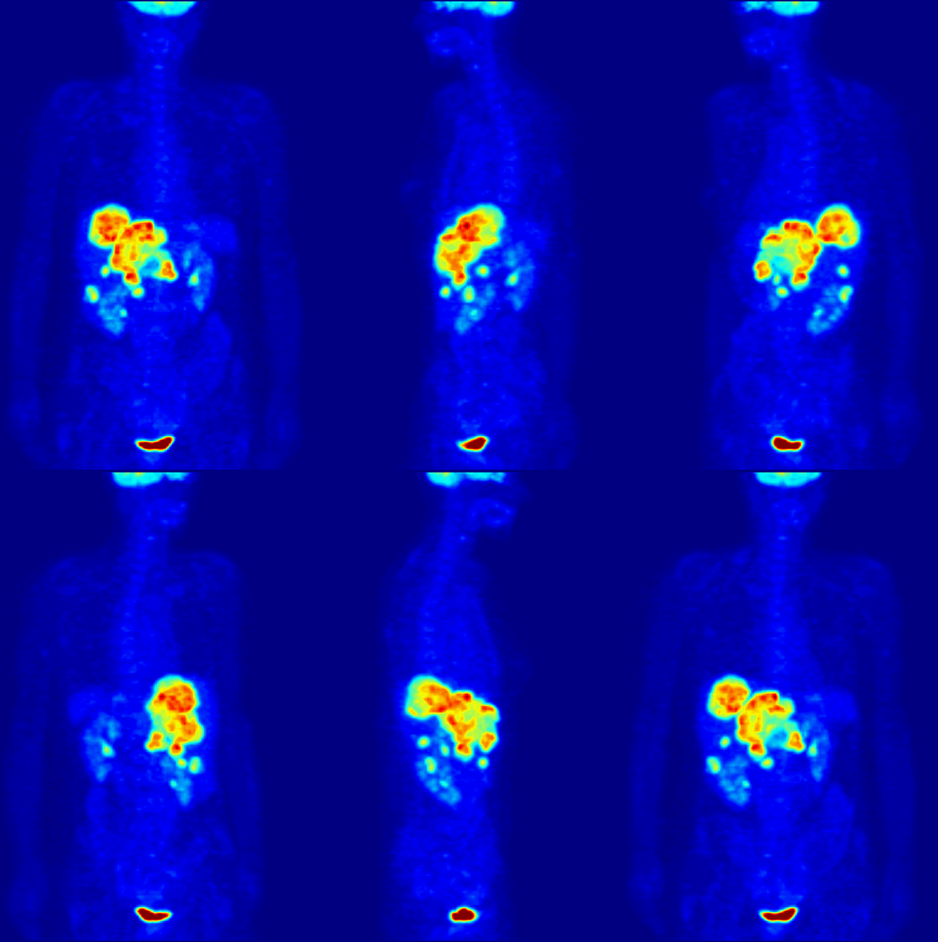
Biodistribution
Biodistribution is a method of tracking where compounds of interest travel in an experimental animal or human subject. For example, in the development of new compounds for PET (positron emission tomography) scanning, a radioactive isotope is chemically joined with a peptide (subunit of a protein). This particular class of isotopes emits positrons (which are antimatter particles, equal in mass to the electron, but with a positive charge). When ejected from the nucleus, positrons encounter an electron, and undergo annihilation which produces two gamma rays travelling in opposite directions. These gamma rays can be measured, and when compared to a standard, quantified. Biodistribution analysis Purpose and results A useful novel radiolabelled compound is one that is suitable either for medical imaging of certain body parts such as brain or tumors (injecting low doses of radioactivity) or for treating tumors (requiring injection of high doses of radioactivity). In both cases, the comp ... [...More Info...] [...Related Items...] OR: [Wikipedia] [Google] [Baidu] |
Positron Emission Tomography
Positron emission tomography (PET) is a functional imaging technique that uses radioactive substances known as radiotracers to visualize and measure changes in metabolic processes, and in other physiological activities including blood flow, regional chemical composition, and absorption. Different tracers are used for various imaging purposes, depending on the target process within the body, such as: * Fluorodeoxyglucose ( 18F">sup>18FDG or FDG) is commonly used to detect cancer; * 18Fodium fluoride">sup>18Fodium fluoride (Na18F) is widely used for detecting bone formation; * Oxygen-15 (15O) is sometimes used to measure blood flow. PET is a common imaging technique, a medical scintillography technique used in nuclear medicine. A radiopharmaceutical—a radioisotope attached to a drug—is injected into the body as a tracer. When the radiopharmaceutical undergoes beta plus decay, a positron is emitted, and when the positron interacts with an ordinary electron, the tw ... [...More Info...] [...Related Items...] OR: [Wikipedia] [Google] [Baidu] |
SPECT
Single-photon emission computed tomography (SPECT, or less commonly, SPET) is a nuclear medicine tomographic imaging technique using gamma rays. It is very similar to conventional nuclear medicine planar imaging using a gamma camera (that is, scintigraphy), but is able to provide true 3D information. This information is typically presented as cross-sectional slices through the patient, but can be freely reformatted or manipulated as required. The technique needs delivery of a gamma-emitting radioisotope (a radionuclide) into the patient, normally through injection into the bloodstream. On occasion, the radioisotope is a simple soluble dissolved ion, such as an isotope of gallium(III). Usually, however, a marker radioisotope is attached to a specific ligand to create a radioligand, whose properties bind it to certain types of tissues. This marriage allows the combination of ligand and radiopharmaceutical to be carried and bound to a place of interest in the body, where th ... [...More Info...] [...Related Items...] OR: [Wikipedia] [Google] [Baidu] |
Medical Genetics
Medical genetics is the branch of medicine that involves the diagnosis and management of hereditary disorders. Medical genetics differs from human genetics in that human genetics is a field of scientific research that may or may not apply to medicine, while medical genetics refers to the application of genetics to medical care. For example, research on the causes and inheritance of genetic disorders would be considered within both human genetics and medical genetics, while the diagnosis, management, and counselling people with genetic disorders would be considered part of medical genetics. In contrast, the study of typically non-medical phenotypes such as the genetics of eye color would be considered part of human genetics, but not necessarily relevant to medical genetics (except in situations such as albinism). ''Genetic medicine'' is a newer term for medical genetics and incorporates areas such as gene therapy, personalized medicine, and the rapidly emerging new medical specia ... [...More Info...] [...Related Items...] OR: [Wikipedia] [Google] [Baidu] |
Applied Genetics
Genetic engineering, also called genetic modification or genetic manipulation, is the modification and manipulation of an organism's genes using technology. It is a set of technologies used to change the genetic makeup of cells, including the transfer of genes within and across species boundaries to produce improved or novel organisms. New DNA is obtained by either isolating and copying the genetic material of interest using recombinant DNA methods or by artificially synthesising the DNA. A construct is usually created and used to insert this DNA into the host organism. The first recombinant DNA molecule was made by Paul Berg in 1972 by combining DNA from the monkey virus SV40 with the lambda virus. As well as inserting genes, the process can be used to remove, or "knock out", genes. The new DNA can either be inserted randomly or targeted to a specific part of the genome. An organism that is generated through genetic engineering is considered to be genetically modified ... [...More Info...] [...Related Items...] OR: [Wikipedia] [Google] [Baidu] |
Choroid Plexus
The choroid plexus, or plica choroidea, is a plexus of cells that arises from the tela choroidea in each of the ventricles of the brain. Regions of the choroid plexus produce and secrete most of the cerebrospinal fluid (CSF) of the central nervous system. The choroid plexus consists of modified ependymal cells surrounding a core of capillaries and loose connective tissue. Multiple cilia on the ependymal cells move to circulate the cerebrospinal fluid. Structure Location There is a choroid plexus in each of the four ventricles. In the lateral ventricles, it is found in the body, and continued in an enlarged amount in the atrium. There is no choroid plexus in the anterior horn. In the third ventricle, there is a small amount in the roof that is continuous with that in the body, via the interventricular foramina, the channels that connect the lateral ventricles with the third ventricle. A choroid plexus is in part of the roof of the fourth ventricle. Microana ... [...More Info...] [...Related Items...] OR: [Wikipedia] [Google] [Baidu] |
Baculoviruses
''Baculoviridae'' is a family of viruses. Arthropods, among the most studied being Lepidoptera, Hymenoptera and Diptera, serve as natural hosts. Currently, 85 species are placed in this family, assigned to four genera. Baculoviruses are known to infect insects, with over 600 host species having been described. Immature (larval) forms of lepidopteran species (moths and butterflies) are the most common hosts, but these viruses have also been found infecting sawflies, and mosquitoes. Although baculoviruses are capable of entering mammalian cells in culture, they are not known to be capable of replication in mammalian or other vertebrate animal cells. Starting in the 1940s, they were used and studied widely as biopesticides in crop fields. Baculoviruses contain a circular, double-stranded DNA (dsDNA) genome ranging from 80 to 180 kbp. Historical influence The earliest records of baculoviruses can be found in the literature from as early as the 16th century in reports of "wilting ... [...More Info...] [...Related Items...] OR: [Wikipedia] [Google] [Baidu] |
Avidin
Avidin is a tetrameric biotin-binding protein produced in the oviducts of birds, reptiles and amphibians and deposited in the whites of their eggs. Dimeric members of the avidin family are also found in some bacteria. In chicken egg white, avidin makes up approximately 0.05% of total protein (approximately 1800 μg per egg). The tetrameric protein contains four identical subunits (homotetramer), each of which can bind to biotin (Vitamin B7, vitamin H) with a high degree of affinity and specificity. The dissociation constant of the avidin-biotin complex is measured to be ''K''D ≈ 10−15 M, making it one of the strongest known non-covalent bonds. In its tetrameric form, avidin is estimated to be 66–69 k Da in size. 10% of the molecular weight is contributed by carbohydrate, composed of four to five mannose and three N-acetylglucosamine residues The carbohydrate moieties of avidin contain at least three unique oligosaccharide structural types that are similar in ... [...More Info...] [...Related Items...] OR: [Wikipedia] [Google] [Baidu] |
Polymerase Chain Reaction
The polymerase chain reaction (PCR) is a method widely used to make millions to billions of copies of a specific DNA sample rapidly, allowing scientists to amplify a very small sample of DNA (or a part of it) sufficiently to enable detailed study. PCR was invented in 1983 by American biochemist Kary Mullis at Cetus Corporation. Mullis and biochemist Michael Smith (chemist), Michael Smith, who had developed other essential ways of manipulating DNA, were jointly awarded the Nobel Prize in Chemistry in 1993. PCR is fundamental to many of the procedures used in genetic testing and research, including analysis of Ancient DNA, ancient samples of DNA and identification of infectious agents. Using PCR, copies of very small amounts of DNA sequences are exponentially amplified in a series of cycles of temperature changes. PCR is now a common and often indispensable technique used in medical laboratory research for a broad variety of applications including biomedical research and forensic ... [...More Info...] [...Related Items...] OR: [Wikipedia] [Google] [Baidu] |
Optical Imaging
Medical optical imaging is the use of light as an investigational imaging technique for medical applications, pioneered by American Physical Chemist Britton Chance. Examples include optical microscopy, spectroscopy, endoscopy, scanning laser ophthalmoscopy, laser Doppler imaging, optical coherence tomography, and transdermal optical imaging. Because light is an electromagnetic wave, similar phenomena occur in X-rays, microwaves, and radio waves. Optical imaging systems may be divided into diffusive and ballistic imaging systems. A model for photon migration in turbid biological media has been developed by Bonner et al. Such a model can be applied for interpretation data obtained from laser Doppler blood-flow monitors and for designing protocols for therapeutic excitation of tissue chromophores. Diffusive optical imaging Diffuse optical imaging (DOI) is a method of imaging using near-infrared spectroscopy (NIRS) or fluorescence-based methods. When used to create 3D ... [...More Info...] [...Related Items...] OR: [Wikipedia] [Google] [Baidu] |
Marker Gene
In biology, a marker gene may have several meanings. In nuclear biology and molecular biology, a marker gene is a gene used to determine if a nucleic acid sequence has been successfully inserted into an organism's DNA. In particular, there are two sub-types of these marker genes: a selectable marker and a marker for screening. In metagenomics and phylogenetics, a marker gene is an orthologous gene group which can be used to delineate between taxonomic lineages. Selectable marker A selectable marker protects the organism from a ''selective agent'' that would normally kill it or prevent its growth. In a transformation reaction, depending on the transformation efficiency, only one in several million to billion cells may take up DNA. Rather than checking every single cell, scientists use a selective agent to kill all cells that do not contain the foreign DNA, leaving only the desired ones. Antibiotics are the most common selective agents. In bacteria, antibiotics are used almost e ... [...More Info...] [...Related Items...] OR: [Wikipedia] [Google] [Baidu] |
Contrast Agent
A contrast agent (or contrast medium) is a substance used to increase the contrast of structures or fluids within the body in medical imaging. Contrast agents absorb or alter external electromagnetism or ultrasound, which is different from radiopharmaceuticals, which emit radiation themselves. In X-ray imaging, contrast agents enhance the radiodensity in a target tissue or structure. In magnetic resonance imaging (MRI), contrast agents shorten (or in some instances increase) the relaxation times of nuclei within body tissues in order to alter the contrast in the image. Contrast agents are commonly used to improve the visibility of blood vessels and the gastrointestinal tract. The types of contrast agent are classified according to their intended imaging modalities. Radiocontrast media For radiography, which is based on X-rays, iodine and barium are the most common types of contrast agent. Various sorts of iodinated contrast agents exist, with variations occurring between the ... [...More Info...] [...Related Items...] OR: [Wikipedia] [Google] [Baidu] |


