|
Biatora Toensbergii
''Biatora toensbergii'' is a species of corticolous (bark-dwelling), crustose lichen in the family Ramalinaceae. It is found in Norway and northwestern North America. Taxonomy The lichen was formally described as a new species in 1995 by the lichenologists Håkon Holien and Christian Printzen. The type specimen was collected by the Norwegian lichenologist Tor Tønsberg in 1981, north of Bjørktjørnane (Nord-Trøndelag, Norway) at an elevation of ; it was found growing on ''Alnus incana'' in a south-facing ravine. The species epithet honours the collector, Tønsberg. Molecular phylogenetics analysis suggests that '' Biatora pycnidiata'' is a closely related species, and that these two species form a clade that has a sister group relationship with a clade containing ''Biatora efflorescens'' and '' Biatora helvola''. Description The thallus of ''Biatora toensbergii'' is , meaning it spreads outwards and can reach up to in diameter. It has a cracked, appearance, resembling a n ... [...More Info...] [...Related Items...] OR: [Wikipedia] [Google] [Baidu] |
Corticolous Lichen
A corticolous lichen is a lichen that grows on bark.Alan Silverside's Lichen Glossary (a-f), Alan Silverside/ref> This is contrasted with lignicolous lichen, which grows on wood that has had the bark stripped from it,Alan Silverside's Lichen Glossary (g-o), Alan Silverside/ref> and saxicolous lichen, which grows on rock.Alan Silverside's Lichen Glossary (p-z), Alan Silverside/ref> Examples of corticolous lichens include the crustose lichen Crustose lichens are lichens that form a crust which strongly adheres to the Substrate (biology), substrate (soil, rock, tree bark, etc.), making separation from the substrate impossible without destruction. The basic structure of crustose lichen ... '' Graphis plumierae'', foliose lichen '' Melanohalea subolivacea'' and the fruticose '' Bryoria fuscescens''.Náttúrufræðistofnun Íslands celandic Institute of Natural History(1996). Válisti 1: Plöntur.' (in Icelandic) Reykjavík: Náttúrufræðistofnun Íslands. See also * Phyllopsora ... [...More Info...] [...Related Items...] OR: [Wikipedia] [Google] [Baidu] |
Medulla (lichenology) zone, but above the lower cortex.Galloway, D.J. (1992). Flora of Australia - ''Lichen Glossary'' The medulla generally has a cottony appearance. It is the widest layer of a heteromerous lichen thallus.
The medulla is a horizontal layer within a lichen thallus. It is a loosely arranged layer of interlaced hyphae below the upper cortex and photobiont A lichen ( , ) is a hybrid colony of algae or cyanobacteria living symbiotically among filaments of multiple fungus species, along with yeasts and bacteria embedded in the cortex or "skin", in a mutualistic relationship. References Fungal morphology and anatomy Lichenology {{lichen-stub ...[...More Info...] [...Related Items...] OR: [Wikipedia] [Google] [Baidu] |
Argopsin
Argopsin, also known as 1-chloropannarin, is a secondary metabolite produced by many lichen species, such as '' Biatora cuprea'' and '' Micarea lignaria''. Argopsin (also known as 1'-chloropannarin) is a chlorinated depsidone compound first isolated from the lichen '' Argopsis friesiana'' by Siegfried Huneck and Elke Mackenzie in 1975. It was independently discovered by both Huneck and Mackenzie's team and by Bodo and Molho in the same year. The structure was confirmed through multiple analytical methods including mass spectrometry, which showed characteristic isotope peaks at mass-to-charge ratios (m/z) of 396, 398, and 400 in a 9:6:1 ratio, indicating the presence of two chlorine atoms. Its UV spectrum matched that of pannarin, and infrared spectroscopy revealed characteristic aldehyde (1725 cm⁻¹) and hydroxyl (3500 cm⁻¹) bands. The researchers synthesized argopsin by chlorinating pannarin in acetic acid, which produced a compound identical to the naturall ... [...More Info...] [...Related Items...] OR: [Wikipedia] [Google] [Baidu] |
Thin-layer Chromatography
Thin-layer chromatography (TLC) is a chromatography technique that separates components in non-volatile mixtures. It is performed on a TLC plate made up of a non-reactive solid coated with a thin layer of adsorbent material. This is called the stationary phase. The sample is deposited on the plate, which is eluted with a solvent or solvent mixture known as the mobile phase (or eluent). This solvent then moves up the plate via capillary action. As with all chromatography, some compounds are more attracted to the mobile phase, while others are more attracted to the stationary phase. Therefore, different compounds move up the TLC plate at different speeds and become separated. To visualize colourless compounds, the plate is viewed under UV light or is stained.Jork, H., Funk, W., Fischer, W., Wimmer, H. (1990): Thin-Layer Chromatography: Reagents and Detection Methods, Volume 1a, VCH, Weinheim, Testing different stationary and mobile phases is often necessary to obtain well-defined an ... [...More Info...] [...Related Items...] OR: [Wikipedia] [Google] [Baidu] |
Spot Test (lichen)
A spot test in lichenology is a spot analysis used to help identify lichens. It is performed by placing a drop of a chemical reagent on different parts of the lichen and noting the colour change (or lack thereof) associated with application of the chemical. The tests are routinely encountered in dichotomous keys for lichen species, and they take advantage of the wide array of lichen products (secondary metabolites) produced by lichens and their uniqueness among taxa. As such, spot tests reveal the presence or absence of chemicals in various parts of a lichen. They were first proposed as a method to help identify species by the Finnish lichenologist William Nylander in 1866. Three common spot tests use either 10% aqueous KOH solution (K test), saturated aqueous solution of bleaching powder or calcium hypochlorite (C test), or 5% alcoholic ''p''-phenylenediamine solution (P test). The colour changes occur due to presence of particular secondary metabolites in the lichen. In ide ... [...More Info...] [...Related Items...] OR: [Wikipedia] [Google] [Baidu] |
Septum
In biology, a septum (Latin language, Latin for ''something that encloses''; septa) is a wall, dividing a Body cavity, cavity or structure into smaller ones. A cavity or structure divided in this way may be referred to as septate. Examples Human anatomy * Interatrial septum, the wall of tissue that is a sectional part of the left and right atria of the heart * Interventricular septum, the wall separating the left and right ventricles of the heart * Lingual septum, a vertical layer of fibrous tissue that separates the halves of the tongue *Nasal septum: the cartilage wall separating the nostrils of the nose * Alveolar septum: the thin wall which separates the Pulmonary alveolus, alveoli from each other in the lungs * Orbital septum, a palpebral ligament in the upper and lower eyelids * Septum pellucidum or septum lucidum, a thin structure separating two fluid pockets in the brain * Uterine septum, a malformation of the uterus * Septum of the penis, Penile septum, a fibrous w ... [...More Info...] [...Related Items...] OR: [Wikipedia] [Google] [Baidu] |
Ascospore
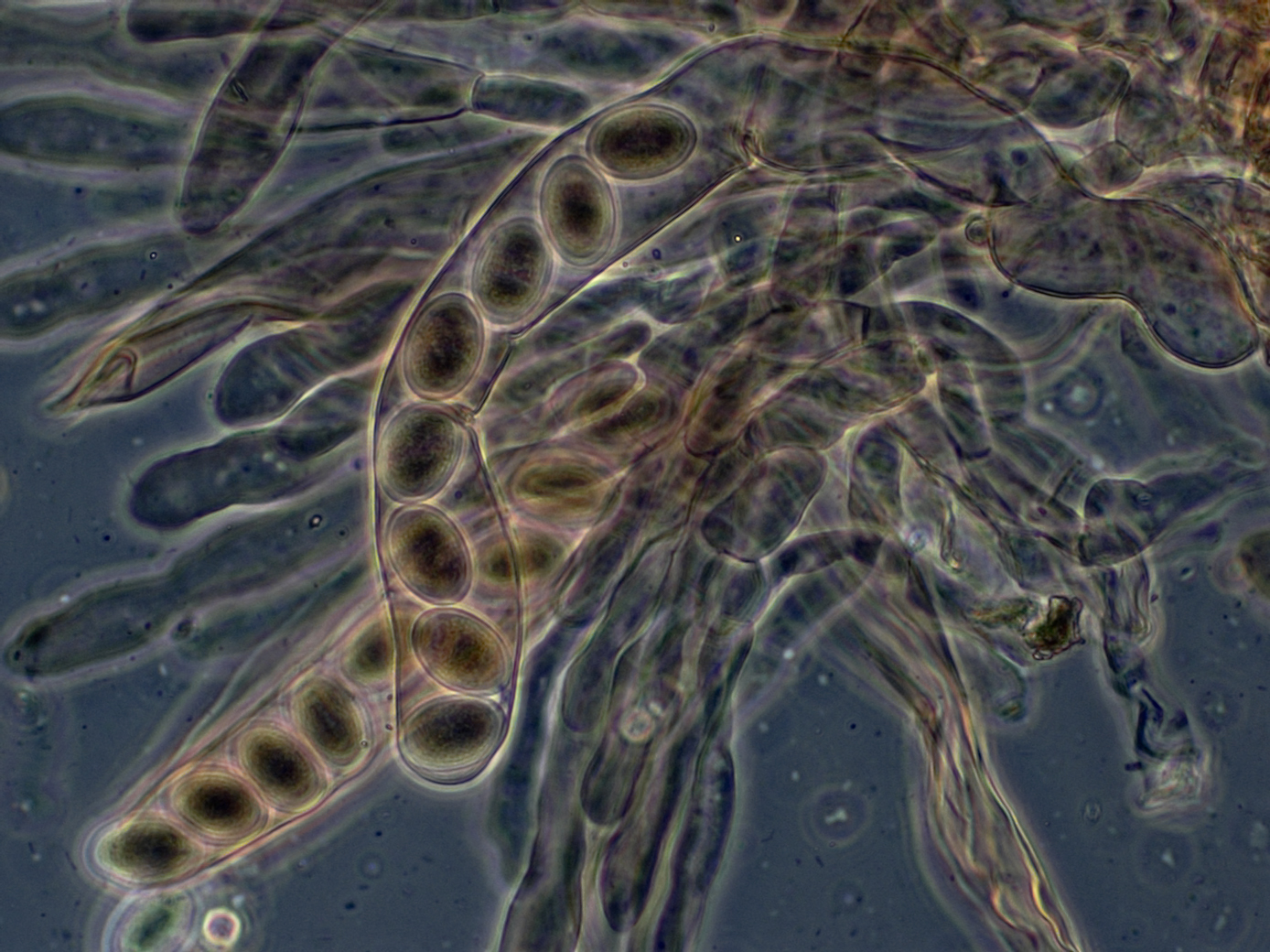
In fungi, an ascospore is the sexual spore formed inside an ascus—the sac-like cell that defines the division Ascomycota, the largest and most diverse Division (botany), division of fungi. After two parental cell nucleus, nuclei fuse, the ascus undergoes meiosis (halving of genetic material) followed by a mitosis (cell division), ordinarily producing eight genetically distinct haploid spores; most yeasts stop at four ascospores, whereas some moulds carry out extra post-meiotic divisions to yield dozens. Many asci build turgor, internal pressure and shoot their spores clear of the calm boundary layer, thin layer of still air enveloping the fruit body, whereas subterranean truffles depend on animals for biological dispersal, dispersal. Ontogeny, Development shapes both form and endurance of ascospores. A hook-shaped crozier aligns the paired nuclei; a double-biological membrane, membrane system then parcels each daughter nucleus, and successive wall layers of β-glucan, chitosan ... [...More Info...] [...Related Items...] OR: [Wikipedia] [Google] [Baidu] |
Biatora
''Biatora'' is a genus of lichens in the family Ramalinaceae. Originally circumscribed in 1817,Fries EM, Sandberg A. (1817). ''Lichenum dianome nova''. Lund. the genus consists of crustose and squamulose lichens with green algal photobionts, biatorine apothecia, colorless, simple to 3-septate ascospores, and bacilliform pycnospores. Description ''Biatora'' species are crustose lichens with a spreading () thallus that may appear thin and somewhat membranous in places. The surface is often cracked () and, in species that grow in association with mosses, may be or warted. The thallus is typically creamy white, dull green, glaucous green, or green-grey and lacks a distinct outer protective layer (). Some species produce soredia, small reproductive that facilitate dispersal. A , the initial fungal layer that some lichens form before developing a full thallus, is absent. The photosynthetic partner () is a alga, a group characterized by spherical to broadly ellipsoidal cells. The ... [...More Info...] [...Related Items...] OR: [Wikipedia] [Google] [Baidu] |
Ascus
An ascus (; : asci) is the sexual spore-bearing cell produced in ascomycete fungi. Each ascus usually contains eight ascospores (or octad), produced by meiosis followed, in most species, by a mitotic cell division. However, asci in some genera or species can occur in numbers of one (e.g. '' Monosporascus cannonballus''), two, four, or multiples of four. In a few cases, the ascospores can bud off conidia that may fill the asci (e.g. '' Tympanis'') with hundreds of conidia, or the ascospores may fragment, e.g. some '' Cordyceps'', also filling the asci with smaller cells. Ascospores are nonmotile, usually single celled, but not infrequently may be coenocytic (lacking a septum), and in some cases coenocytic in multiple planes. Mitotic divisions within the developing spores populate each resulting cell in septate ascospores with nuclei. The term ocular chamber, or oculus, refers to the epiplasm (the portion of cytoplasm not used in ascospore formation) that is surrounded by the ... [...More Info...] [...Related Items...] OR: [Wikipedia] [Google] [Baidu] |
Paraphyses
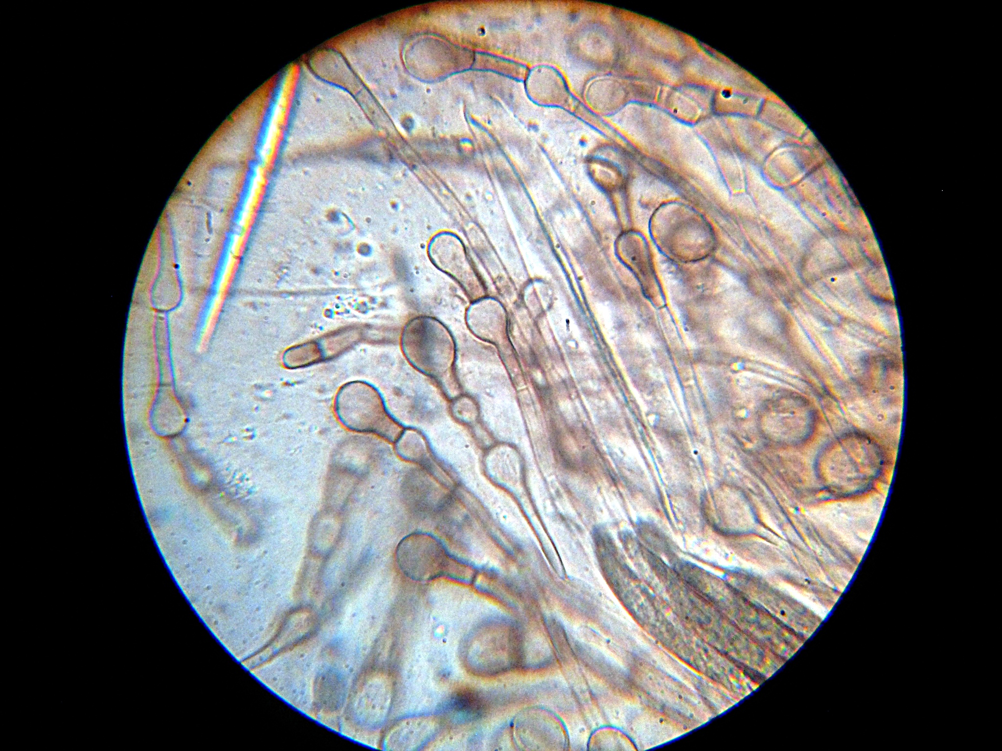
Paraphyses are erect sterile filament-like support structures occurring among the reproductive apparatuses of fungi, ferns, bryophytes and some thallophytes. The singular form of the word is paraphysis. In certain fungi, they are part of the fertile spore-bearing layer. More specifically, paraphyses are sterile filamentous hyphal end cells composing part of the hymenium of Ascomycota and Basidiomycota interspersed among either the asci or basidia respectively, and not sufficiently differentiated to be called cystidia A cystidium (: cystidia) is a relatively large cell found on the sporocarp of a basidiomycete (for example, on the surface of a mushroom gill), often between clusters of basidia. Since cystidia have highly varied and distinct shapes that are o ..., which are specialized, swollen, often protruding cells. The tips of paraphyses may contain the pigments which colour the hymenium. In ferns and mosses, they are filament-like structures that are found on sporangi ... [...More Info...] [...Related Items...] OR: [Wikipedia] [Google] [Baidu] |
Hypha
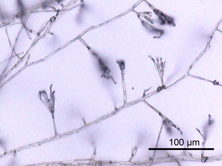
A hypha (; ) is a long, branching, filamentous structure of a fungus, oomycete, or actinobacterium. In most fungi, hyphae are the main mode of vegetative growth, and are collectively called a mycelium. Structure A hypha consists of one or more cells surrounded by a tubular cell wall. In most fungi, hyphae are divided into cells by internal cross-walls called "septa" (singular septum). Septa are usually perforated by pores large enough for ribosomes, mitochondria, and sometimes nuclei to flow between cells. The major structural polymer in fungal cell walls is typically chitin, in contrast to plants and oomycetes that have cellulosic cell walls. Some fungi have aseptate hyphae, meaning their hyphae are not partitioned by septa. Hyphae have an average diameter of 4–6 μm. Growth Hyphae grow at their tips. During tip growth, cell walls are extended by the external assembly and polymerization of cell wall components, and the internal production of new cell membrane. ... [...More Info...] [...Related Items...] OR: [Wikipedia] [Google] [Baidu] |


