|
Bascom Palmer Eye Institute
Bascom Palmer Eye Institute is the University of Miami School of Medicine's ophthalmic care, research, and education center. The institute is based in the Health District of Miami, Florida, and has been ranked consistently as the best eye hospital and vision research center in the nation. Bascom Palmer Eye Institute faculty and staff treat patients from around the world at the institute's multi-location facilities, including its flagship location in Miami and at satellite facilities elsewhere in Miami-Dade County, Broward County, Palm Beach County, and Collier County in South Florida. The institute's clinical faculty treats more than 250,000 patients annually, provides 24-hour emergency care, and is the only community-based ophthalmic care center for indigent and low-income patients of Miami-Dade County. History Ophthalmology at the University of Miami School of Medicine began in 1955 and attained departmental status in 1959. Bascom Palmer Eye Institute was founded seven yea ... [...More Info...] [...Related Items...] OR: [Wikipedia] [Google] [Baidu] |
Miami
Miami is a East Coast of the United States, coastal city in the U.S. state of Florida and the county seat of Miami-Dade County, Florida, Miami-Dade County in South Florida. It is the core of the Miami metropolitan area, which, with a population of 6.14 million, is the second-largest metropolitan area in the Southeastern United States, Southeast after Atlanta metropolitan area, Atlanta, and the Metropolitan statistical area#United States, ninth-largest in the United States. With a population of 442,241 as of the 2020 United States census, 2020 census, Miami is the List of municipalities in Florida, second-most populous city in Florida, after Jacksonville, Florida, Jacksonville. Miami has the List of tallest buildings in the United States#Cities with the most skyscrapers, third-largest skyline in the U.S. with over List of tallest buildings in Miami, 300 high-rises, 70 of which exceed . Miami is a major center and leader in finance, commerce, culture, arts, and internation ... [...More Info...] [...Related Items...] OR: [Wikipedia] [Google] [Baidu] |
Glaucoma
Glaucoma is a group of eye diseases that can lead to damage of the optic nerve. The optic nerve transmits visual information from the eye to the brain. Glaucoma may cause vision loss if left untreated. It has been called the "silent thief of sight" because the loss of vision usually occurs slowly over a long period of time. A major risk factor for glaucoma is increased pressure within the eye, known as Intraocular pressure, intraocular pressure (IOP). It is associated with old age, a family history of glaucoma, and certain medical conditions or the use of some medications. The word ''glaucoma'' comes from the Ancient Greek word (), meaning 'gleaming, blue-green, gray'. Of the different types of glaucoma, the most common are called open-angle glaucoma and closed-angle glaucoma. Inside the eye, a liquid called Aqueous humour, aqueous humor helps to maintain shape and provides nutrients. The aqueous humor normally drains through the trabecular meshwork. In open-angle glaucoma, ... [...More Info...] [...Related Items...] OR: [Wikipedia] [Google] [Baidu] |
Vitreous Membrane
The vitreous membrane (or hyaloid membrane or vitreous cortex) is a layer of collagen separating the vitreous humour from the rest of the eye. At least two parts have been identified anatomically. The posterior hyaloid membrane separates the rear of the vitreous from the retina. It is a false anatomical membrane. The anterior hyaloid membrane separates the front of the vitreous from the lens (anatomy), lens. Andres Bernal, Jean-Marie Parel, Fabrice MannsEvidence for posterior zonular fiber attachment on the anterior hyaloid membrane "Investigative Ophthalmology and Visual Science" 2006, 47, 4708-4713. Bernal et al. describe it "as a delicate structure in the form of a thin layer that runs from the pars plana to the posterior lens, where it shares its attachment with the posterior zonule via Weigert's ligament, also known as Egger's line". References External links Image at ivy-rose.co.uk Human eye anatomy {{eye-stub ... [...More Info...] [...Related Items...] OR: [Wikipedia] [Google] [Baidu] |
HIV/AIDS
The HIV, human immunodeficiency virus (HIV) is a retrovirus that attacks the immune system. Without treatment, it can lead to a spectrum of conditions including acquired immunodeficiency syndrome (AIDS). It is a Preventive healthcare, preventable disease. It can be managed with treatment and become a manageable chronic health condition. While there is no cure or vaccine for HIV, Management of HIV/AIDS, antiretroviral treatment can slow the course of the disease, and if used before significant disease progression, can extend the life expectancy of someone living with HIV to a nearly standard level. An HIV-positive person on treatment can expect to live a normal life, and die with the virus, not of it. Effective #Treatment, treatment for HIV-positive people (people living with HIV) involves a life-long regimen of medicine to suppress the virus, making the viral load undetectable. Treatment is recommended as soon as the diagnosis is made. An HIV-positive person who has an ... [...More Info...] [...Related Items...] OR: [Wikipedia] [Google] [Baidu] |
Acute Retinal Necrosis
Acute retinal necrosis (ARN) is a medical inflammatory condition of the eye. The condition presents itself as a necrotizing retinitis. The inflammation onset is due to certain herpes viruses, varicella zoster virus (VZV), herpes simplex virus (HSV-1 and HSV-2) and Epstein–Barr virus (EBV). People with the condition usually display redness of the eye, white or off-white colored patches that are patches of retinal necrosis. ARN can progress into other conditions such as uveitis, detachment of the retina, and ultimately can lead to blindness. The disease was first characterized in 1971, in Japan. Akira Urayama and his colleagues had six patients whose cases showed signs of acute necrotizing retinitis, retinal arteritis, choroiditis, and late-onset retinal detachment. The combination of the conditions was given the name acute retinal necrosis. The first reports of ARN came about in 1971. It is unclear whether it was previously just reported as something else. Urayama and his coll ... [...More Info...] [...Related Items...] OR: [Wikipedia] [Google] [Baidu] |
Herpes Simplex
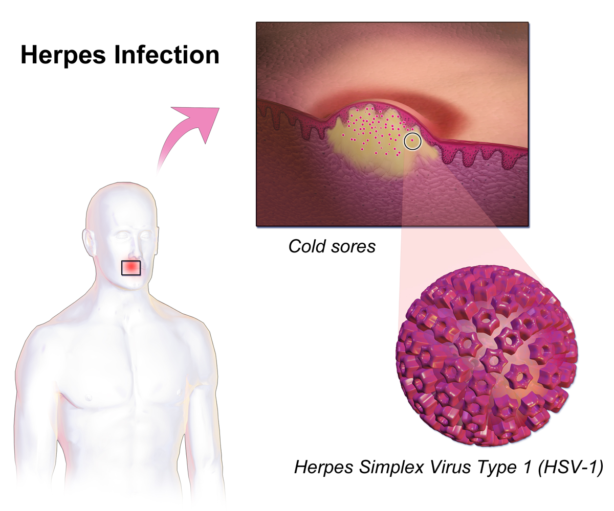
Herpes simplex, often known simply as herpes, is a viral disease, viral infection caused by the herpes simplex virus. Herpes infections are categorized by the area of the body that is infected. The two major types of herpes are Cold sore, oral herpes and genital herpes, though Herpes simplex#Types of herpes, other forms also exist. Oral herpes involves the face or mouth. It may result in small blisters in groups, often called cold sores or fever blisters, or may just cause a sore throat. Genital herpes involves the genitalia. It may have minimal symptoms or form blisters that break open and result in small ulcers. These typically heal over two to four weeks. Tingling or shooting pains may occur before the blisters appear. Herpes cycles between periods of active disease followed by periods without symptoms. The first episode is often more severe and may be associated with fever, muscle pains, swollen lymph nodes and headaches. Over time, episodes of active disease decrease in ... [...More Info...] [...Related Items...] OR: [Wikipedia] [Google] [Baidu] |
Limbal Stem Cell
Limbal stem cells, also known as corneal epithelial stem cells, are unipotent stem cells located in the basal epithelial layer of the corneal limbus. They form the border between the cornea and the sclera. Characteristics of limbal stem cells include a slow turnover rate, high proliferative potential, clonogenicity, expression of stem cell markers, as well as the ability to regenerate the entire corneal epithelium. Limbal stem cell proliferation has the role of maintaining the cornea; for example, by replacing cells that are lost via tears. Additionally, these cells also prevent the conjunctival epithelial cells from migrating onto the surface of the cornea. Medical conditions and treatments Damage to the limbus can lead to limbal stem cell deficiency (LSCD); this may be primary - related to an insufficient stromal microenvironment to support stem cell functions, such as aniridia, and other congenital conditions, or secondary – caused by external factors that destroy the limbal s ... [...More Info...] [...Related Items...] OR: [Wikipedia] [Google] [Baidu] |
Diabetic Retinopathy
Diabetic retinopathy (also known as diabetic eye disease) is a medical condition in which damage occurs to the retina due to diabetes. It is a leading cause of blindness in developed countries and one of the lead causes of sight loss in the world, even though there are many new therapies and improved treatments for helping people live with diabetes. Diabetic retinopathy affects up to 80 percent of those who have had both Type 1 diabetes, type 1 and Type 2 diabetes, type 2 diabetes for 20 years or more. In at least 90% of new cases, progression to more aggressive forms of sight threatening retinopathy and maculopathy could be reduced with proper treatment and monitoring of the eyes. The longer a person has diabetes, the higher their chances of developing diabetic retinopathy. Each year in the United States, diabetic retinopathy accounts for 12% of all new cases of blindness. It is also the leading cause of blindness in people aged 20 to 64. Signs and symptoms Nearly all peopl ... [...More Info...] [...Related Items...] OR: [Wikipedia] [Google] [Baidu] |
Vitreous Body
The vitreous body (''vitreous'' meaning "glass-like"; , ) is the clear gel that fills the space between the Lens (vision), lens and the retina of the eye, eyeball (the vitreous chamber) in humans and other vertebrates. It is often referred to as the vitreous humor (also spelled humour), from Latin meaning liquid, or simply "the vitreous". Vitreous fluid or "liquid vitreous" is the liquid component of the vitreous gel, found after a vitreous detachment. It is not to be confused with the aqueous humor, the other fluid in the eye that is found between the cornea and lens. Structure The vitreous humor is a transparent, colorless, gelatinous mass that fills the space in the eye between the lens and the retina. It is surrounded by a layer of collagen called the vitreous membrane (or hyaloid membrane or vitreous cortex) separating it from the rest of the eye. It makes up four-fifths of the volume of the eyeball. The vitreous humour is fluid-like near the centre, and gel-like near the ed ... [...More Info...] [...Related Items...] OR: [Wikipedia] [Google] [Baidu] |
Vitrectomy
Vitrectomy is a surgery to remove some or all of the vitreous humor from the Human eye, eye. Anterior vitrectomy entails removing small portions of the vitreous humor from the front structures of the eye—often because these are tangled in an intraocular lens or other structures. ''Pars plana'' vitrectomy is a general term for a group of operations accomplished in the deeper part of the eye, all of which involve removing some or all of the vitreous humor—the eye's clear internal jelly. Even before the modern era, some surgeons performed crude vitrectomies. For instance, Dutch surgeon Anton Nuck (1650–1692) claimed to have removed vitreous by suction in a young man with an inflamed eye. In Boston, John Collins Warren (surgeon, born 1778), John Collins Warren (1778–1856) performed a crude limited vitrectomy for angle closure glaucoma. Anesthesia for vitrectomy Options for anesthesia for vitrectomy are general anaesthesia, local anesthesia, Topical anesthetic, topical a ... [...More Info...] [...Related Items...] OR: [Wikipedia] [Google] [Baidu] |
Retina
The retina (; or retinas) is the innermost, photosensitivity, light-sensitive layer of tissue (biology), tissue of the eye of most vertebrates and some Mollusca, molluscs. The optics of the eye create a focus (optics), focused two-dimensional image of the visual world on the retina, which then processes that image within the retina and sends nerve impulses along the optic nerve to the visual cortex to create visual perception. The retina serves a function which is in many ways analogous to that of the photographic film, film or image sensor in a camera. The neural retina consists of several layers of neurons interconnected by Chemical synapse, synapses and is supported by an outer layer of pigmented epithelial cells. The primary light-sensing cells in the retina are the photoreceptor cells, which are of two types: rod cell, rods and cone cell, cones. Rods function mainly in dim light and provide monochromatic vision. Cones function in well-lit conditions and are responsible fo ... [...More Info...] [...Related Items...] OR: [Wikipedia] [Google] [Baidu] |
Angiography
Angiography or arteriography is a medical imaging technique used to visualize the inside, or lumen, of blood vessels and organs of the body, with particular interest in the arteries, veins, and the heart chambers. Modern angiography is performed by injecting a radio-opaque contrast agent into the blood vessel and imaging using X-ray based techniques such as fluoroscopy. With time-of-flight (TOF) magnetic resonance it is no longer necessary to use a contrast. The word itself comes from the Greek words ἀνγεῖον ''angeion'' 'vessel' and γράφειν ''graphein'' 'to write, record'. The film or image of the blood vessels is called an ''angiograph'', or more commonly an ''angiogram''. Though the word can describe both an arteriogram and a venogram, in everyday usage the terms angiogram and arteriogram are often used synonymously, whereas the term venogram is used more precisely. The term angiography has been applied to radionuclide angiography and newer vascular ima ... [...More Info...] [...Related Items...] OR: [Wikipedia] [Google] [Baidu] |


