|
Balloon Cell Nevus
Balloon cell nevus is a benign nevus. It appears like a melanocytic nevus. Histologically it is characterized by swollen, pale, polyhedral melanocytes, with pale cytoplasm and a central nucleus. It is different to balloon cell melanoma, which has larger nuclei and is structured like a melanoma. It was first described by Judalaewitsch in 1901. Signs and symptoms Balloon cell nevi can affect the skin, choroid, and conjunctiva. Usually, they appear as elevated, mobile, hyperpigmented masses in the ocular adnexa. Diagnosis The characteristics of balloon cells include their relatively large sizes, small, round nuclei positioned in the center, and largely transparent cytoplasm. Examining under a microscope is particularly crucial when it comes to balloon cell nevi. Progressive vacuolization of melanocytes or nevus cells, caused by the enlargement as well as eventual destruction of melanosomes, results in the formation of balloon cells. See also * Pseudomelanoma * Skin les ... [...More Info...] [...Related Items...] OR: [Wikipedia] [Google] [Baidu] |
Nevus
Nevus () is a nonspecific medical terminology, medical term for a visible, circumscribed, chronic (medicine), chronic lesion of the skin or mucosa. The term originates from , which is Latin for "birthmark"; however, a nevus can be either congenital (present at birth) or acquired. Common terms (''mole'', ''birthmark'', ''beauty mark'', etc.) are used to describe nevi, but these terms do not distinguish specific types of nevi from one another. Classification The term ''nevus'' is applied to a number of conditions caused by Neoplasm, neoplasias and hyperplasias of melanocytes, as well as a number of pigmentation disorders, both hypermelanotic (containing increased melanin, the pigment responsible for skin color) and hypomelanotic (containing decreased melanin). Suspicious skin moles which are multi-colored or pink may be a finding in skin cancer. Increased melanin Usually acquired * Melanocytic nevus ** Melanocytic nevi can be categorized based on the location of melanocytic ... [...More Info...] [...Related Items...] OR: [Wikipedia] [Google] [Baidu] |
Ocular Adnexa
The accessory visual structures (or adnexa of eye, ocular adnexa, etc.) are the protecting and supporting structures ( adnexa) of the eye, including the eyebrow, eyelids, and lacrimal apparatus. The eyebrows, eyelids, eyelashes, lacrimal gland and drainage apparatus all play a crucial role with regards to globe protection, lubrication, and minimizing the risk of ocular infection. The adnexal structures also help to keep the cornea moist and clean. One source defines "ocular adnexa" as the orbit, conjunctiva, and eyelids. The orbit and extraocular muscles allow for the smooth movement of the eyeball. Eyebrow The eyebrow is an area of thick, short hairs above the eye. The main function is to prevent sweat, water, and other debris falling into the eye, but they are also important to human communication and facial expressions. Eyelid An eyelid is a thin fold of skin that covers and protects the eye. The levator palpebrae superioris muscle helps in the movement of eyeli ... [...More Info...] [...Related Items...] OR: [Wikipedia] [Google] [Baidu] |
Pseudomelanoma
Pseudomelanoma (also known as a "recurrent melanocytic nevus", and "recurrent nevus") is a cutaneous condition in which melanotic skin lesions clinically resemble a superficial spreading melanoma at the site of a recent shave removal of a melanocytic nevus. Problem with the recurrent nevus The melanocytes left behind in the wound regrow in an abnormal pattern. Rather than the even and regular lace like network, the pigments tends to grow in streaks of varying width within the scar. This is often accompanied by scarring, inflammation, and blood vessel changes – giving both the clinical and histologic impression of a melanoma or a severe dysplastic nevus. When the patient is reexamined years later without the assistance of the original biopsy report, the physician will often require the removal of the scar with the recurrent nevus to assure that a melanoma is not missed. Saucerization biopsy Also known as "scoop", "scallop", or "shave" excisional biopsy, or "shave excision" ... [...More Info...] [...Related Items...] OR: [Wikipedia] [Google] [Baidu] |
Melanosome
A melanosome is an organelle found in animal cells and is the site for synthesis, storage and transport of melanin, the most common light-absorbing pigment found in the animal kingdom. Melanosomes are responsible for color and photoprotection in animal cells and tissues. Melanosomes are synthesised in the skin in melanocyte cells, as well as the eye in choroidal melanocytes and retinal pigment epithelial (RPE) cells. In lower vertebrates, they are found in melanophores or chromatophores. Structure Melanosomes are relatively large organelles, measuring up to 500 nm in diameter. They are bound by a bilipid membrane and are, in general, rounded, sausage-like, or cigar-like in shape. The shape is constant for a given species and cell type. They have a characteristic ultrastructure on electron microscopy, which varies according to the maturity of the melanosome, and for research purposes a numeric staging system is sometimes used. Synthesis of melanin Melanosomes are dep ... [...More Info...] [...Related Items...] OR: [Wikipedia] [Google] [Baidu] |
Nevus Cell
Nevus cells are a variant of melanocytes. They are larger than typical melanocytes, do not have dendrites, and have more abundant cytoplasm with coarse granules. They are usually located at the dermoepidermal junction or in the dermis of the skin. Dermal nevus cells can be further classified: type A (epithelioid) dermal nevus cells mature into type B (lymphocytoid) dermal nevus cells which mature further into type C (neuroid) dermal nevus cells, through a process involving downwards migration. Nevus cells are the primary component of a melanocytic nevus. Nevus cells can also be found in lymph nodes and the thymus. See also *List of human cell types derived from the germ layers This is a list of Cell (biology), cells in humans derived from the three embryonic germ layers – ectoderm, mesoderm, and endoderm. Cells derived from ectoderm Surface ectoderm Skin * Trichocyte (human), Trichocyte * Keratinocyte Anterior pi ... References Skin anatomy {{anatomy-stub ... [...More Info...] [...Related Items...] OR: [Wikipedia] [Google] [Baidu] |
Melanocyte
Melanocytes are melanin-producing neural-crest, neural crest-derived cell (biology), cells located in the bottom layer (the stratum basale) of the skin's epidermis (skin), epidermis, the middle layer of the eye (the uvea), the inner ear, vaginal epithelium, meninges, bones, and heart found in many mammals and birds. Melanin is a dark pigment primarily responsible for skin color. Once synthesized, melanin is contained in special organelles called melanosomes which can be transported to nearby keratinocytes to induce pigmentation. Thus darker skin tones have more melanosomes present than lighter skin tones. Functionally, melanin serves as protection against Ultraviolet, UV radiation. Melanocytes also have a role in the immune system. Function Through a process called melanogenesis, melanocytes produce melanin, which is a pigment found in the human skin, skin, human eye, eyes, hair, nasal cavity, and inner ear. This melanogenesis leads to a long-lasting pigmentation, which i ... [...More Info...] [...Related Items...] OR: [Wikipedia] [Google] [Baidu] |
Vacuolization
Vacuolization is the formation of vacuoles or vacuole-like structures, within or adjacent to cells. Perinuclear vacuolization of epidermal keratinocytes is most likely inconsequential when not observed in combination with other pathologic findings. In dermatopathology "vacuolization" often refers specifically to vacuoles in the basal cell-basement membrane zone area, where it is an unspecific sign of disease.Kumar, Vinay; Fausto, Nelso; Abbas, Abul (2004) ''Robbins & Cotran Pathologic Basis of Disease'' (7th ed.). Saunders. Page 1230. . It may be a sign of for example vacuolar interface dermatitis, which in turn has many causes. It is one of the components of koilocytosis, which may be present in potentially pre- cancerous cervical, oral and anal lesions. See also * Skin lesion * Skin disease A skin condition, also known as cutaneous condition, is any medical condition that affects the integumentary system—the organ system that encloses the body and includes skin, ... [...More Info...] [...Related Items...] OR: [Wikipedia] [Google] [Baidu] |
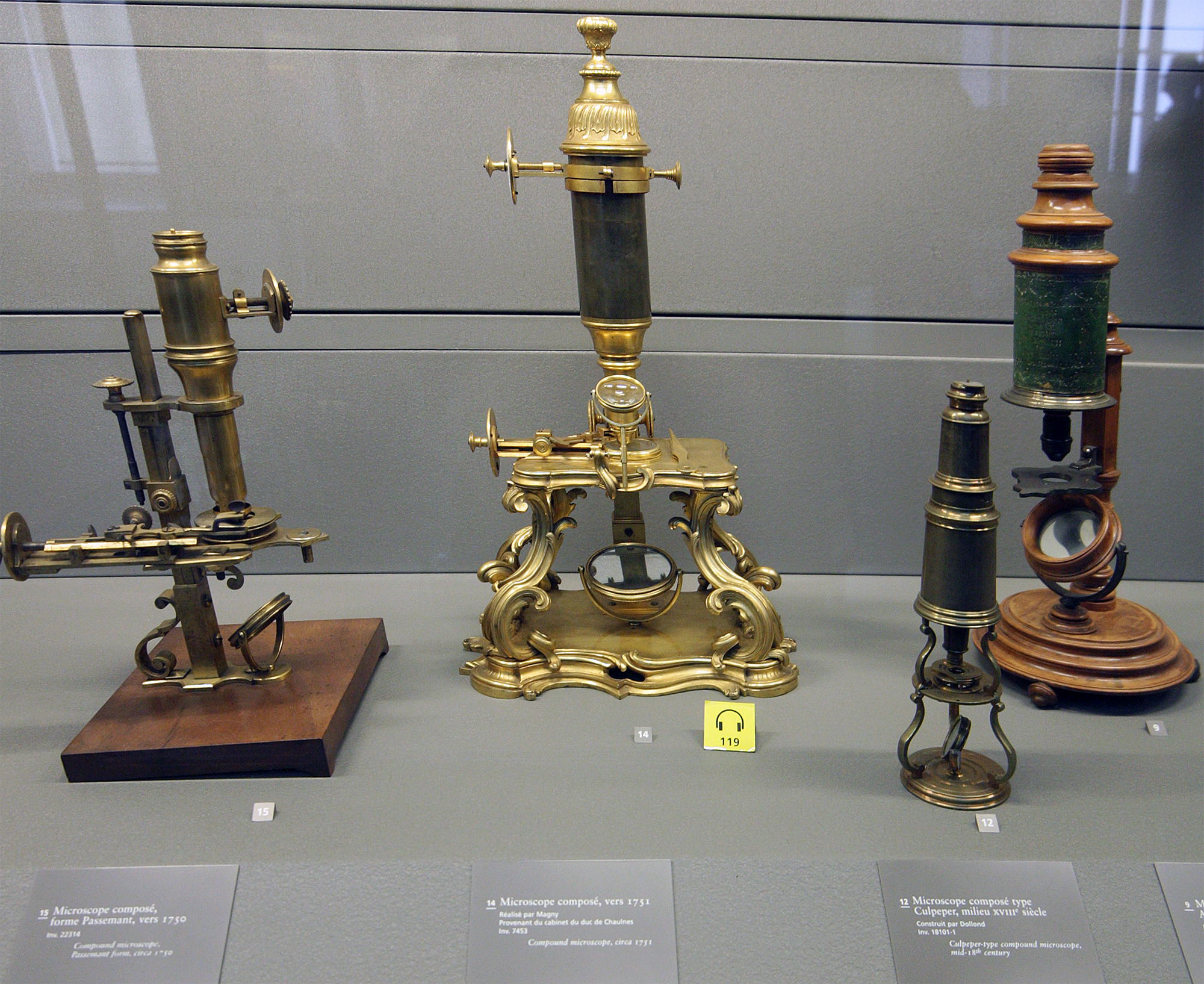
Microscope
A microscope () is a laboratory equipment, laboratory instrument used to examine objects that are too small to be seen by the naked eye. Microscopy is the science of investigating small objects and structures using a microscope. Microscopic means being invisible to the eye unless aided by a microscope. There are many types of microscopes, and they may be grouped in different ways. One way is to describe the method an instrument uses to interact with a sample and produce images, either by sending a beam of light or electrons through a sample in its optical path, by detecting fluorescence, photon emissions from a sample, or by scanning across and a short distance from the surface of a sample using a probe. The most common microscope (and the first to be invented) is the optical microscope, which uses lenses to refract visible light that passed through a microtome, thinly sectioned sample to produce an observable image. Other major types of microscopes are the fluorescence micro ... [...More Info...] [...Related Items...] OR: [Wikipedia] [Google] [Baidu] |
Hyperpigmentation
Hyperpigmentation, also known as the dark spots or circles on the skin, is the darkening of an area of Human skin, skin or nail (anatomy), nails caused by increased melanin. Causes Hyperpigmentation can be caused by sun damage, inflammation, or other skin injuries, including those related to acne vulgaris.James, William; Berger, Timothy; Elston, Dirk (2005). ''Andrews' Diseases of the Skin: Clinical Dermatology''. (10th ed.). Saunders. . People with darker skin tones are more prone to hyperpigmentation, especially with excess sun exposure. Many forms of hyperpigmentation are caused by an excess production of melanin. Hyperpigmentation can be diffuse or focal, affecting such areas as the face and the back of the hands. Melanin is produced by melanocytes at the lower layer of the epidermis. Melanin is a class of pigment responsible for producing color in the body in places such as the eyes, skin, and hair. The process of melanin synthesis (melanogenesis) starts with the oxidation ... [...More Info...] [...Related Items...] OR: [Wikipedia] [Google] [Baidu] |
Melanocytic Nevus
A melanocytic nevus (also known as nevocytic nevus, nevus-cell nevus, and commonly as a mole) is usually a Malignancy, noncancerous condition of pigment-producing Human skin, skin cells. It is a type of melanocytic tumor that contains nevus cells. A mole can be either subdermal (under the skin) or a pigmented growth on the skin, formed mostly of a type of cell known as a melanocyte. The high concentration of the body's pigmenting agent, melanin, is responsible for their dark color. Moles are a member of the family of skin lesions known as nevi (singular "nevus"), occurring commonly in humans. Some sources equate the term "mole" with "melanocytic nevus", but there are also sources that equate the term "mole" with any nevus form. The majority of moles appear during the first 2 decades of a person's life, with about 1 in every 100 babies being born with moles. Acquired moles are a form of Benign tumor, benign neoplasm, while congenital moles, or congenital nevi, are considered a min ... [...More Info...] [...Related Items...] OR: [Wikipedia] [Google] [Baidu] |
Conjunctiva
In the anatomy of the eye, the conjunctiva (: conjunctivae) is a thin mucous membrane that lines the inside of the eyelids and covers the sclera (the white of the eye). It is composed of non-keratinized, stratified squamous epithelium with goblet cells, stratified columnar epithelium and stratified cuboidal epithelium (depending on the zone). The conjunctiva is highly Angiogenesis, vascularised, with many microvessels easily accessible for imaging studies. Structure The conjunctiva is typically divided into three parts: Blood supply Blood to the bulbar conjunctiva is primarily derived from the ophthalmic artery. The blood supply to the palpebral conjunctiva (the eyelid) is derived from the external carotid artery. However, the circulations of the bulbar conjunctiva and palpebral conjunctiva are linked, so both bulbar conjunctival and palpebral conjunctival vessels are supplied by both the ophthalmic artery and the external carotid artery, to varying extents. Nerve supply Se ... [...More Info...] [...Related Items...] OR: [Wikipedia] [Google] [Baidu] |
Choroid
The choroid, also known as the choroidea or choroid coat, is a part of the uvea, the vascular layer of the eye. It contains connective tissues, and lies between the retina and the sclera. The human choroid is thickest at the far extreme rear of the eye (at 0.2 mm), while in the outlying areas it narrows to 0.1 mm. The choroid provides oxygen and nourishment to the outer layers of the retina. Along with the ciliary body and iris, the choroid forms the uveal tract. The structure of the choroid is generally divided into four layers (classified in order of furthest away from the retina to closest): *Haller's layer – outermost layer of the choroid consisting of larger diameter blood vessels; * Sattler's layer – layer of medium diameter blood vessels; * Choriocapillaris – layer of capillaries; and * Bruch's membrane (synonyms: Lamina basalis, Complexus basalis, Lamina vitra) – innermost layer of the choroid. Blood supply There are two circulations of the eye: ... [...More Info...] [...Related Items...] OR: [Wikipedia] [Google] [Baidu] |


