|
Arterial Aneurysm
An aneurysm is an outward bulging, likened to a bubble or balloon, caused by a localized, abnormal, weak spot on a blood vessel wall. Aneurysms may be a result of a hereditary condition or an acquired disease. Aneurysms can also be a nidus (starting point) for clot formation (thrombosis) and embolization. As an aneurysm increases in size, the risk of rupture increases, which could lead to uncontrolled bleeding. Although they may occur in any blood vessel, particularly lethal examples include aneurysms of the circle of Willis in the brain, aortic aneurysms affecting the thoracic aorta, and abdominal aortic aneurysms. Aneurysms can arise in the heart itself following a heart attack, including both ventricular and atrial septal aneurysms. There are congenital atrial septal aneurysms, a rare heart defect. Etymology The word is from Greek: ἀνεύρυσμα, aneurysma, "dilation", from ἀνευρύνειν, aneurynein, "to dilate". Classification Aneurysms are classified by ... [...More Info...] [...Related Items...] OR: [Wikipedia] [Google] [Baidu] |
Vascular Surgery
Vascular surgery is a surgical subspecialty in which vascular diseases involving the arteries, veins, or lymphatic vessels, are managed by medical therapy, minimally-invasive catheter procedures and surgical reconstruction. The specialty evolved from general surgery, general and cardiac surgery, cardiovascular surgery where it refined the management of just the vessels, no longer treating the heart or other organs. Modern vascular surgery includes open surgery techniques, endovascular (minimally invasive) techniques and medical management of vascular diseases - unlike the parent specialities. The vascular surgeon is trained in the diagnosis and management of diseases affecting all parts of the vascular system excluding the coronaries and intracranial vasculature. Vascular surgeons also are called to assist other physicians to carry out surgery near vessels, or to salvage vascular injuries that include hemorrhage control, dissection, occlusion or simply for safe exposure of vascul ... [...More Info...] [...Related Items...] OR: [Wikipedia] [Google] [Baidu] |
Atherosclerosis
Atherosclerosis is a pattern of the disease arteriosclerosis, characterized by development of abnormalities called lesions in walls of arteries. This is a chronic inflammatory disease involving many different cell types and is driven by elevated blood levels of cholesterol. These lesions may lead to narrowing of the arterial walls due to buildup of atheromatous plaques. At the onset, there are usually no symptoms, but if they develop, symptoms generally begin around middle age. In severe cases, it can result in coronary artery disease, stroke, peripheral artery disease, or kidney disorders, depending on which body part(s) the affected arteries are located in the body. The exact cause of atherosclerosis is unknown and is proposed to be multifactorial. Risk factors include dyslipidemia, abnormal cholesterol levels, elevated levels of inflammatory biomarkers, high blood pressure, diabetes, smoking (both active and passive smoking), obesity, genetic factors, family history, lifes ... [...More Info...] [...Related Items...] OR: [Wikipedia] [Google] [Baidu] |
Common Iliac Artery
The common iliac artery is a large artery of the abdomen paired on each side. It originates from the aortic bifurcation at the level of the 4th lumbar vertebra. It ends in front of the sacroiliac joint, one on either side, and each bifurcates into the external and internal iliac arteries. Structure The common iliac artery are about 4 cm long in adults and more than a centimeter in diameter. It begins as a branch of the aorta. This is at the level of the fourth lumbar vertebra. It runs inferolaterally, along the medial border of the psoas muscles. It bifurcates into the external iliac artery and the internal iliac artery at the pelvic brim, in front of the sacroiliac joints. The common iliac artery, and all of its branches, exist as paired structures (that is to say, there is one on the left side and one on the right). The distribution of the common iliac artery is basically the pelvis and lower limb (as the femoral artery) on the corresponding side. Relations Both ... [...More Info...] [...Related Items...] OR: [Wikipedia] [Google] [Baidu] |
Abdominal Aorta
In human anatomy, the abdominal aorta is the largest artery in the abdominal cavity. As part of the aorta, it is a direct continuation of the descending aorta (of the thorax). Structure The abdominal aorta begins at the level of the diaphragm, crossing it via the aortic hiatus, technically behind the diaphragm, at the vertebral level of T12. It travels down the posterior wall of the abdomen, anterior to the vertebral column. It thus follows the curvature of the lumbar vertebrae, that is, convex anteriorly. The peak of this convexity is at the level of the third lumbar vertebra (L3). It runs parallel to the inferior vena cava, which is located just to the right of the abdominal aorta, and becomes smaller in diameter as it gives off branches. This is thought to be due to the large size of its principal branches. At the 11th rib, the diameter is 122mm long and 55mm wide and this is because of the constant pressure. The abdominal aorta is clinically divided into 2 segments: # Th ... [...More Info...] [...Related Items...] OR: [Wikipedia] [Google] [Baidu] |
Aortic Arch
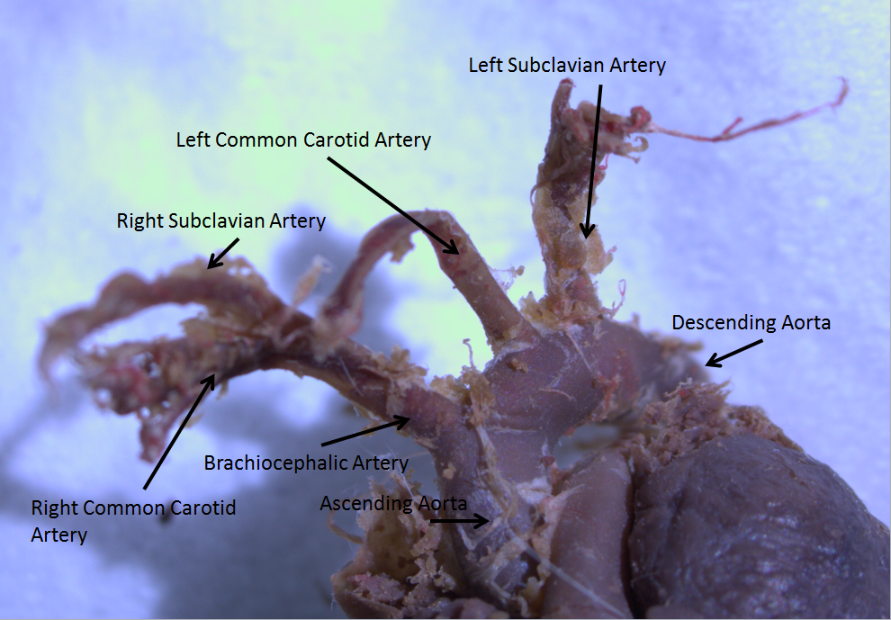
The aortic arch, arch of the aorta, or transverse aortic arch () is the part of the aorta between the ascending and descending aorta. The arch travels backward, so that it ultimately runs to the left of the trachea. Structure The aorta begins at the level of the upper border of the second/third sternocostal articulation of the right side, behind the ventricular outflow tract and pulmonary trunk. The right atrial appendage overlaps it. The first few centimeters of the ascending aorta and pulmonary trunk lies in the same pericardial sheath and runs at first upward, arches over the pulmonary trunk, right pulmonary artery, and right main bronchus to lie behind the right second coastal cartilage. The right lung and sternum lies anterior to the aorta at this point. The aorta then passes posteriorly and to the left, anterior to the trachea, and arches over left main bronchus and left pulmonary artery, and reaches to the left side of the T4 vertebral body. Apart from T4 vertebral ... [...More Info...] [...Related Items...] OR: [Wikipedia] [Google] [Baidu] |
Endovascular Coiling
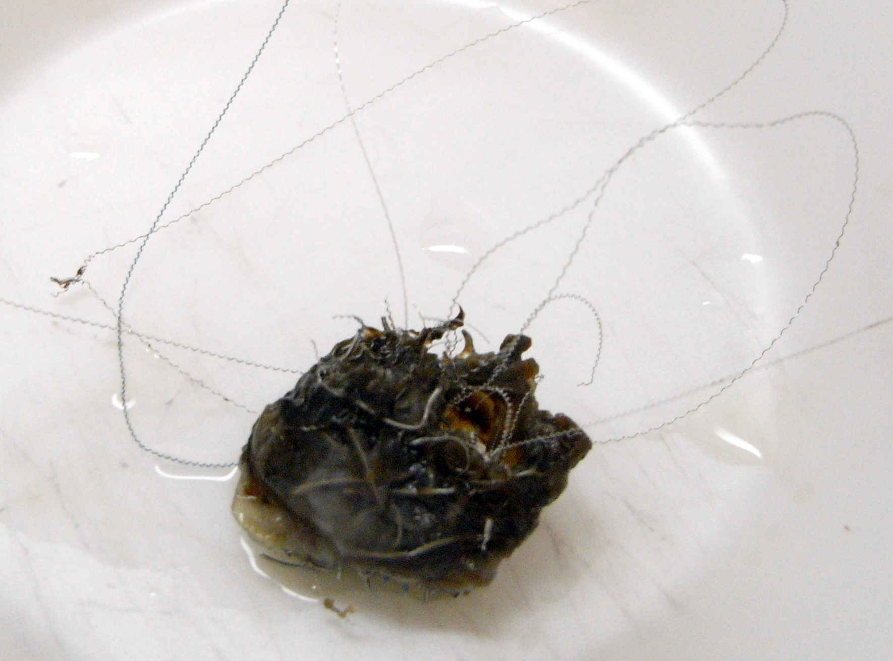
Endovascular coiling is an Interventional neuroradiology, endovascular treatment for intracranial aneurysms and bleeding throughout the body. The procedure reduces blood circulation to an aneurysm or blood vessel through the implantation of detachable platinum wires, with the clinician inserting one or more into the blood vessel or aneurysm until it is determined that blood flow is no longer occurring within the space. It is one of two main treatments for cerebral aneurysms, the other being surgical clipping. Medical uses Endovascular coiling is typically used to treat Intracranial aneurysm, cerebral aneurysms. The main goal is prevention of rupture in unruptured aneurysms, and prevention of rebleeding in ruptured aneurysms by limiting blood circulation to the aneurysm space. Clinically, packing density is recommended to be 20-30% or more of the aneurysm's volume, typically requiring deployment of multiple wires. Higher volumes may be difficult due to the delicate nature of the a ... [...More Info...] [...Related Items...] OR: [Wikipedia] [Google] [Baidu] |
Arterial Aneurysm
An aneurysm is an outward bulging, likened to a bubble or balloon, caused by a localized, abnormal, weak spot on a blood vessel wall. Aneurysms may be a result of a hereditary condition or an acquired disease. Aneurysms can also be a nidus (starting point) for clot formation (thrombosis) and embolization. As an aneurysm increases in size, the risk of rupture increases, which could lead to uncontrolled bleeding. Although they may occur in any blood vessel, particularly lethal examples include aneurysms of the circle of Willis in the brain, aortic aneurysms affecting the thoracic aorta, and abdominal aortic aneurysms. Aneurysms can arise in the heart itself following a heart attack, including both ventricular and atrial septal aneurysms. There are congenital atrial septal aneurysms, a rare heart defect. Etymology The word is from Greek: ἀνεύρυσμα, aneurysma, "dilation", from ἀνευρύνειν, aneurynein, "to dilate". Classification Aneurysms are classified by ... [...More Info...] [...Related Items...] OR: [Wikipedia] [Google] [Baidu] |
Coronary Angiogram
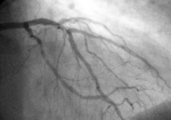
A coronary catheterization is a minimally invasive procedure to access the coronary circulation and blood filled chambers of the heart using a catheter. It is performed for both diagnostic and interventional (treatment) purposes. Coronary catheterization is one of the several cardiology diagnostic tests and procedures. Specifically, through the injection of a liquid radiocontrast agent and illumination with X-rays, angiocardiography allows the recognition of Vascular occlusion, occlusion, stenosis, restenosis, thrombosis or aneurysmal enlargement of the coronary artery lumen (anatomy), lumens; heart chamber size; heart muscle contraction performance; and some aspects of heart valve function. Important internal heart and lung blood pressures, not measurable from outside the body, can be accurately measured during the test. The relevant problems that the test deals with most commonly occur as a result of advanced atherosclerosis – atheroma activity within the wall of the coronary ... [...More Info...] [...Related Items...] OR: [Wikipedia] [Google] [Baidu] |
Percutaneous
{{More citations needed, date=January 2021 In surgery, a percutaneous procedurei.e. Granger et al., 2012 is any medical procedure or method where access to inner organs or other tissue is done via needle-puncture of the skin, rather than by using an "open" approach where inner organs or tissue are exposed (typically with the use of a scalpel). The percutaneous approach is commonly used in vascular procedures such as angioplasty and stenting. This involves a needle catheter getting access to a blood vessel, followed by the introduction of a wire through the lumen (pathway) of the needle. It is over this wire that other catheters can be placed into the blood vessel. This technique is known as the modified Seldinger technique. More generally, "percutaneous", via its Latin roots means, 'by way of the skin'. An example would be percutaneous drug absorption from topical medications. More often, percutaneous is typically used in reference to placement of medical devices using ... [...More Info...] [...Related Items...] OR: [Wikipedia] [Google] [Baidu] |
Physical Trauma
Injury is physiology, physiological damage to the living tissue of any organism, whether Injury in humans, in humans, Injury in animals, in other animals, or Injury in plants, in plants. Injuries can be caused in many ways, including mechanically with penetrating trauma, penetration by sharp objects such as Tooth, teeth or blunt trauma, with blunt objects, by heat or cold, or by venoms and biotoxins. Injury prompts an Inflammation, inflammatory response in many taxa of animals; this prompts wound healing. In both plants and animals, substances are often released to help to occlude the wound, limiting loss of fluids and the entry of pathogens such as bacteria. Many organisms secrete antimicrobial chemicals which limit wound infection; in addition, animals have a variety of immune responses for the same purpose. Both plants and animals have regrowth mechanisms which may result in complete or partial healing over the injury. Cells too can Cell damage, repair damage to a certain de ... [...More Info...] [...Related Items...] OR: [Wikipedia] [Google] [Baidu] |
Thrombus
A thrombus ( thrombi) is a solid or semisolid aggregate from constituents of the blood (platelets, fibrin, red blood cells, white blood cells) within the circulatory system during life. A blood clot is the final product of the blood coagulation step in hemostasis in or out of the circulatory system. There are two components to a thrombus: aggregated platelets and red blood cells that form a plug, and a mesh of cross-linked fibrin protein. The substance making up a thrombus is sometimes called cruor. A thrombus is a healthy response to injury intended to stop and prevent further bleeding, but can be harmful in thrombosis, when a clot obstructs blood flow through a healthy blood vessel in the circulatory system. In the microcirculation consisting of the very small and smallest blood vessels the capillaries, tiny thrombi known as microclots can obstruct the flow of blood in the capillaries. This can cause a number of problems particularly affecting the pulmonary alveolus, alveoli ... [...More Info...] [...Related Items...] OR: [Wikipedia] [Google] [Baidu] |
Pseudoaneurysm
A pseudoaneurysm, also known as a false aneurysm, is a locally contained hematoma outside an artery or the heart due to damage to the vessel wall. The injury passes through all three layers of the arterial wall, causing a leak, which is contained by a new, weak "wall" formed by the products of the clotting cascade. A pseudoaneurysm (PSA) does not contain any layer of the vessel wall. This differentiates it from a true aneurysm, which is contained by all three layers of the vessel wall, and a dissecting aneurysm, which has a breach in the innermost layer of an artery and subsequent dissection/separation of the tunica intima from the tunica media. A pseudoaneurysm, being associated with a vessel, can be pulsatile; it may be confused with a true aneurysm or dissecting aneurysm. The most common presentation of pseudoaneurysm is femoral artery pseudoaneurysm following access for an endovascular procedure, and this event may complicate up to 8% of vascular interventional proc ... [...More Info...] [...Related Items...] OR: [Wikipedia] [Google] [Baidu] |


