|
Aplasia
Aplasia (; from Greek ''a'', "not", "no" + ''plasis'', "formation") is a birth defect where an organ or tissue is wholly or largely absent. It is caused by a defect in a developmental process. Aplastic anemia is the failure of the body to produce blood cells. It may occur at any time, and has multiple causes. __TOC__ Types Pure red cell aplasia Pure red cell aplasia (PRCA) is caused by the selective destruction or inhibition of erythroid progenitor or precursor cells. It is characterized by anemia and reticulocytopenia and can be chronic or acute. Diamond–Blackfan anemia is a type of PRCA that occurs at birth. PRCA can be acquired as a primary disorder or as a result of another disorder. Immunosuppressive drugs, particularly corticosteroids, will usually result in a temporary or permanent remission. The final outcome is primarily determined by the underlying disorder. Aplasia cutis congenita Aplasia cutis congenita is a condition in which some or large portions of ... [...More Info...] [...Related Items...] OR: [Wikipedia] [Google] [Baidu] |
Aplasia Cutis Congenita
Aplasia cutis congenita is a rare disorder characterized by congenital absence of skin. Ilona J. Frieden classified ACC in 1986 into 9 groups on the basis of location of the lesions and associated congenital anomalies.Moss C, Shahidulla H. Naevi and other developmental defects. In: Burns T, Breathnach S, Cox N, Griffiths C, editors. Rook's Textbook of Dermatology. 8th ed. United Kingdom (UK): Wiley-Blackwell Publication; 2010. p. 18, 18.98-18. 106. The scalp is the most commonly involved area with lesser involvement of trunk and extremities. Frieden classified ACC with fetus papyraceus as type 5. This type presents as truncal ACC with symmetrical absence of skin in stellate or butterfly pattern with or without involvement of proximal limbs.Meena N, Saxena AK, Sinha S, Dixit N. Aplasia cutis congenita with fetus papyraceus. Indian J Paediatr Dermatol 2015;16:48-9. It is the most common congenital cicatricial alopecia, and is a congenital focal absence of epidermis with or without evi ... [...More Info...] [...Related Items...] OR: [Wikipedia] [Google] [Baidu] |
Reticulocytopenia
Reticulocytopenia is the medical term for an abnormal decrease in circulating red blood cell precursors ( reticulocytes) that can lead to anemia due to resulting low red blood cell (erythrocyte) production. Reticulocytopenia may be an isolated finding or it may not be associated with abnormalities in other hematopoietic cell lineages such as those that produce white blood cells (leukocytes) or platelets (thrombocytes), a decrease in all three of these lineages is referred to as pancytopenia. With isolated reticulocytopenia, the main cause is Parvovirus B19 infection of reticulocytes leading to transient anemia. In patients who rely on frequent red cell regeneration e.g. sickle cell disease, a reticulocytopenia can lead to a severe anemia due to the cessation in red cell production (erythropoiesis), referred to as aplastic crisis. If pancytopenia is present, bone marrow failure must be considered and evaluation for bone marrow failure syndromes or aplastic anemia must be pursued. T ... [...More Info...] [...Related Items...] OR: [Wikipedia] [Google] [Baidu] |
Birth Defect
A birth defect is an abnormal condition that is present at birth, regardless of its cause. Birth defects may result in disabilities that may be physical, intellectual, or developmental. The disabilities can range from mild to severe. Birth defects are divided into two main types: structural disorders in which problems are seen with the shape of a body part and functional disorders in which problems exist with how a body part works. Functional disorders include metabolic and degenerative disorders. Some birth defects include both structural and functional disorders. Birth defects may result from genetic or chromosomal disorders, exposure to certain medications or chemicals, or certain infections during pregnancy. Risk factors include folate deficiency, drinking alcohol or smoking during pregnancy, poorly controlled diabetes, and a mother over the age of 35 years old. Many birth defects are believed to involve multiple factors. Birth defects may be visible at birth or dia ... [...More Info...] [...Related Items...] OR: [Wikipedia] [Google] [Baidu] |
Pure Red Cell Aplasia
Pure red cell aplasia (PRCA) or erythroblastopenia refers to a type of aplastic anemia affecting the precursors to red blood cells but usually not to white blood cells. In PRCA, the bone marrow ceases to produce red blood cells. There are multiple etiologies that can cause PRCA. The condition has been first described by Paul Kaznelson in 1922. Signs and symptoms Signs and symptoms may include: * Pale appearance * Rapid heart rate * Fatigue Causes Causes of PRCA include: Diagnosis Treatment PRCA is considered an autoimmune disease as it will respond to immunosuppressant treatment such as cyclosporin in many patients, though this approach is not without risk. It has also been shown to respond to treatments with rituximab and tacrolimus Tacrolimus, sold under the brand name Prograf among others, is an immunosuppressive drug. After Allotransplantation, allogenic organ transplant, the risk of organ Transplant rejection, rejection is moderate. To lower the risk of or ... [...More Info...] [...Related Items...] OR: [Wikipedia] [Google] [Baidu] |
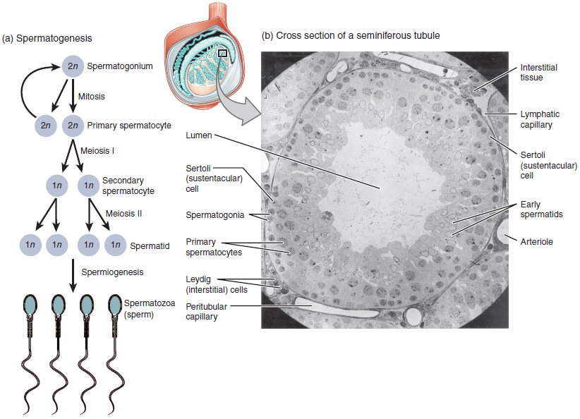
Seminiferous Tubule
Seminiferous tubules are located within the testicles, and are the specific location of meiosis, and the subsequent creation of male gametes, namely spermatozoa. Structure The epithelium of the tubule consists of a type of sustentacular cells known as Sertoli cells, which are tall, columnar type cells that line the tubule. In between the Sertoli cells are spermatogenic cells, which differentiate through meiosis to sperm cells. Sertoli cells function to nourish the developing sperm cells. They secrete androgen-binding protein, a binding protein which increases the concentration of testosterone. There are two types: convoluted and straight, convoluted toward the lateral side, and straight as the tubule comes medially to form ducts that will exit the testis. The seminiferous tubules are formed from the testis cords that develop from the primitive gonadal cords, formed from the gonadal ridge. Function Spermatogenesis, the process for producing spermatozoa, takes place in ... [...More Info...] [...Related Items...] OR: [Wikipedia] [Google] [Baidu] |
Sertoli Cell
Sertoli cells are a type of sustentacular "nurse" cell found in human testes which contribute to the process of spermatogenesis (the production of sperm) as a structural component of the seminiferous tubules. They are activated by follicle-stimulating hormone (FSH) secreted by the adenohypophysis and express FSH receptor on their membranes. History Sertoli cells are named after Enrico Sertoli, an Italian physiologist who discovered them while studying medicine at the University of Pavia, Italy. He published a description of his eponymous cell in 1865. The cell was discovered by Sertoli with a Belthle microscope which had been purchased in 1862. In the 1865 publication, his first description used the terms "tree-like cell" or "stringy cell"; most importantly, he referred to these as "mother cells". Other scientists later used Enrico's family name to label these cells in publications, beginning in 1888. As of 2006, two textbooks that are devoted specifically to the Sertoli cell ... [...More Info...] [...Related Items...] OR: [Wikipedia] [Google] [Baidu] |
Blood–testis Barrier
The blood–testis barrier is a physical barrier between the blood vessels and the seminiferous tubules of the animal testes. The name "blood-testis barrier" is misleading as it is not a blood-organ barrier in a strict sense, but is formed between Sertoli cells of the seminiferous tubule and isolates the further developed stages of germ cells from the blood. A more correct term is the Sertoli cell barrier (SCB). Structure The walls of seminiferous tubules are lined with primitive germ layer cells and by Sertoli cells. The barrier is formed by tight junctions, adherens junctions and gap junctions between the Sertoli cells, which are sustentacular cells (supporting cells) of the seminiferous tubules, and divides the seminiferous tubule into a basal compartment (outer side of the tubule, in contact with blood and lymph) and an endoluminal compartment (inner side of the tubule, isolated from blood and lymph). The tight junctions are formed by intercellular adhesion molecules in betwe ... [...More Info...] [...Related Items...] OR: [Wikipedia] [Google] [Baidu] |
Follicle-stimulating Hormone
Follicle-stimulating hormone (FSH) is a gonadotropin, a glycoprotein polypeptide hormone. FSH is synthesized and secreted by the gonadotropic cells of the anterior pituitary gland and regulates the development, growth, puberty, pubertal maturation, and reproductive processes of the body. FSH and luteinizing hormone (LH) work together in the reproductive system. Structure FSH is a 35.5 kDa glycoprotein protein dimer, heterodimer, consisting of two polypeptide units, alpha and beta. Its structure is similar to those of luteinizing hormone (LH), thyroid-stimulating hormone (TSH), and human chorionic gonadotropin (hCG). The chorionic gonadotropin alpha, alpha subunits of the glycoproteins LH, FSH, TSH, and hCG are identical and consist of 96 amino acids, while the beta subunits vary. Both subunits are required for biological activity. FSH has a beta subunit of 111 amino acids (FSH β), which confers its specific biologic action, and is responsible for interaction with the follicl ... [...More Info...] [...Related Items...] OR: [Wikipedia] [Google] [Baidu] |
Greek Language
Greek (, ; , ) is an Indo-European languages, Indo-European language, constituting an independent Hellenic languages, Hellenic branch within the Indo-European language family. It is native to Greece, Cyprus, Italy (in Calabria and Salento), southern Albania, and other regions of the Balkans, Caucasus, the Black Sea coast, Asia Minor, and the Eastern Mediterranean. It has the list of languages by first written accounts, longest documented history of any Indo-European language, spanning at least 3,400 years of written records. Its writing system is the Greek alphabet, which has been used for approximately 2,800 years; previously, Greek was recorded in writing systems such as Linear B and the Cypriot syllabary. The Greek language holds a very important place in the history of the Western world. Beginning with the epics of Homer, ancient Greek literature includes many works of lasting importance in the European canon. Greek is also the language in which many of the foundational texts ... [...More Info...] [...Related Items...] OR: [Wikipedia] [Google] [Baidu] |
Azoospermia
Azoospermia is the medical condition of a man whose semen contains no sperm. It is associated with male infertility, but many forms are amenable to medical treatment. In humans, azoospermia affects about 1% of the male population and may be seen in up to 20% of male infertility situations in Canada. In a non-pathological context, azoospermia is also the intended result of a vasectomy. Classification Azoospermia can be classified into three major types. Many conditions listed may also cause various degrees of oligospermia rather than azoospermia. Pretesticular and testicular azoospermia are known as non-obstructive azoospermia, whereas post-testicular azoospermia is considered obstructive. Pretesticular Inadequate stimulation of normal testicles and the genital tract characterizes pretesticular azoospermia. Typically, follicle-stimulating hormone (FSH) levels are low (hypogonadotropic), commensurate with inadequate stimulation of the testes to produce sperm. Examples include ... [...More Info...] [...Related Items...] OR: [Wikipedia] [Google] [Baidu] |
Spermatogenesis
Spermatogenesis is the process by which haploid spermatozoa develop from germ cells in the seminiferous tubules of the testicle. This process starts with the Mitosis, mitotic division of the stem cells located close to the basement membrane of the tubules. These cells are called Spermatogonial Stem Cells, spermatogonial stem cells. The mitotic division of these produces two types of cells. Type A cells replenish the stem cells, and type B cells differentiate into primary spermatocytes. The primary spermatocyte divides meiotically (Meiosis I) into two secondary spermatocytes; each secondary spermatocyte divides into two equal haploid spermatids by Meiosis II. The spermatids are transformed into spermatozoa (sperm) by the process of spermiogenesis. These develop into mature spermatozoa, also known as sperm, sperm cells. Thus, the primary spermatocyte gives rise to two cells, the secondary spermatocytes, and the two secondary spermatocytes by their subdivision produce four spermatoz ... [...More Info...] [...Related Items...] OR: [Wikipedia] [Google] [Baidu] |


