|
Ansa Lenticularis
The ansa lenticularis (''ansa lentiformis'' in older texts) is a part of the brain, making up the superior layer of the substantia innominata. Its fibers, derived from the medullary lamina of the lentiform nucleus, pass medially to end in the thalamus and subthalamic region, while others are said to end in the tegmentum and red nucleus. It is classified by NeuroNames as part of the subthalamus The subthalamus or ventral thalamus is a part of the diencephalon. Its most prominent structure is the subthalamic nucleus. The subthalamus connects to the globus pallidus, a subcortical nucleus of the basal ganglia. Structure The subthalamus .... References External links * {{Authority control Thalamic connections Basal ganglia connections ... [...More Info...] [...Related Items...] OR: [Wikipedia] [Google] [Baidu] |
Substantia Innominata
The substantia innominata, also innominate substance or substantia innominata of Meynert (Latin for unnamed substance), is a series of layers in the human brain consisting partly of gray and partly of white matter, which lies below the anterior part of the thalamus and lentiform nucleus. It is included as part of the anterior perforated substance (as it appears to be perforated by many holes which are actually blood vessels). It is part of the basal forebrain structures and includes the nucleus basalis. A portion of the substantia innominata, below the globus pallidus is considered as part of the extended amygdala. Layers It consists of three layers, superior, middle, and inferior. * The ''superior layer'' is named the ansa lenticularis, and its fibers, derived from the medullary lamina of the lentiform nucleus, pass medially to end in the thalamus and subthalamic region, while others are said to end in the tegmentum and red nucleus. * The ''middle layer'' consists of nerve cel ... [...More Info...] [...Related Items...] OR: [Wikipedia] [Google] [Baidu] |
Lentiform Nucleus
The lentiform nucleus (or lentiform complex, lenticular nucleus, or lenticular complex) are the putamen (laterally) and the globus pallidus (medially), collectively. Due to their proximity, these two structures were formerly considered one, however, the two are separated by a thin layer of white matter—the external medullary lamina—and are functionally and connectionally distinct. The lentiform nucleus is a large, lens-shaped mass of gray matter just lateral to the internal capsule. It forms part of the basal ganglia. With the caudate nucleus, it forms the dorsal striatum. Structure When divided horizontally, it exhibits, to some extent, the appearance of a biconvex lens, while a coronal section of its central part presents a somewhat triangular outline. It is shorter than the caudate nucleus and does not extend as far forward. Relations It is deep/medial to the insular cortex, with which it is coextensive; the two are separated by intervening structures. It is lateral to ... [...More Info...] [...Related Items...] OR: [Wikipedia] [Google] [Baidu] |
Thalamus
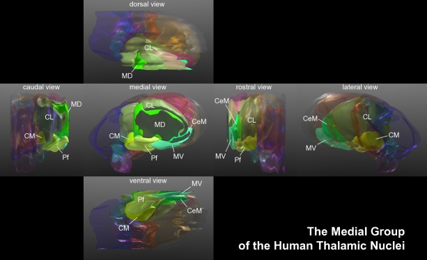
The thalamus (: thalami; from Greek language, Greek Wikt:θάλαμος, θάλαμος, "chamber") is a large mass of gray matter on the lateral wall of the third ventricle forming the wikt:dorsal, dorsal part of the diencephalon (a division of the forebrain). Nerve fibers project out of the thalamus to the cerebral cortex in all directions, known as the thalamocortical radiations, allowing hub (network science), hub-like exchanges of information. It has several functions, such as the relaying of sensory neuron, sensory and motor neuron, motor signals to the cerebral cortex and the regulation of consciousness, sleep, and alertness. Anatomically, the thalami are paramedian symmetrical structures (left and right), within the vertebrate brain, situated between the cerebral cortex and the midbrain. It forms during embryonic development as the main product of the diencephalon, as first recognized by the Swiss embryologist and anatomist Wilhelm His Sr. in 1893. Anatomy The thalami ar ... [...More Info...] [...Related Items...] OR: [Wikipedia] [Google] [Baidu] |
Tegmentum
The tegmentum (from Latin for "covering") is a general area within the brainstem. The tegmentum is the ventral part of the midbrain and the tectum is the dorsal part of the midbrain. It is located between the ventricular system and distinctive basal or ventral structures at each level. It forms the floor of the midbrain (mesencephalon) whereas the tectum forms the ceiling. It is a multisynaptic network of neurons that is involved in many subconscious homeostatic and reflexive pathways. It is a motor center that relays inhibitory signals to the thalamus and basal nuclei preventing unwanted body movement. The tegmentum area includes various different structures, such as the rostral end of the reticular formation, several nuclei controlling eye movements, the periaqueductal gray matter, the red nucleus, the substantia nigra, and the ventral tegmental area. The tegmentum is the location of several cranial nerve nuclei. The nuclei of CN III and IV are located in the tegmentum port ... [...More Info...] [...Related Items...] OR: [Wikipedia] [Google] [Baidu] |
Red Nucleus
The red nucleus or nucleus ruber is a structure in the rostral midbrain involved in motor coordination. The red nucleus is pale pink, which is believed to be due to the presence of iron in at least two different forms: hemoglobin and ferritin. The structure is located in the midbrain tegmentum next to the substantia nigra and comprises caudal magnocellular and rostral parvocellular components. The red nucleus and substantia nigra are subcortical centers of the extrapyramidal motor system. Function In a vertebrate without a significant corticospinal tract, gait is mainly controlled by the red nucleus. However, in primates, where the corticospinal tract is dominant, the rubrospinal tract may be regarded as vestigial in motor function. Therefore, the red nucleus is less important in primates than in many other mammals. Nevertheless, the crawling of babies is controlled by the red nucleus, as is arm swinging in typical walking. The red nucleus may play an additional role ... [...More Info...] [...Related Items...] OR: [Wikipedia] [Google] [Baidu] |
NeuroNames
''NeuroNames'' is an integrated nomenclature for structures in the brain and spinal cord of the four species most studied by neuroscientists: human, macaque, rat and mouse. It offers a standard, controlled vocabulary of common names for structures, which is suitable for unambiguous neuroanatomical indexing of information in digital databases. Terms in the standard vocabulary have been selected for ease of pronunciation, mnemonic value, and frequency of use in recent neuroscientific publications. Structures and their relations to each other are defined in terms of the standard vocabulary. Currently NeuroNames contains standard names, synonyms and definitions of some 2,500 neuroanatomical entities. The nomenclature is maintained by the University of Washington and is the core component of a tool called "BrainInfo". BrainInfo helps one identify structures in the brain. One can either search by a structure name or locate the structure in a brain atlas and get information such as it ... [...More Info...] [...Related Items...] OR: [Wikipedia] [Google] [Baidu] |
Subthalamus
The subthalamus or ventral thalamus is a part of the diencephalon. Its most prominent structure is the subthalamic nucleus. The subthalamus connects to the globus pallidus, a subcortical nucleus of the basal ganglia. Structure The subthalamus is located ventral to the thalamus, medial to the internal capsule and lateral to the hypothalamus. It is a region formed by several grey matter nuclei and their associated white matter structures, namely: *The subthalamic nucleus, whose neurons contain glutamate and have excitatory effects over neurons of globus pallidus and substantia nigra * Zona incerta, located between fields of Forel H1 and H2. It is continuous with the thalamic reticular nucleus and receives input from the precentral cortex. * Subthalamic fasciculus, formed by fibers that connect the globus pallidus with the subthalamic nucleus * Fields of Forel * Ansa lenticularis During development the subthalamus is continuous with the hypothalamus, but is separated by whi ... [...More Info...] [...Related Items...] OR: [Wikipedia] [Google] [Baidu] |
Thalamic Connections
The thalamus (: thalami; from Greek θάλαμος, "chamber") is a large mass of gray matter on the lateral wall of the third ventricle forming the dorsal part of the diencephalon (a division of the forebrain). Nerve fibers project out of the thalamus to the cerebral cortex in all directions, known as the thalamocortical radiations, allowing hub-like exchanges of information. It has several functions, such as the relaying of sensory and motor signals to the cerebral cortex and the regulation of consciousness, sleep, and alertness. Anatomically, the thalami are paramedian symmetrical structures (left and right), within the vertebrate brain, situated between the cerebral cortex and the midbrain. It forms during embryonic development as the main product of the diencephalon, as first recognized by the Swiss embryologist and anatomist Wilhelm His Sr. in 1893. Anatomy The thalami are paired structures of gray matter about four centimetres long and ovoid in appearance, located in ... [...More Info...] [...Related Items...] OR: [Wikipedia] [Google] [Baidu] |


