|
Ankycorbin
Ankycorbin is an ankyrin repeat and Coiled coil, coiled-coil domain containing protein that in humans is encoded by the ''RAI14'' gene. It is expressed in a variety of human tissues and is thought to play a role in actin regulation of ectoplasmic specialization, establishment of sperm polarity and sperm adhesion. It may also promote the integrity of Sertoli cell tight junctions at the blood testis barrier. Gene Location RAI14 contains 15 exons and is located on the plus strand of chromosome 5, chromosome number 5 at position 5p13.2. It spans 5,068 base pairs from position 34,656,328 to 34,832,612. Gene Neighborhood Genes TTC23L and LOC105374721 neighbor RAI14 on chromosome 5. Expression RAI14 is expressed within a wide range of human tissues. Some areas of the highest expression by TPM (transcripts per million) include tissues of the endometrium, smooth muscle, cervix, cervix, testis, and spleen. Within the human brain, RAI14 expression is abundant in the area around the ... [...More Info...] [...Related Items...] OR: [Wikipedia] [Google] [Baidu] |
RAI14 Expression Via The Human Protein Atlas
Ankycorbin is a protein that in humans is encoded by the RAI14 gene. Ankycorbin has been associated with the cortical actin cytoskeleton structures in terminal web, cell-cell adhesion sites as well as stress fibre Stress fibers are contractile actin bundles found in non-muscle cells. They are composed of actin (microfilaments) and non-muscle myosin II (NMMII), and also contain various crosslinking proteins, such as α-actinin, to form a highly regulated ...s. References Further reading * * * * {{gene-5-stub ... [...More Info...] [...Related Items...] OR: [Wikipedia] [Google] [Baidu] |
RAI14
Ankycorbin is a protein that in humans is encoded by the RAI14 gene. Ankycorbin has been associated with the cortical actin cytoskeleton structures in terminal web, cell-cell adhesion sites as well as stress fibre Stress fibers are contractile actin bundles found in non-muscle cells. They are composed of actin (microfilaments) and non-muscle myosin II (NMMII), and also contain various crosslinking proteins, such as α-actinin, to form a highly regulated ...s. References Further reading * * * * {{gene-5-stub ... [...More Info...] [...Related Items...] OR: [Wikipedia] [Google] [Baidu] |
RAI14 Expression Within Human Brain
Ankycorbin is a protein that in humans is encoded by the RAI14 gene. Ankycorbin has been associated with the cortical actin cytoskeleton structures in terminal web, cell-cell adhesion sites as well as stress fibre Stress fibers are contractile actin bundles found in non-muscle cells. They are composed of actin (microfilaments) and non-muscle myosin II (NMMII), and also contain various crosslinking proteins, such as α-actinin, to form a highly regulated ...s. References Further reading * * * * {{gene-5-stub ... [...More Info...] [...Related Items...] OR: [Wikipedia] [Google] [Baidu] |
Ankyrin Repeat
The ankyrin repeat is a 33-residue motif in proteins consisting of two alpha helices separated by loops, first discovered in signaling proteins in yeast Cdc10 and ''Drosophila'' Notch. Domains consisting of ankyrin tandem repeats mediate protein–protein interactions and are among the most common structural motifs in known proteins. They appear in bacterial, archaeal, and eukaryotic proteins, but are far more common in eukaryotes. Ankyrin repeat proteins, though absent in most viruses, are common among poxviruses. Most proteins that contain the motif have four to six repeats, although its namesake ankyrin contains 24, and the largest known number of repeats is 34, predicted in a protein expressed by '' Giardia lamblia''. Ankyrin repeats typically fold together to form a single, linear solenoid structure called ankyrin repeat domains. These domains are one of the most common protein–protein interaction platforms in nature. They occur in a large number of functionally div ... [...More Info...] [...Related Items...] OR: [Wikipedia] [Google] [Baidu] |
Dalmatian Pelican
The Dalmatian pelican (''Pelecanus crispus'') is the largest member of the pelican family, and perhaps the world's largest freshwater bird, although rivaled in weight and length by the largest swans. They are elegant soaring birds, with wingspans rivaling those of the great albatrosses, and their flocks fly in graceful synchrony. With a range spanning across much of Central Eurasia, from the Mediterranean in the West to the Taiwan Strait in the East, and from the Persian Gulf in the South to Siberia in the North, it is a short-to-medium-distance migrant between breeding and overwintering areas. No subspecies are known to exist over its wide range, but based on size differences, a Pleistocene paleosubspecies, ''P. c. palaeocrispus,'' has been described from fossils recovered at Binagady, Azerbaijan. As with other pelicans, the males are larger than the females, and likewise their diet is mainly fish. Their curly nape feathers, grey legs and silvery-white plumage are dist ... [...More Info...] [...Related Items...] OR: [Wikipedia] [Google] [Baidu] |
Seminiferous Tubule
Seminiferous tubules are located within the testes, and are the specific location of meiosis, and the subsequent creation of male gametes, namely spermatozoa. Structure The epithelium of the tubule consists of a type of sustentacular cells known as Sertoli cells, which are tall, columnar type cells that line the tubule. In between the Sertoli cells are spermatogenic cells, which differentiate through meiosis to sperm cells. Sertoli cells function to nourish the developing sperm cells. They secrete androgen-binding protein, a binding protein which increases the concentration of testosterone. There are two types: convoluted and straight, convoluted toward the lateral side, and straight as the tubule comes medially to form ducts that will exit the testis. The seminiferous tubules are formed from the testis cords that develop from the primitive gonadal cords, formed from the gonadal ridge. Function Spermatogenesis, the process for producing spermatozoa, takes place in th ... [...More Info...] [...Related Items...] OR: [Wikipedia] [Google] [Baidu] |
Blood–testis Barrier
The blood–testis barrier is a physical barrier between the blood vessels and the seminiferous tubules of the animal testes. The name "blood-testis barrier" is misleading in that it is not a blood-organ barrier in a strict sense, but is formed between Sertoli cells of the seminiferous tubule and as such isolates the further developed stages of germ cells from the blood. A more correct term is the "Sertoli cell barrier" (SCB). Structure The walls of seminiferous tubules are lined with primitive germ layer cells and by Sertoli cells. The barrier is formed by tight junctions, adherens junctions and gap junctions between the Sertoli cells, which are sustentacular cells (supporting cells) of the seminiferous tubules, and divides the seminiferous tubule into a basal compartment (outer side of the tubule, in contact with blood and lymph) and an endoluminal compartment (inner side of the tubule, isolated from blood and lymph). The tight junctions are formed by intercellular adhes ... [...More Info...] [...Related Items...] OR: [Wikipedia] [Google] [Baidu] |
Sertoli Cell
Sertoli cells are a type of sustentacular "nurse" cell found in human testes which contribute to the process of spermatogenesis (the production of sperm) as a structural component of the seminiferous tubules. They are activated by follicle-stimulating hormone (FSH) secreted by the adenohypophysis and express FSH receptor on their membranes. History Sertoli cells are named after Enrico Sertoli, an Italian physiologist who discovered them while studying medicine at the University of Pavia, Italy. He published a description of his eponymous cell in 1865. The cell was discovered by Sertoli with a Belthle microscope which had been purchased in 1862. In the 1865 publication, his first description used the terms "tree-like cell" or "stringy cell"; most importantly, he referred to these as "mother cells". Other scientists later used Enrico's family name to label these cells in publications, beginning in 1888. As of 2006, two textbooks that are devoted specifically to the Sertoli cel ... [...More Info...] [...Related Items...] OR: [Wikipedia] [Google] [Baidu] |
N-terminus
The N-terminus (also known as the amino-terminus, NH2-terminus, N-terminal end or amine-terminus) is the start of a protein or polypeptide, referring to the free amine group (-NH2) located at the end of a polypeptide. Within a peptide, the amine group is bonded to the carboxylic group of another amino acid, making it a chain. That leaves a free carboxylic group at one end of the peptide, called the C-terminus, and a free amine group on the other end called the N-terminus. By convention, peptide sequences are written N-terminus to C-terminus, left to right (in LTR writing systems). This correlates the translation direction to the text direction, because when a protein is translated from messenger RNA, it is created from the N-terminus to the C-terminus, as amino acids are added to the carboxyl end of the protein. Chemistry Each amino acid has an amine group and a carboxylic group. Amino acids link to one another by peptide bonds which form through a dehydration reaction ... [...More Info...] [...Related Items...] OR: [Wikipedia] [Google] [Baidu] |
Ankyrin Repeat
The ankyrin repeat is a 33-residue motif in proteins consisting of two alpha helices separated by loops, first discovered in signaling proteins in yeast Cdc10 and ''Drosophila'' Notch. Domains consisting of ankyrin tandem repeats mediate protein–protein interactions and are among the most common structural motifs in known proteins. They appear in bacterial, archaeal, and eukaryotic proteins, but are far more common in eukaryotes. Ankyrin repeat proteins, though absent in most viruses, are common among poxviruses. Most proteins that contain the motif have four to six repeats, although its namesake ankyrin contains 24, and the largest known number of repeats is 34, predicted in a protein expressed by '' Giardia lamblia''. Ankyrin repeats typically fold together to form a single, linear solenoid structure called ankyrin repeat domains. These domains are one of the most common protein–protein interaction platforms in nature. They occur in a large number of functionally div ... [...More Info...] [...Related Items...] OR: [Wikipedia] [Google] [Baidu] |
Five Prime Untranslated Region
The 5′ untranslated region (also known as 5′ UTR, leader sequence, transcript leader, or leader RNA) is the region of a messenger RNA (mRNA) that is directly upstream from the initiation codon. This region is important for the regulation of translation of a transcript by differing mechanisms in viruses, prokaryotes and eukaryotes. While called untranslated, the 5′ UTR or a portion of it is sometimes translated into a protein product. This product can then regulate the translation of the main coding sequence of the mRNA. In many organisms, however, the 5′ UTR is completely untranslated, instead forming a complex secondary structure to regulate translation. The 5′ UTR has been found to interact with proteins relating to metabolism, and within the 5′ UTR. In addition, this region has been involved in transcription regulation, such as the sex-lethal gene in ''Drosophila''. Regulatory elements within 5′ UTRs have also been linked to mRNA export. General structure ... [...More Info...] [...Related Items...] OR: [Wikipedia] [Google] [Baidu] |
Protein Isoform
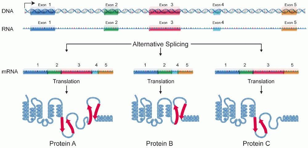
A protein isoform, or "protein variant", is a member of a set of highly similar proteins that originate from a single gene or gene family and are the result of genetic differences. While many perform the same or similar biological roles, some isoforms have unique functions. A set of protein isoforms may be formed from alternative splicings, variable promoter usage, or other post-transcriptional modifications of a single gene; post-translational modifications are generally not considered. (For that, see Proteoforms.) Through RNA splicing mechanisms, mRNA has the ability to select different protein-coding segments (exons) of a gene, or even different parts of exons from RNA to form different mRNA sequences. Each unique sequence produces a specific form of a protein. The discovery of isoforms could explain the discrepancy between the small number of protein coding regions genes revealed by the human genome project and the large diversity of proteins seen in an organism: differen ... [...More Info...] [...Related Items...] OR: [Wikipedia] [Google] [Baidu] |


