|
Anatomical Simulation
Anatomy () is the branch of morphology concerned with the study of the internal structure of organisms and their parts. Anatomy is a branch of natural science that deals with the structural organization of living things. It is an old science, having its beginnings in prehistoric times. Anatomy is inherently tied to developmental biology, embryology, comparative anatomy, evolutionary biology, and phylogeny, as these are the processes by which anatomy is generated, both over immediate and long-term timescales. Anatomy and physiology, which study the structure and function of organisms and their parts respectively, make a natural pair of related disciplines, and are often studied together. Human anatomy is one of the essential basic sciences that are applied in medicine, and is often studied alongside physiology. Anatomy is a complex and dynamic field that is constantly evolving as discoveries are made. In recent years, there has been a significant increase in the use of advan ... [...More Info...] [...Related Items...] OR: [Wikipedia] [Google] [Baidu] |
Macroscopic
The macroscopic scale is the length scale on which objects or phenomena are large enough to be visible with the naked eye, without magnifying optical instruments. It is the opposite of microscopic. Overview When applied to physical phenomena and bodies, the macroscopic scale describes things as a person can directly perceive them, without the aid of magnifying devices. This is in contrast to observations ( microscopy) or theories ( microphysics, statistical physics) of objects of geometric lengths smaller than perhaps some hundreds of micrometres. A macroscopic view of a ball is just that: a ball. A microscopic view could reveal a thick round skin seemingly composed entirely of puckered cracks and fissures (as viewed through a microscope) or, further down in scale, a collection of molecules in a roughly spherical shape (as viewed through an electron microscope). An example of a physical theory that takes a deliberately macroscopic viewpoint is thermodynamics. An exampl ... [...More Info...] [...Related Items...] OR: [Wikipedia] [Google] [Baidu] |
Magnetic Resonance Imaging
Magnetic resonance imaging (MRI) is a medical imaging technique used in radiology to generate pictures of the anatomy and the physiological processes inside the body. MRI scanners use strong magnetic fields, magnetic field gradients, and radio waves to form images of the organs in the body. MRI does not involve X-rays or the use of ionizing radiation, which distinguishes it from computed tomography (CT) and positron emission tomography (PET) scans. MRI is a medical application of nuclear magnetic resonance (NMR) which can also be used for imaging in other NMR applications, such as NMR spectroscopy. MRI is widely used in hospitals and clinics for medical diagnosis, staging and follow-up of disease. Compared to CT, MRI provides better contrast in images of soft tissues, e.g. in the brain or abdomen. However, it may be perceived as less comfortable by patients, due to the usually longer and louder measurements with the subject in a long, confining tube, although ... [...More Info...] [...Related Items...] OR: [Wikipedia] [Google] [Baidu] |
Ultrasound Imaging
Medical ultrasound includes diagnostic techniques (mainly imaging) using ultrasound, as well as therapeutic applications of ultrasound. In diagnosis, it is used to create an image of internal body structures such as tendons, muscles, joints, blood vessels, and internal organs, to measure some characteristics (e.g., distances and velocities) or to generate an informative audible sound. The usage of ultrasound to produce visual images for medicine is called medical ultrasonography or simply sonography, or echography. The practice of examining pregnant women using ultrasound is called obstetric ultrasonography, and was an early development of clinical ultrasonography. The machine used is called an ultrasound machine, a sonograph or an echograph. The visual image formed using this technique is called an ultrasonogram, a sonogram or an echogram. Ultrasound is composed of sound waves with frequencies greater than 20,000 Hz, which is the approximate upper threshold of hu ... [...More Info...] [...Related Items...] OR: [Wikipedia] [Google] [Baidu] |
Radiography
Radiography is an imaging technology, imaging technique using X-rays, gamma rays, or similar ionizing radiation and non-ionizing radiation to view the internal form of an object. Applications of radiography include medical ("diagnostic" radiography and "therapeutic radiography") and industrial radiography. Similar techniques are used in airport security, (where "body scanners" generally use backscatter X-ray). To create an image in conventional radiography, a beam of X-rays is produced by an X-ray generator and it is projected towards the object. A certain amount of the X-rays or other radiation are absorbed by the object, dependent on the object's density and structural composition. The X-rays that pass through the object are captured behind the object by a X-ray detector, detector (either photographic film or a digital detector). The generation of flat two-dimensional images by this technique is called Projection radiography, projectional radiography. In computed tomography (C ... [...More Info...] [...Related Items...] OR: [Wikipedia] [Google] [Baidu] |
Medical Imaging
Medical imaging is the technique and process of imaging the interior of a body for clinical analysis and medical intervention, as well as visual representation of the function of some organs or tissues (physiology). Medical imaging seeks to reveal internal structures hidden by the skin and bones, as well as to diagnose and treat disease. Medical imaging also establishes a database of normal anatomy and physiology to make it possible to identify abnormalities. Although imaging of removed organ (anatomy), organs and Tissue (biology), tissues can be performed for medical reasons, such procedures are usually considered part of pathology instead of medical imaging. Measurement and recording techniques that are not primarily designed to produce images, such as electroencephalography (EEG), magnetoencephalography (MEG), electrocardiography (ECG), and others, represent other technologies that produce data susceptible to representation as a parameter graph versus time or maps that contain ... [...More Info...] [...Related Items...] OR: [Wikipedia] [Google] [Baidu] |
Cadaver
A cadaver, often known as a corpse, is a Death, dead human body. Cadavers are used by medical students, physicians and other scientists to study anatomy, identify disease sites, determine causes of death, and provide tissue (biology), tissue to repair a defect in a living human being. Students in medical school study and dissect cadavers as a part of their education. Others who study cadavers include archaeologists and arts students. In addition, a cadaver may be used in the development and evaluation of surgical instruments. The term ''cadaver'' is used in courts of law (and, to a lesser extent, also by media outlets such as newspapers) to refer to a dead body, as well as by recovery teams searching for bodies in natural disasters. The word comes from the Latin word ''cadere'' ("to fall"). Related terms include ''cadaverous'' (resembling a cadaver) and ''cadaveric spasm'' (a muscle spasm causing a dead body to twitch or jerk). A cadaver graft (also called “postmortem graft”) ... [...More Info...] [...Related Items...] OR: [Wikipedia] [Google] [Baidu] |
History Of Anatomy
The history of anatomy spans from the earliest examinations of sacrifice, sacrificial victims to the advanced studies of the human body conducted by modern scientists. Written descriptions of human organs and parts can be traced back thousands of years to ancient Ancient Egyptian anatomical studies, Egyptian papyri, where attention to the body was necessitated by their highly elaborate Ancient Egyptian funerary practices, burial practices. Theoretical considerations of the structure and function of the human body did not develop until far later, in ancient Greece. Ancient Greek philosophers, like Alcmaeon of Croton, Alcmaeon and Empedocles, and ancient Greek doctors, like Hippocrates and Hippocratic Corpus, his school, paid attention to the causes of life, disease, and different functions of the body. Aristotle advocated dissection of animals as part of his program for understanding the Four causes, causes of biological Aristotle's theory of universals, forms. During the Hellenist ... [...More Info...] [...Related Items...] OR: [Wikipedia] [Google] [Baidu] |
Cell Biology
Cell biology (also cellular biology or cytology) is a branch of biology that studies the structure, function, and behavior of cells. All living organisms are made of cells. A cell is the basic unit of life that is responsible for the living and functioning of organisms. Cell biology is the study of the structural and functional units of cells. Cell biology encompasses both prokaryotic and eukaryotic cells and has many subtopics which may include the study of cell metabolism, cell communication, cell cycle, biochemistry, and cell composition. The study of cells is performed using several microscopy techniques, cell culture, and cell fractionation. These have allowed for and are currently being used for discoveries and research pertaining to how cells function, ultimately giving insight into understanding larger organisms. Knowing the components of cells and how cells work is fundamental to all biological sciences while also being essential for research in biomedical fiel ... [...More Info...] [...Related Items...] OR: [Wikipedia] [Google] [Baidu] |
Histology
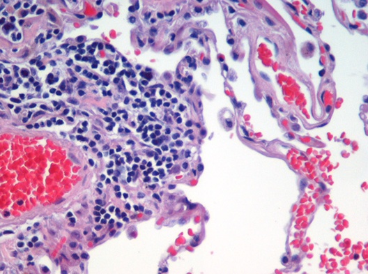
Histology, also known as microscopic anatomy or microanatomy, is the branch of biology that studies the microscopic anatomy of biological tissue (biology), tissues. Histology is the microscopic counterpart to gross anatomy, which looks at larger structures visible without a microscope. Although one may divide microscopic anatomy into ''organology'', the study of organs, ''histology'', the study of tissues, and ''cytology'', the study of cell (biology), cells, modern usage places all of these topics under the field of histology. In medicine, histopathology is the branch of histology that includes the microscopic identification and study of diseased tissue. In the field of paleontology, the term paleohistology refers to the histology of fossil organisms. Biological tissues Animal tissue classification There are four basic types of animal tissues: muscle tissue, nervous tissue, connective tissue, and epithelial tissue. All animal tissues are considered to be subtypes of these ... [...More Info...] [...Related Items...] OR: [Wikipedia] [Google] [Baidu] |
Tissue (biology)
In biology, tissue is an assembly of similar cells and their extracellular matrix from the same embryonic origin that together carry out a specific function. Tissues occupy a Biological organisation#Levels, biological organizational level between cell (biology), cells and a complete organ (biology), organ. Accordingly, organs are formed by the functional grouping together of multiple tissues. The English word "tissue" Morphological derivation, derives from the French word "", the past participle of the verb tisser, "to weave". The study of tissues is known as histology or, in connection with disease, as histopathology. Xavier Bichat is considered as the "Father of Histology". Plant histology is Studied Space Shuttle designs, studied in both plant anatomy and Plant physiology, physiology. The classical tools for studying tissues are the Microtome#Applications, paraffin block in which tissue is embedded and then sectioned, the staining, histological stain, and the Microscope, o ... [...More Info...] [...Related Items...] OR: [Wikipedia] [Google] [Baidu] |


