|
Ambolestes
''Ambolestes'' is an extinct genus of eutherian mammal from the Early Cretaceous of China. It includes a single species, ''Ambolestes zhoui'', known from a single complete skeleton recovered from the Yixian Formation (126 Ma), part of the fossiliferous Jehol biota. ''Ambolestes'' is one of the most basal eutherians, presenting a combination of features from both early eutherians (stem-placentals) and early metatherians (stem-marsupials). This is responsible for the generic name of ''Ambolestes'': "''ambo''" is Latin for "both", while "-''lestes''" (Greek for "robber") is a popular suffix for fossil mammals. The species name honors influential Jehol paleontologist Zhou Zhonghe. Description ''Ambolestes'' was a fairly small mammal, with an estimated mass of 34–44 g (about the size of a modern mouse opossum, ''Marmosa''). It was likely similar in appearance and habits to other putative Yixian Formation therians, such as ''Eomaia'' and ''Sinodelphys''. There are several simila ... [...More Info...] [...Related Items...] OR: [Wikipedia] [Google] [Baidu] |
Eutherian
Eutheria (from Greek , 'good, right' and , 'beast'; ), also called Pan-Placentalia, is the clade consisting of placental mammals and all therian mammals that are more closely related to placentals than to marsupials. Eutherians are distinguished from non-eutherians by various phenotypic traits of the feet, ankles, jaws and teeth. All extant eutherians lack epipubic bones, which are present in all other living mammals (marsupials and monotremes). This allows for expansion of the abdomen during pregnancy, though epipubic bones are present in many primitive eutherians. Eutheria was named in 1872 by Theodore Gill; in 1880, Thomas Henry Huxley defined it to encompass a more broadly defined group than Placentalia. The earliest unambiguous eutherians are known from the Early Cretaceous Yixian Formation of China, dating around 120 million years ago. Two tribosphenic mammals, '' Durlstodon'' and '' Durlstotherium'' from the Berriasian age (~145–140 million years ago) of the Ear ... [...More Info...] [...Related Items...] OR: [Wikipedia] [Google] [Baidu] |
Eomaia
''Eomaia'' ("dawn mother") is a genus of extinct fossil mammals containing the single species ''Eomaia scansoria'', discovered in rocks that were found in the Yixian Formation, Liaoning Province, China, and dated to the Barremian Age of the Lower Cretaceous about . The single fossil specimen of this species is in length and virtually complete. An estimate of the body weight is . It is exceptionally well-preserved for a 125-million-year-old specimen. Although the fossil's skull is squashed flat, its teeth, tiny foot bones, cartilages and even its fur are visible. Description The ''Eomaia'' fossil shows clear traces of hair. However, this is not the earliest clear evidence of hair in the mammalian lineage, as fossils of '' Volaticotherium'', and the docodont ''Castorocauda'', discovered in rocks dated to about , also have traces of fur. ''Eomaia scansoria'' possessed several features in common with placental mammals that distinguish them from metatherians, the group that include ... [...More Info...] [...Related Items...] OR: [Wikipedia] [Google] [Baidu] |
Early Cretaceous
The Early Cretaceous (geochronology, geochronological name) or the Lower Cretaceous (chronostratigraphy, chronostratigraphic name) is the earlier or lower of the two major divisions of the Cretaceous. It is usually considered to stretch from 143.1 Megaannum#SI prefix multipliers, Ma to 100.5 Ma. Geology Proposals for the exact age of the Barremian–Aptian boundary ranged from 126 to 117 Ma until recently (as of 2019), but based on drillholes in Svalbard the defining Anoxic event#Cretaceous, early Aptian Oceanic Anoxic Event 1a (OAE1a) was dated to 123.1±0.3 Ma, limiting the possible range for the boundary to c. 122–121 Ma. There is a possible link between this anoxic event and a series of Early Cretaceous large igneous provinces (LIP). The Ontong Java Plateau, Ontong Java-Manihiki Plateau, Manihiki-Hikurangi Plateau, Hikurangi large igneous province, emplaced in the South Pacific at c. 120 Ma, is by far the largest LIP in Earth's history. The Onto ... [...More Info...] [...Related Items...] OR: [Wikipedia] [Google] [Baidu] |
Premolar
The premolars, also called premolar Tooth (human), teeth, or bicuspids, are transitional teeth located between the Canine tooth, canine and Molar (tooth), molar teeth. In humans, there are two premolars per dental terminology#Quadrant, quadrant in the permanent teeth, permanent set of teeth, making eight premolars total in the mouth. They have at least two Cusp (dentistry), cusps. Premolars can be considered transitional teeth during chewing, or mastication. They have properties of both the canines, that lie anterior and molars that lie Posterior (anatomy), posterior, and so food can be transferred from the canines to the premolars and finally to the molars for grinding, instead of directly from the canines to the molars. Human anatomy The premolars in humans are the maxillary first premolar, maxillary second premolar, mandibular first premolar, and the mandibular second premolar. Premolar teeth by definition are permanent teeth Anatomical terms of location#Proximal and distal, ... [...More Info...] [...Related Items...] OR: [Wikipedia] [Google] [Baidu] |
Middle Ear
The middle ear is the portion of the ear medial to the eardrum, and distal to the oval window of the cochlea (of the inner ear). The mammalian middle ear contains three ossicles (malleus, incus, and stapes), which transfer the vibrations of the eardrum into waves in the fluid and membranes of the inner ear. The hollow space of the middle ear is also known as the tympanic cavity and is surrounded by the tympanic part of the temporal bone. The auditory tube (also known as the Eustachian tube or the pharyngotympanic tube) joins the tympanic cavity with the nasal cavity ( nasopharynx), allowing pressure to equalize between the middle ear and throat. The primary function of the middle ear is to efficiently transfer acoustic energy from compression waves in air to fluid–membrane waves within the cochlea. Structure Ossicles The middle ear contains three tiny bones known as the ossicles: '' malleus'', ''incus'', and '' stapes''. The ossicles were given their Latin names f ... [...More Info...] [...Related Items...] OR: [Wikipedia] [Google] [Baidu] |
Ectotympanic
The ectotympanic, or tympanicum, is a bony structure found in all mammals, located on the tympanic part of the temporal bone, which holds the tympanic membrane (eardrum) in place. In catarrhine primates (including humans), it takes a tube-shape. Its position and attachment to the skull vary between primates, and can be either inside or outside the auditory bulla. It is homologous with the angular bone of non-mammalian tetrapods A tetrapod (; from Ancient Greek τετρα- ''(tetra-)'' 'four' and πούς ''(poús)'' 'foot') is any four- limbed vertebrate animal of the clade Tetrapoda (). Tetrapods include all extant and extinct amphibians and amniotes, with the lat .... When the latter is present, it contacts the entotympanic. References External links webref: Anthropology * * Skull bones Mammal anatomy {{Vertebrate anatomy-stub ... [...More Info...] [...Related Items...] OR: [Wikipedia] [Google] [Baidu] |
Trapezium (bone)
The trapezium bone (greater multangular bone) is a carpal bone in the hand. It forms the radial border of the carpal tunnel. Structure The trapezium is distinguished by a deep groove on its anterior surface. It is situated at the radial side of the carpus, between the scaphoid and the first metacarpal bone (the metacarpal bone of the thumb). It is homologous with the first distal carpal of reptiles and amphibians. Surfaces The trapezium is an irregular-shaped carpal bone found within the hand. The trapezium is found within the distal row of carpal bones, and is directly adjacent to the metacarpal bone of the thumb. On its ulnar surface are found the trapezoid and scaphoid bones. The '' superior surface'' is directed upward and medialward; medially it is smooth, and articulates with the scaphoid; laterally it is rough and continuous with the lateral surface. The '' inferior surface'' is oval, concave from side to side, convex from before backward, so as to form a saddle- ... [...More Info...] [...Related Items...] OR: [Wikipedia] [Google] [Baidu] |
Mandibular Angle
__NOTOC__ The angle of the mandible (a.k.a. gonial angle, Masseteric Tuberosity, and Masseteric Insertion) is located at the posterior border at the junction of the lower border of the ramus of the mandible. The angle of the mandible, which may be either inverted or everted, is marked by rough, oblique ridges on each side, for the attachment of the masseter laterally, and the pterygoideus internus (medial pterygoid muscle) medially; the stylomandibular ligament is attached to the angle between these muscles. The forensic term for the midpoint of the mandibular angle is the gonion. The gonion is a cephalometric landmark located at the lowest, posterior, and lateral point on the angle. This site is at the apex of the maximum curvature of the mandible, where the ascending ramus becomes the body of the mandible. The mandibular angle has been named as a forensic tool for gender determination, but some studies have called into question whether there is any significant sex differe ... [...More Info...] [...Related Items...] OR: [Wikipedia] [Google] [Baidu] |
Triquetral Bone
The triquetral bone (; also called triquetrum, pyramidal, three-faced, and formerly cuneiform bone) is located in the wrist on the medial side of the proximal row of the carpus between the lunate and pisiform bones. It is on the ulnar side of the hand, but does not directly articulate with the ulna. Instead, it is connected to and articulates with the ulna through the Triangular fibrocartilage discManaster, B. J., Julia Crim "Imaging Anatomy: Musculoskeletal E-Book" Elsevier Health Sciences, 2016, p. 326. and ligament, which forms part of the ulnocarpal joint capsule. It connects with the pisiform, hamate, and lunate bones. It is the 2nd most commonly fractured carpal bone. Structure The triquetral is one of the eight carpal bones of the hand. It is a three-faced bone found within the proximal row of carpal bones. Situated beneath the pisiform, it is one of the carpal bones that form the carpal arch, within which lies the carpal tunnel. The triquetral bone may be distingui ... [...More Info...] [...Related Items...] OR: [Wikipedia] [Google] [Baidu] |
Hamate
The hamate bone (from Latin language, Latin wiktionary:hamatus, hamatus, "hooked"), or unciform bone (from Latin language, Latin ''wikt:uncus, uncus'', "hook"), Latin os hamatum and occasionally abbreviated as just hamatum, is a bone in the human wrist readily distinguishable by its wedge shape and a hook-like process ("hamulus") projecting from its Radioulnar, palmar surface. Structure The hamate is an irregularly shaped carpal bone found within the hand. The hamate is found within the distal row of carpal bones, and abuts the metacarpals of the little finger and ring finger. Adjacent to the hamate on the ulnar side, and slightly proximal and ulnar to it, is the pisiform bone. Adjacent on the radial side is the capitate, and proximal is the lunate bone. Surfaces The hamate bone has six surfaces: * The ''superior'', the apex of the wedge, is narrow, convex, smooth, and articulates with the lunate bone, lunate. * The ''inferior'' articulates with the fourth and fifth metacarpal ... [...More Info...] [...Related Items...] OR: [Wikipedia] [Google] [Baidu] |
Scaphoid
The scaphoid bone is one of the carpal bones of the wrist. It is situated between the hand and forearm on the thumb side of the wrist (also called the lateral or radial side). It forms the radial border of the carpal tunnel. The scaphoid bone is the largest bone of the proximal row of wrist bones, its long axis being from above downward, lateralward, and forward. It is approximately the size and shape of a medium cashew nut. Structure The scaphoid is situated between the proximal and distal rows of carpal bones. It is located on the radial side of the wrist, adjacent to the styloid process of the radius. It articulates with the radius, lunate, trapezoid, trapezium, and capitate. Over 80% of the bone is covered in articular cartilage. Bone The palmar surface of the scaphoid is concave, and forming a distal tubercle, giving attachment to the transverse carpal ligament. The proximal surface is triangular, smooth and convex. The lateral surface is narrow and gives attachm ... [...More Info...] [...Related Items...] OR: [Wikipedia] [Google] [Baidu] |
Canine Tooth
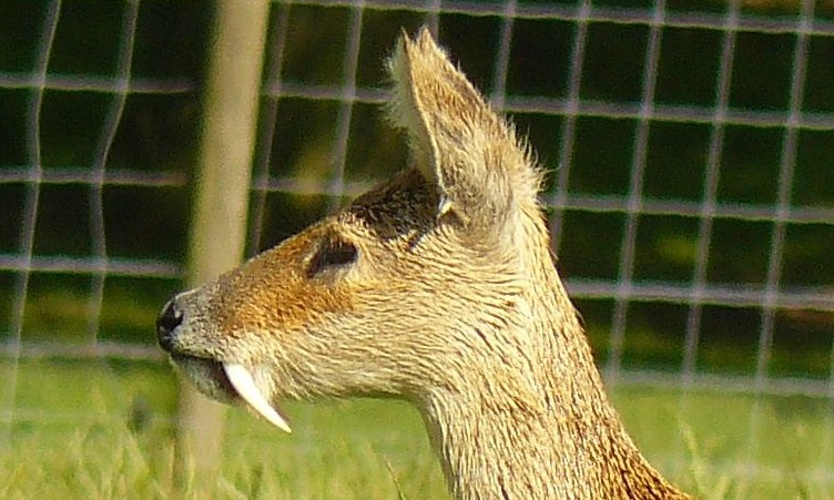
In mammalian oral anatomy, the canine teeth, also called cuspids, dogteeth, eye teeth, vampire teeth, or fangs, are the relatively long, pointed teeth. In the context of the upper jaw, they are also known as '' fangs''. They can appear more flattened, however, causing them to resemble incisors and leading them to be called ''incisiform''. They developed and are used primarily for firmly holding food in order to tear it apart, and occasionally as weapons. They are often the largest teeth in a mammal's mouth. Individuals of most species that develop them normally have four, two in the upper jaw and two in the lower, separated within each jaw by incisors; humans and dogs are examples. In most species, canines are the anterior-most teeth in the maxillary bone. The four canines in humans are the two upper maxillary canines and the two lower mandibular canines. They are specially prominent in dogs (Canidae), hence the name. Details There are generally four canine teeth: two ... [...More Info...] [...Related Items...] OR: [Wikipedia] [Google] [Baidu] |


